 Tools
Tools
 Bioprosthetic Aortic Valve Replacement
Introduction
Native heart valves are remarkably durable, functioning under the relentless mechanical stress of approximately 80 beats per minute for 7 decades and exceeding 3 billion cardiac cycles over a lifetime. Despite this extraordinary endurance, clinically significant valvular heart disease affects nearly 2% of the population and remains a major cause of morbidity and mortality worldwide.[1] The evolution of valvular heart disease management reflects decades of surgical innovation and multidisciplinary collaboration, progressing from early closed digital commissurotomies and the first ball-and-cage prostheses to modern valve replacement techniques. In contrast, advances in preventive medicine, such as the recognition that prompt treatment of streptococcal pharyngitis prevents rheumatic heart disease, have further transformed patient outcomes.
Bioprosthetic aortic valve replacement has become a cornerstone in the treatment of severe aortic stenosis, aortic regurgitation, and destructive aortic valve endocarditis requiring surgical intervention. Surgical aortic valve replacement (SAVR) with a bioprosthetic valve utilizes highly processed bovine or porcine tissue leaflets mounted on a supporting frame, providing excellent hemodynamic performance without the lifelong anticoagulation required for mechanical prostheses. Appropriate patient selection, understanding the advantages and limitations of bioprosthetic versus mechanical valves, meticulous operative technique, and comprehensive perioperative management are essential to achieving optimal outcomes. This activity reviews the indications, evaluation, surgical principles, postoperative management, complications, and interprofessional care required for successful bioprosthetic aortic valve replacement.
Anatomy and Physiology
Register For Free And Read The Full Article
 Search engine and full access to all medical articles
- 10 free questions in your specialty
- Free CME/CE Activities
- Free daily question in your email
- Save favorite articles to your dashboard
- Emails offering discounts
Learn more about a Subscription to StatPearls Point-of-Care
Anatomy and Physiology
When considering the replacement of an aortic valve, it is essential to consider the form and function of the native valve. The aortic valve separates 2 remarkably different pressure zones over a height of approximately 12 to 18 mm. The ventricular side contracts in systole while the aortic side expands; then, in diastole, the ventricular side relaxes while the aortic side contracts somewhat due to aortic elasticity. The aortic valve must completely separate and insulate these 2 pressure zones, then it should be able to rapidly reverse that separation and equalize the pressure gradient, oscillating between these 2 states fluidly. The aortic valve must also protect the coronary ostia and promote blood flow to them.[2]
Tracing flow from the left ventricle, the first landmark is the ventriculo-aortic junction (VAJ), where ventricular myocardium transitions to aortic tissue; in cross-sectional views, it corresponds to the line joining the nadirs of the 3 cusps. More distally, the cusp hinge lines ascend and attach to the aortic wall; this attachment plane is termed the basal ring or surgical aortic annulus. This “annulus” is not a discrete, uniform ring; it is mildly ovoid and irregular because each cusp differs in height, angulation, and insertion, producing variable separation between the VAJ and the basal ring. This creates regions of “ventricle within the sinus,” conferring additional root compliance and allowing approximately 18% to 20% systolic expansion.[3]
Beyond the valve leaflets, the superior margin of cusp attachment defines the sinotubular junction (STJ), which demarcates the terminus of the aortic root and the onset of the tubular ascending aorta. The lumen expands modestly from the ventriculo-aortic junction (VAJ) to the STJ, then narrows as it transitions into the ascending aorta (see Image. Anatomic Landmarks of the Aortic Root). Posterior to each cusp, this expansion forms a sinus of Valsalva. The right sinus gives rise to the right coronary artery and the left sinus to the left coronary artery, whereas the posterior (noncoronary) sinus lacks a coronary origin. Coronary ostial variants occur in approximately 1% of individuals, commonly including separate left anterior descending (LAD) and circumflex (LCx) ostia or an LCx arising from the right coronary system.[3]
The geometry of these sinuses allows eddy currents to form between the bulged wall of the aortic root and the valve leaflets, preventing the valve from getting stuck on the aortic wall and thus keeping it slightly open for blood flow into the coronaries and in a ready position to close instantly at the end of systole.[2] The valve leaflets themselves are covered by endocardial endothelial cells and interspersed with valve interstitial cells that are quiescently resting through the 3 layers: elastin-rich ventricularis towards the ventricle, proteoglycan-rich spongiosa in the middle, and collagen-rich fibrinosa towards the aorta.[4]
The aortic valve has several surgically relevant anatomic relationships that must be considered to avoid injury. The noncoronary and the left coronary cusps of the aortic valve are structurally contiguous with the strongest fibrous tissue of the heart, the central fibrous body. Still, as such, they are also structurally continuous with the anterior leaflet of the mitral valve. Directly underneath the junction of the right coronary cusp and the noncoronary cusp is the ventricular membranous septum and the atrioventricular node. The junction of the right and left coronary cusps is externally continuous with the pulmonary artery. Quite proximal to the ostium of the right coronary is the branch point of the sinoatrial nodal artery. A network of tiny coronary veins may be present around the ostium of the left coronary.[3]
The pathophysiology of the aortic valve stems from either stenosis or insufficiency (regurgitation), and both are failures of the valve to insulate the 2 pressure zones, the heart and the aorta, from each other. A stenotic valve (with less than half the normal 3 to 5 cm² surface area) exposes the ventricle to a pressure gradient, leading to ventricular wall stress. Wall stress sets the heart up for a compensatory cycle of hypertrophy that minimizes wall stress at the ultimate expense of impaired coronary blood flow, fibrosis, and further wall stress.[5] An insufficient (or regurgitant) valve promotes equalization of pressure between the heart and the aorta, leading to wall stress and a compensatory cycle of diastolic relaxation, some hypertrophy, and ultimately impaired coronary blood flow and forward failure.[6]
Aortic stenosis is the most common valvular pathology requiring correction in high-income countries, with most cases due to senile degenerative calcification of normal trileaflet valves or degeneration of congenital bicuspid valves.[5][7] In low- and middle-income countries, rheumatic heart disease is still common and accounts for most valvular pathology. Rheumatic heart disease most commonly afflicts the mitral valve, but to a lesser extent, it also can cause aortic stenosis.[8]
Aortic insufficiency needing correction is less common than stenosis and may be caused by myxomatous disease, connective tissue disorders, aortic dissection, or infective endocarditis.[9] When a degenerative valve is so heavily calcified that it no longer opens well and fails to close properly, both stenosis and insufficiency are often present.[5][7] In broad, oversimplified terms, aortic stenosis is correctable by aortic valve replacement, while isolated aortic insufficiency may be correctable by aortic valve replacement or repair.
Indications
One of the difficulties with valvular heart disease is determining when patients need an intervention. For example, in aortic stenosis, only half of the patients have symptoms on presentation, yet half of them may die within 2 years.[5] Thus, the indicated timing for intervention is crucial. The American College of Cardiology (ACC) and the American Heart Association (AHA) published consensus guidelines in 2014, updated in 2017, which serve as standard indications for the management of valvular heart disease, as summarized below.[9][10]
Valvular heart disease is staged A through D, based on echocardiography parameters:
- Stage A
- At-risk features (ie, bicuspid valve), while asymptomatic with normal parameters
- Stage B
- Asymptomatic with moderate parameters
- Stage C1
- Severe parameters yet still asymptomatic due to preserved ejection fraction
- Stage C2
- Severe parameters yet still asymptomatic at rest despite reduced ejection fraction
- Stage D
- Symptomatic with or without severe parameters
Severe stenosis is defined by aortic valve area less than 1.0 cm2, mean pressure gradient of 40 mm Hg or more, and aortic jet velocity of 4.0 m/sec or more. Severe parameters for insufficiency include regurgitant jet width of 65% or more of the left ventricular outflow tract, vena contracta greater than 0.6 cm, holodiastolic aortic flow reversal, effective regurgitant orifice of 0.3 cm2 or greater, regurgitant fraction of 50% or more, and regurgitant volume of 60 mL/beat or greater. In general, patients with aortic valve stage A receive a screening echocardiogram; patients with stage B disease receive echocardiogram surveillance every 3 to 5 years; patients with stage C disease receive echocardiogram surveillance every 0.5 to 1 year with possible intervention; and patients with stage D disease receive an intervention. A heart valve team should assess any patient considered for intervention in a multidisciplinary fashion. Class I and class II indications for intervention for aortic stenosis and aortic insufficiency are summarized below.
Aortic Stenosis Class I
- Stage D (severe parameters + symptomatic)
- Stage C or D (severe parameters regardless of symptoms) if already indicated for another heart surgery
- Stage C or D (severe parameters regardless of symptoms) if ejection fraction <50%
Aortic Stenosis Class II
- Stage C with very severe parameters (jet velocity >5.0 cm/sec)
- Stage C with decreased exercise tolerance
- Stage D without severe parameters (low-flow/low-gradient), but low-dose dobutamine stress produces high-flow/high-gradient parameters.
- Stage D without severe parameters, but anatomic and clinical evidence suggest stenosis is the underlying cause of symptoms.
- Stage B if already indicated for another heart surgery
- Stage C if the low-risk patient and jet velocity is increasing >0.3 cm/sec/year
Aortic Insufficiency Class I
- Stage D (severe parameters + symptomatic)
- Stage C or D (severe parameters regardless of symptoms) if already indicated for another heart surgery
- Stage C or D (severe parameters regardless of symptoms) if ejection fraction <50%
Aortic Insufficiency Class II
- Stage C with pronounced left ventricular dilation (>50 mm at end-diastole)
- Stage B if already indicated for another heart surgery
Once a patient meets the indications for an intervention, the heart valve team will assess the patient’s operative and interventional risk using a combination of Society of Thoracic Surgeons (STS) risk calculators, frailty indices, and a patient-specific accounting of organ failures and comorbidities to determine whether SAVR or transcatheter aortic valve replacement (TAVR) is appropriate.
For patients with stage B or C disease indications above (ie, asymptomatic patients), SAVR is the only indicated intervention at this time. For patients with stage D disease, either SAVR or TAVR may be an option, depending on the above risk. For low risk (<4% operative mortality), SAVR is indicated. For intermediate risk (4%–8% operative mortality), either SAVR may be offered as a class I recommendation or TAVR as a class II recommendation. For high-risk patients (>8% operative mortality), SAVR and TAVR have equivalent class I recommendations. For prohibitive surgical risk (>50% mortality or ≥3 organ systems already failed), TAVR carries the class I recommendation, and SAVR should not be offered.[10]
Once SAVR is chosen, the team must decide between valve replacement and repair. Again, in broad terms, stenosis is correctable by replacement, while isolated insufficiency may be correctable by replacement or repair. Some groups have also shown success in repairing bicuspid disease.[11] Whether repair is an option depends on the valve's quality and the availability of specific surgical expertise. The Aortic Valve Repair versus Mechanical Valve Replacement for Root Aneurysm study, originally called Conservation Aortique Valvulaire dans les Insuffisances Aortiques et les Anévrismes de la Racine aortique (CAVIAAR), has revealed that in the first 4 years after surgery, standardized aortic valve repair is at least as safe as mechanical valve replacement and is associated with fewer valve-related deaths and major bleeding, without more reoperations.[12]
Once a replacement is chosen, there are 3 options: a mechanical valve, a stentless pulmonary autograft/homograft (Ross procedure), or a bioprosthetic valve. Each option varies in requirements for anticoagulation and long-term durability. This variability should be clearly communicated to the patient in accordance with the ACC/AHA guidelines, and the decision-making process should be shared with the patient.
In contrast, previously, patient age heavily influenced the choice.[10] Age still plays a role in the decision process: the historically demonstrated long-term durability of mechanical valves is favored for patients under 50. In contrast, bioprosthetic valves are favored for patients over 70. The Ross procedure is preferred for young patients with a contraindication to anticoagulation; otherwise, all other patients with a contraindication to anticoagulation are recommended to have a bioprosthetic valve.[10][13]
Other variables must be considered when selecting a replacement valve. Pre-existing renal disease, hyperparathyroidism, and young age all increase the risk of calcium deposition and bioprosthetic structural deterioration.[14] Lack of access to regular international normalized ratio (INR) assays favors the use of bioprosthetic valves. Comorbid conditions requiring anticoagulation, high risk for reoperation (eg, prior chest radiation or porcelain aorta), and a small aortic root size (if a future valve-in-valve TAVR procedure is needed) all favor a mechanical prosthesis.[10]
Not to be omitted in a discussion of valvular disease, surgical treatment of infective endocarditis cannot be over-generalized; it must be tailored to the full scenario of the particular patient, ie, how much should be debrided and when to perform the debridement. The ACC/AHA offers some basic guidelines.[9] A full, culture data driven course of antibiotics should be the first-line treatment until 1 of the following scenarios pushes the multidisciplinary team toward early surgical source control: deteriorating heart failure, highly virulent or resistant organisms, new heart block, annular abscess, relapsing infection, persistent bacteremia for 5 to 7 days, presence of a prosthetic valve infection, infected defibrillator, pacemaker, or leads, recurrent emboli, persistent vegetations despite adequate antibiotic therapy, or mobile vegetation of more than 1 cm. In the guidelines, it is not mandatory to obtain negative blood cultures before proceeding with valve replacement. Still, given the need to implant a foreign body that can be seeded by infection, judicious clinical judgment is warranted.
Contraindications
There are no absolute contraindications to bioprosthetic SAVR when surgery is lifesaving; however, several patient-specific factors may favor alternative treatment strategies or valve types. Patients with prohibitive or excessively high operative risk, as determined by multidisciplinary heart team assessment incorporating surgical risk scores, frailty, comorbidities, and anatomic considerations, are generally better served with TAVR or medical management rather than SAVR.[10]
Pregnancy presents unique challenges in the management of severe aortic valve disease. Cardiac surgery requiring cardiopulmonary bypass during pregnancy carries substantial maternal and fetal risks and should be avoided whenever possible. Even in patients with stage D aortic stenosis or regurgitation, intervention during pregnancy is generally reserved for severe, refractory heart failure or other life-threatening complications.
Women with severe symptomatic aortic valve disease should ideally undergo definitive valve intervention before conception.[9] Valve selection also requires careful consideration in women of childbearing potential. Bioprosthetic valves are associated with accelerated structural valve degeneration in younger patients and may deteriorate more rapidly during pregnancy, potentially necessitating earlier reintervention.[14] Conversely, mechanical prostheses require lifelong anticoagulation; although warfarin is teratogenic during the first trimester, several anticoagulation strategies can optimize maternal and fetal outcomes throughout pregnancy.
In patients undergoing surgery for active infective endocarditis, minimizing the implantation of prosthetic material is often desirable. Although bioprosthetic valve replacement is not contraindicated, homograft aortic root replacement may be preferred in selected patients with extensive periannular infection, root abscess, or destructive endocarditis, as homografts may offer greater resistance to recurrent infection.[15] Neurologic complications also influence operative timing. When feasible, valve surgery should generally be delayed for approximately 2 to 3 weeks after an ischemic stroke and approximately 4 weeks after an intracranial hemorrhage to reduce the risk of neurologic deterioration. However, emergent surgery may still be required in patients with uncontrolled infection or hemodynamic instability.[15]
Bioprosthetic valves may also be unacceptable for patients with personal, cultural, ethical, or religious objections to the use of porcine or bovine tissue-derived implants. In these circumstances, alternative prosthetic options should be discussed through shared decision-making while respecting patient values and preferences.[16][17] Patients with a limited life expectancy unrelated to valvular disease or those unlikely to derive meaningful functional benefit from valve replacement because of advanced frailty, severe irreversible comorbidities, or poor overall prognosis may not be appropriate candidates for bioprosthetic SAVR. In these cases, the risks of surgery may outweigh the anticipated benefits, and palliative or conservative management should be considered through multidisciplinary discussion.
Preparation
Preoperative preparation for bioprosthetic SAVR begins with a comprehensive evaluation to confirm the diagnosis, assess disease severity, define the underlying pathology, and determine operative risk. At a minimum, all patients should undergo a thorough history and physical examination, chest radiography, and transthoracic echocardiography (TTE) with 2-dimensional and Doppler imaging to characterize valve morphology, quantify stenosis or regurgitation, assess left ventricular size and function, and identify associated valvular or structural abnormalities.[9] Additional imaging is obtained as clinically indicated. Transesophageal echocardiography (TEE) provides superior visualization of valve anatomy, prosthetic planning, and infective endocarditis.
Computed tomography angiography (CTA) is useful for evaluating the aortic root, ascending aorta, annular dimensions, calcification burden, and peripheral vasculature. At the same time, cardiac magnetic resonance imaging (CMR) may better quantify ventricular function or regurgitant volume when echocardiographic findings are inconclusive. Coronary angiography by cardiac catheterization is routinely performed in patients with suspected or known coronary artery disease or when coronary anatomy must be defined before surgery.[9]
Routine preoperative laboratory testing includes a complete blood count, comprehensive metabolic panel, coagulation studies, blood type and crossmatch, and screening for active infection. Pulmonary function testing, carotid duplex ultrasonography, frailty assessment, and dental evaluation may be appropriate in selected patients, particularly those with advanced age, multiple comorbidities, or planned prosthetic valve implantation. Optimization of heart failure, arrhythmias, hypertension, diabetes mellitus, renal dysfunction, and nutritional status should occur before surgery whenever feasible. Anticoagulants and antiplatelet agents are managed according to current perioperative guidelines, balancing thromboembolic and bleeding risks.
A multidisciplinary heart team, including cardiac surgeons, cardiologists, cardiac anesthesiologists, imaging specialists, and other appropriate healthcare professionals, should review each case to confirm the indication for surgery, determine procedural risk, and develop an individualized operative plan. An essential component of preoperative preparation is shared decision-making regarding prosthetic valve selection. Patients should receive counseling on the advantages and disadvantages of bioprosthetic versus mechanical valves, including durability, the likelihood of future reintervention, anticoagulation requirements, age, comorbidities, pregnancy considerations, and personal preferences before the final valve type is selected.[9]
Technique or Treatment
The standard approach to perform aortic valve replacement is through a median sternotomy using cardiopulmonary bypass (CPB). With the advent of TAVR, minimally invasive SAVR approaches, such as through a right anterior minithoracotomy or with robotic surgery platforms, are also being performed, albeit with an hour-long CPB time.[18] CPB arterial cannulation is most commonly through the ascending aorta, and venous cannulation is through the right atrium; peripheral femoral cannulation has also been used.
The left ventricle is typically vented through the right superior pulmonary vein. Cardioplegia is commonly given antegrade through the coronary ostia once they are exposed, with care taken to properly identify and perfuse the ostia, especially in cases of bicuspid valves where anomalies may be present, such as a high right coronary origin. Supplemental retrograde cardioplegia through the coronary sinus might be suitable for hypertrophied hearts with aortic stenosis or severe aortic insufficiency. Redosing of cardioplegia must be performed periodically.[3]
With details varying by center-specific practices, after 300 IU/kg of heparin has been administered, the ascending aorta is cross-clamped, CPB is established, the patient is cooled to 30 °C, and cardioplegia is administered; the specific steps of aortic valve replacement can then begin. The entirety of the work is performed through an aortotomy at the level of the sinotubular junction or 1 cm above; the aortotomy may be transverse or oblique, extending down into the noncoronary sinus.[18] Retraction sutures may be placed at each commissure and at the distal ascending aorta to aid exposure, and excision of the native valves can begin.
The excision of native valves usually begins with the right cusp and can proceed either clockwise or counterclockwise. Valves are excised sharply with scissors or a blade, debriding the annulus of residual calcium deposits, which are carefully removed from the ventricle, so that none should become trapped in the coronary ostia or embolize to the brain after the aortic cross-clamp has been removed. The mitral valve may now be inspected from the aortic side as a matter of routine surgical practice.[18]
After selecting a valve (see the paragraph below), use the corresponding commercial valve sizer to select the best-fit valve. The sizer, at the simplest level, is a disk attached to a probe that matches the desired valve size. Since the sinotubular junction is typically where the aorta tapers, this area usually limits the introduction of the sizer and valve.
In cases where the basal ring/surgical aortic annulus is too narrow, an enlargement procedure may be used, as discussed below. When the appropriately sized valve is chosen, the valve is mounted on the valve introducer, introduced into the field, and implantation is begun. Although some surgeons have explored sutureless valve implantation, most valves are sutured into place with a 2-0 permanent braided suture, such as polyester in alternating colors like teal and white, to better identify which suture goes where in a tight space.[19]
When selecting a suitable bioprosthetic valve, the surgeon considers multiple variables. The most obvious variable is size. Prostheses range from 19 to 31 mm; however, sizes do not correspond across manufacturers; a 21 mm in one brand may be a 23 mm in another.[14]
Using a valve too small for the patient may leave a residual pressure gradient, preventing the full benefits of ventricular remodeling. The largest size that the patient’s annulus can accommodate is chosen, represented by the effective valve orifice area (EOA). When the EOA indexed to the patient’s body surface area (iEOA) is too small, this is called patient-prosthesis mismatch (PPM).[14]
To avoid PPM, the iEOA for aortic valve replacements should be more than 0.85 cm/m. Severe PPM is defined as iEOA 0.65 cm/m or less. All of this may be calculated preoperatively from imaging, then fine-tuned with interoperative measurements. More subtle are the differences in hemodynamic performance and durability of valve prostheses. Durability and hemodynamic performance have historically been regarded as superior in mechanical compared to bioprosthetic valves, but this superiority has decreased with newer bioprosthetic models.[14] On the other hand, insights from the German Aortic Valve Registry (GARY) revealed that aortic valve replacement with biological or mechanical prostheses had similar 5-year survival and reoperation outcomes in a propensity-matched cohort, but a significantly higher stroke rate after mechanical aortic valve replacement.[20]
Supra-annular bioprosthetic valves tend to have better hemodynamics than annular and infra-annular bioprosthetics. Stentless bioprosthetic valves lack a metal stent, so they lack an intrinsic gradient and are considered to have better hemodynamic performance, but they also require more time to implant properly.[14] Whether porcine or bovine pericardial, all bioprosthetics are subject to calcific deterioration.
Early bioprosthetics were treated with high-pressure glutaraldehyde fixation to add stability and reduce xenograft antigenicity, but this was found to expose excessive collagen and phospholipids, leading to later calcium binding and resultant deterioration. Most modern treatments favor low- or zero-pressure fixation, and many commercially available models employ additional proprietary anticalcification chemical treatments to reduce calcification. Some argue for additional immunologic and atherosclerotic components in calcific bioprosthetic degeneration.[14] Accurately tailoring the valve choice and intervention technique to the individual patient may yield a superior outcome compared with a generic approach.[21]
Continuous, interrupted, or horizontal mattress technique may be used, with or without pledgets, to reinforce weak annuli. The suture is commonly passed through the annulus from the ventricular to the aortic side, then through the sewing ring surrounding the valve. If pledgets are used, placing the pledgets below the annulus may allow the valve to sit on top, accommodating a larger valve.
Suturing may begin at any cusp and proceed in either direction as long as it is systematic. Once all sutures are placed with equal spacing, crossing none, the valve is then held up straight with the introducer, and the valve and introducer are “parachuted” down by the surgeon and assistant, pulling all of the sutures slowly but steadily. The introducer is then removed, the sutures are tied or secured with a grommet application device, and the tails are cut.
The valve is confirmed to sit well with no gaps that could lead to paravalvular leaks, and the coronary ostia are confirmed patent. Retraction sutures are cut, the aortotomy is reapproximated, and the aortotomy is closed with running polypropylene suture in 2 layers. Trendelenburg positioning, CPB circuit manipulation, venting, and TEE are all used to de-air the heart and safely remove the aortic cross-clamp.
TEE is also used to confirm excellent valve function and the absence of paravalvular leak. Chest tubes and pacing wires are placed, hemostasis is achieved, and the patient is weaned off CPB; heparin is reversed, and the patient is decannulated in the usual fashion. The chest is closed.[18] The introduction of sutureless aortic valves has the potential to drastically change these steps, with reduced CPB time and improved valve gradients. However, it has not improved early survival or hospital stay compared to conventional aortic valve repair.[22]
In a small annulus that is still at risk of PPM, despite trialing different valve sizes from different manufacturers with better iEOA, 1 of 4 aortic enlargement procedures may be performed. The Nicks-Nunez procedure enlarges the aorta posteriorly by incising the aorta between the left and noncoronary sinuses vertically down into the aortic root, then closing the incision with a patch.[23] The Rittenhous-Manouguian procedure enlarges the aorta posteriorly by incising the noncoronary sinus down into the annulus and through the anterior leaflet of the mitral valve, followed by patching.[24] The Konno-Rasten procedure enlarges the aorta anteriorly by incising between the right and left sinuses through the left ventricular wall; the aortotomy is closed with a patch, and the ventriculotomy is closed with 2 layers of the patch.[25] The "Bo Yang" Y incision aortic annular enlargement enlarges the aorta posteriorly by incising the aorta between the left and noncoronary sinus, with extension horizontally to roughly the midpoint of each sinus, followed by a patching technique which must be followed carefully to achieve good hemostasis and noncanted positioning of the prosthetic valve.[26]
Postoperatively, valve replacement patients recover similarly to other cardiac surgery patients. Patients with severe ventricular hypertrophy may experience a temporary exacerbation of poor cardiac compliance after CPB so that they may require increased preload and maintenance of central venous pressure at 15 to 18 mm Hg in the immediate postoperative period. Postop valve patients also need to execute the anticoagulation plan made preoperatively. Although a bioprosthetic valve requires less anticoagulation than a mechanical valve, some still advocate maintenance anticoagulation with an international normalized ratio target of 2.5 for at least 3 months while the valve is undergoing endothelialization.
Anticoagulation may be extended to a goal of 6 months for further stroke risk reduction. Indefinite daily aspirin 75 to 100 mg is recommended.[10] Valve surveillance echocardiography is recommended for bioprosthetic valves before discharge, at 6 to 12 months, at 5 years, and when clinical suspicion arises.[14]
Complications
Given the variety of valves on the market, it was recognized early that a common language was needed to compare products and outcomes. The American Association of Thoracic Surgeons (AATS) and the STS maintain an Ad Hoc Liaison Committee for Standardizing Definitions of Prosthetic Heart Valve Morbidity, which updated its definition in 1996.[27] The guidelines recognize hospital mortality before a patient’s discharge as distinct from 30-day mortality, also known as operative mortality. Valve-related mortality is mortality due to one of the below morbid categories not related to progressive heart failure.
The recognized morbid categories are as follows. Structural valve deterioration is defined as any intrinsic change in the valve that leads to stenosis or insufficiency, including calcification, fracture, tear, and suture disruption, but excludes prosthetic valve endocarditis and thrombotic dysfunction, which constitute their own mutually exclusive categories. Nonstructural dysfunction is not intrinsic to the valve and can lead to stenosis or insufficiency, including obstruction from improper placement, a leak from improper sizing, and hemolytic anemia (an indicator of the leak). A bleeding event is an event that leads to hospitalization, transfusion, or death, but does not require taking anticoagulation.[27]
An old study reporting 10-year follow-up of 1 particular mechanical valve, now unavailable, gives representative rates of these complications for the aortic valve position.[28] They are:
- Structural valve deterioration: 0 events/patient years
- Nonstructural dysfunction: 0.2 to 0.8 events/patient years
- Thrombotic events: 0.0 to 0.2 events/patient years
- Embolic events: 1.4 to 2.5 events/patient years
- Bleeding events: 0.8 to 2.5 events/patient years
- Endocarditis events: 0.4 to 0.7 events/patient years
- Reoperation: 0.3 to 1.8 events/patient years
By comparison, representative statistics for bioprosthetic valves at 12 years in the aortic position include 87% freedom from valve-related mortality, 84% freedom from reoperation, 93% freedom from structural valve deterioration explantation for patients aged over 60, and 76% freedom from structural valve deterioration explantation for patients aged under 60.[29] Results from a 25-year study of another bioprosthetic valve, albeit in the mitral position, also report representative outcomes: thromboembolism 0.5%/valve-year, bleeding event 0.7%/valve-year, endocarditis 0.4%/valve-year, and structural valve deterioration 2.3%/valve-year.[30] Structural valve deterioration is the most common cause of reoperation in bioprosthetic valves, especially after 7 or 8 years. Freedom from structural valve deterioration at 10 years is reported to be 70% to 90%, and at 15 years, 50% to 80%.[14]
Clinical Significance
The clinical significance of surgical aortic valve replacement and bioprosthetic valves should be seen in the context of aortic valvular disease, which, if uncorrected, is dreadful. Severe asymptomatic aortic stenosis carries a 5-year survival rate quoted between 38% and 83%, and with the advent of symptoms, a patient faces a sudden cardiac death risk of 2% per month.[5] The natural history for aortic insufficiency is less stark but still concerning, with 6- and10-year survival rates at 75% and 60%, respectively, for asymptomatic patients, while symptomatic patients face a mortality risk of 10% per year.[6]
When juxtaposed with SAVR mortality rates, this natural history shows it is a life-saving surgery. Thirty-day mortality for isolated SAVR is reported to be 3.4%.[31] Fifteen-year all-cause mortality from a trial in the late 1970s is quoted as 66% for mechanical valves and 79% for bioprosthetic valves.[32] Given improvements in SAVR technology and after-care since the 1970s, modern long-term follow-up is anticipated to be even better.
Survivorship is not the only clinically significant benefit. The left ventricle begins to remodel and regress its hypertrophy 18 months through 5 years after surgery, and 70% to 90% of patients with heart failure symptoms find symptomatic regression to New York Heart Association class I levels, remaining stable up through 10 years postoperatively.[33][34]
Enhancing Healthcare Team Outcomes
Optimal outcomes following bioprosthetic aortic valve replacement depend on coordinated care from an experienced multidisciplinary Heart Team that includes cardiac surgeons, cardiologists, cardiac anesthesiologists, advanced practice providers, nurses, perfusionists, pharmacists, rehabilitation specialists, and primary care clinicians. Complex healthcare interventions such as cardiac valve replacement demand excellence not only in technical proficiency but also in nontechnical domains, including communication, human factors, teamwork, safety culture, and optimization of the operative environment. Increasing evidence demonstrates that attention to these factors is essential for achieving high-quality surgical outcomes.[35]
Effective communication during preoperative evaluation ensures appropriate patient selection, risk stratification, prosthesis choice, and optimization of comorbid conditions. Shared decision-making is essential, as patients should understand the benefits and limitations of bioprosthetic versus mechanical valves, anticipated valve durability, anticoagulation requirements, and the potential need for future reintervention. Standardized perioperative protocols, preoperative briefings, and structured handoffs improve procedural efficiency, reduce errors, and enhance patient safety.
Following surgery, coordinated postoperative management is critical to minimizing complications and promoting recovery. Nurses provide continuous monitoring for bleeding, arrhythmias, infection, hemodynamic instability, and neurologic changes while reinforcing patient education regarding wound care, activity restrictions, and recognition of warning signs. Pharmacists optimize medication therapy, including antithrombotic management, heart failure medications, and antimicrobial stewardship when indicated. Advanced practice providers coordinate follow-up, surveillance echocardiography, cardiac rehabilitation referrals, and management of chronic cardiovascular risk factors. Close communication among all team members facilitates early recognition of prosthetic valve dysfunction or other complications, ensuring timely intervention and improving long-term patient-centered outcomes, patient safety, and overall team performance.
Media
(Click Image to Enlarge)
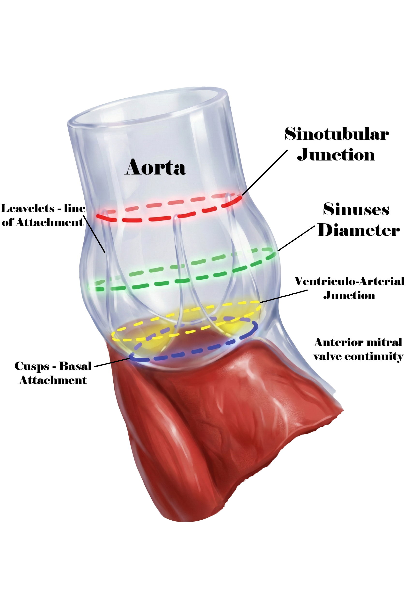
Anatomic Landmarks of the Aortic Root. This illustration demonstrates the key anatomic landmarks of the aortic root that are critical for aortic valve surgery and bioprosthetic valve implantation. Shown are the ventriculo-arterial junction (virtual basal ring), the basal attachment of the aortic valve cusps, the sinuses of Valsalva, the leaflet line of attachment, the sinotubular junction, and the ascending aorta. The figure also highlights the fibrous continuity between the anterior mitral valve leaflet and the aortic root (aorto-mitral curtain).
Mohamed HMM Alahmadi, MBBS, MS-CS, CHPE, MBA
References
Nkomo VT, Gardin JM, Skelton TN, Gottdiener JS, Scott CG, Enriquez-Sarano M. Burden of valvular heart diseases: a population-based study. Lancet (London, England). 2006 Sep 16:368(9540):1005-11 [PubMed PMID: 16980116]
Underwood MJ, El Khoury G, Deronck D, Glineur D, Dion R. The aortic root: structure, function, and surgical reconstruction. Heart (British Cardiac Society). 2000 Apr:83(4):376-80 [PubMed PMID: 10722531]
Kunihara T. Anatomy of the aortic root: implications for aortic root reconstruction. General thoracic and cardiovascular surgery. 2017 Sep:65(9):488-499. doi: 10.1007/s11748-017-0792-y. Epub 2017 Jun 27 [PubMed PMID: 28656518]
Combs MD, Yutzey KE. Heart valve development: regulatory networks in development and disease. Circulation research. 2009 Aug 28:105(5):408-21. doi: 10.1161/CIRCRESAHA.109.201566. Epub [PubMed PMID: 19713546]
Level 3 (low-level) evidenceGénéreux P, Stone GW, O'Gara PT, Marquis-Gravel G, Redfors B, Giustino G, Pibarot P, Bax JJ, Bonow RO, Leon MB. Natural History, Diagnostic Approaches, and Therapeutic Strategies for Patients With Asymptomatic Severe Aortic Stenosis. Journal of the American College of Cardiology. 2016 May 17:67(19):2263-2288. doi: 10.1016/j.jacc.2016.02.057. Epub 2016 Apr 2 [PubMed PMID: 27049682]
Bekeredjian R, Grayburn PA. Valvular heart disease: aortic regurgitation. Circulation. 2005 Jul 5:112(1):125-34 [PubMed PMID: 15998697]
Thaden JJ, Nkomo VT, Enriquez-Sarano M. The global burden of aortic stenosis. Progress in cardiovascular diseases. 2014 May-Jun:56(6):565-71. doi: 10.1016/j.pcad.2014.02.006. Epub 2014 Mar 2 [PubMed PMID: 24838132]
Level 3 (low-level) evidenceSeckeler MD, Hoke TR. The worldwide epidemiology of acute rheumatic fever and rheumatic heart disease. Clinical epidemiology. 2011 Feb 22:3():67-84. doi: 10.2147/CLEP.S12977. Epub 2011 Feb 22 [PubMed PMID: 21386976]
Nishimura RA, Otto CM, Bonow RO, Carabello BA, Erwin JP 3rd, Guyton RA, O'Gara PT, Ruiz CE, Skubas NJ, Sorajja P, Sundt TM 3rd, Thomas JD, ACC/AHA Task Force Members. 2014 AHA/ACC Guideline for the Management of Patients With Valvular Heart Disease: a report of the American College of Cardiology/American Heart Association Task Force on Practice Guidelines. Circulation. 2014 Jun 10:129(23):e521-643. doi: 10.1161/CIR.0000000000000031. Epub 2014 Mar 3 [PubMed PMID: 24589853]
Level 1 (high-level) evidenceNishimura RA, Otto CM, Bonow RO, Carabello BA, Erwin JP 3rd, Fleisher LA, Jneid H, Mack MJ, McLeod CJ, O'Gara PT, Rigolin VH, Sundt TM 3rd, Thompson A. 2017 AHA/ACC Focused Update of the 2014 AHA/ACC Guideline for the Management of Patients With Valvular Heart Disease: A Report of the American College of Cardiology/American Heart Association Task Force on Clinical Practice Guidelines. Circulation. 2017 Jun 20:135(25):e1159-e1195. doi: 10.1161/CIR.0000000000000503. Epub 2017 Mar 15 [PubMed PMID: 28298458]
Level 1 (high-level) evidenceAicher D, Kunihara T, Abou Issa O, Brittner B, Gräber S, Schäfers HJ. Valve configuration determines long-term results after repair of the bicuspid aortic valve. Circulation. 2011 Jan 18:123(2):178-85. doi: 10.1161/CIRCULATIONAHA.109.934679. Epub 2011 Jan 3 [PubMed PMID: 21200006]
Level 2 (mid-level) evidenceLansac E, Di Centa I, Danial P, Bouchot O, Arnaud-Crozat E, Hacini R, Doguet F, Demaria R, Verhoye JP, Jouan J, Chatel D, Lopez S, Folliguet T, Leprince P, Langanay T, Latremouille C, Fayad G, Fleury JP, Monin JL, Mankoubi L, Noghin M, Berrebi A, Pousset S, Laubriet-Jazayeri A, Lafourcade A, Marcault E, Kindo M, Payot L, Bergoend E, Hoffart CJ, Debauchez M, Tubach F. Aortic valve repair versus mechanical valve replacement for root aneurysm: the CAVIAAR multicentric study. European journal of cardio-thoracic surgery : official journal of the European Association for Cardio-thoracic Surgery. 2022 Jul 11:62(2):. pii: ezac283. doi: 10.1093/ejcts/ezac283. Epub [PubMed PMID: 35583290]
Watkins AR, El-Andari R, Fialka NM, Kang JJ, Hong Y, Bozso SJ, Jonker D, Moon M, Nagendran J, Nagendran J. Long-term outcomes following aortic valve replacement in bioprosthetic vs mechanical valves. Heart & lung : the journal of critical care. 2025 Jan-Feb:69():87-93. doi: 10.1016/j.hrtlng.2024.09.016. Epub 2024 Oct 5 [PubMed PMID: 39369559]
Pibarot P, Dumesnil JG. Prosthetic heart valves: selection of the optimal prosthesis and long-term management. Circulation. 2009 Feb 24:119(7):1034-48. doi: 10.1161/CIRCULATIONAHA.108.778886. Epub [PubMed PMID: 19237674]
Gillinov AM, Shah RV, Curtis WE, Stuart RS, Cameron DE, Baumgartner WA, Greene PS. Valve replacement in patients with endocarditis and acute neurologic deficit. The Annals of thoracic surgery. 1996 Apr:61(4):1125-9; discussion 1130 [PubMed PMID: 8607669]
Level 2 (mid-level) evidenceEriksson A, Burcharth J, Rosenberg J. Animal derived products may conflict with religious patients' beliefs. BMC medical ethics. 2013 Dec 1:14():48. doi: 10.1186/1472-6939-14-48. Epub 2013 Dec 1 [PubMed PMID: 24289542]
Level 3 (low-level) evidenceEasterbrook C, Maddern G. Porcine and bovine surgical products: Jewish, Muslim, and Hindu perspectives. Archives of surgery (Chicago, Ill. : 1960). 2008 Apr:143(4):366-70; discussion 370. doi: 10.1001/archsurg.143.4.366. Epub [PubMed PMID: 18427024]
Level 3 (low-level) evidenceTavakoli R, Leprince P, Gassmann M, Jamshidi P, Yamani N, Amour J, Lebreton G. Technique and Patient Selection Criteria of Right Anterior Mini-Thoracotomy for Minimal Access Aortic Valve Replacement. Journal of visualized experiments : JoVE. 2018 Mar 26:(133):. doi: 10.3791/57323. Epub 2018 Mar 26 [PubMed PMID: 29630054]
Parness S, Womble JT, Hester TE, Tasoudis P, Merlo AE. Aortic Valve Replacement in the Current Era. Journal of clinical medicine. 2025 Feb 21:14(5):. doi: 10.3390/jcm14051447. Epub 2025 Feb 21 [PubMed PMID: 40094917]
Vogt F, Santarpino G, Fujita B, Frerker C, Bauer T, Beckmann A, Bekeredjian R, Bleiziffer S, Möllmann H, Walther T, Beyersdorf F, Hamm C, Böning A, Baldus S, Ensminger S, Fischlein T, Eckner D, GARY Executive Board. Surgical aortic valve replacement in patients aged 50-69 years-insights from the German Aortic Valve Registry (GARY). European journal of cardio-thoracic surgery : official journal of the European Association for Cardio-thoracic Surgery. 2022 Jun 15:62(1):. pii: ezac286. doi: 10.1093/ejcts/ezac286. Epub [PubMed PMID: 35567484]
De Backer O, Wong I, Wilkins B, Carranza CL, Søndergaard L. Patient-Tailored Aortic Valve Replacement. Frontiers in cardiovascular medicine. 2021:8():658016. doi: 10.3389/fcvm.2021.658016. Epub 2021 Apr 21 [PubMed PMID: 33969021]
Sever K, Konukoglu O, Yildirim O, Kilercik H, Mansuroglu D. Sutureless Aortic Valve Replacement. Journal of the College of Physicians and Surgeons--Pakistan : JCPSP. 2021 Aug:31(8):891-896. doi: 10.29271/jcpsp.2021.08.891. Epub [PubMed PMID: 34320703]
Nicks R, Cartmill T, Bernstein L. Hypoplasia of the aortic root. The problem of aortic valve replacement. Thorax. 1970 May:25(3):339-46 [PubMed PMID: 5452289]
Manouguian S, Seybold-Epting W. Patch enlargement of the aortic valve ring by extending the aortic incision into the anterior mitral leaflet. New operative technique. The Journal of thoracic and cardiovascular surgery. 1979 Sep:78(3):402-12 [PubMed PMID: 470420]
Rastan H, Koncz J. Aortoventriculoplasty: a new technique for the treatment of left ventricular outflow tract obstruction. The Journal of thoracic and cardiovascular surgery. 1976 Jun:71(6):920-7 [PubMed PMID: 131885]
Level 3 (low-level) evidenceDo-Nguyen CC, Chen SA, Yang B. Y-incision aortic root enlargement - Common mistakes and update of early outcomes. Asian cardiovascular & thoracic annals. 2026 Mar:34(3):303-307. doi: 10.1177/02184923261428546. Epub 2026 Mar 4 [PubMed PMID: 41781359]
Edmunds LH Jr, Clark RE, Cohn LH, Grunkemeier GL, Miller DC, Weisel RD. Guidelines for reporting morbidity and mortality after cardiac valvular operations. European journal of cardio-thoracic surgery : official journal of the European Association for Cardio-thoracic Surgery. 1996:10(9):812-6 [PubMed PMID: 8905288]
Akins CW. Long-term results with the Medtronic-Hall valvular prosthesis. The Annals of thoracic surgery. 1996 Mar:61(3):806-13 [PubMed PMID: 8619697]
Jamieson WR, Riess FC, Raudkivi PJ, Metras J, Busse EF, Goldstein J, Fradet GJ. Medtronic Mosaic porcine bioprosthesis: assessment of 12-year performance. The Journal of thoracic and cardiovascular surgery. 2011 Aug:142(2):302-7.e2. doi: 10.1016/j.jtcvs.2010.08.090. Epub 2011 Feb 1 [PubMed PMID: 21277591]
Bourguignon T, Bouquiaux-Stablo AL, Loardi C, Mirza A, Candolfi P, Marchand M, Aupart MR. Very late outcomes for mitral valve replacement with the Carpentier-Edwards pericardial bioprosthesis: 25-year follow-up of 450 implantations. The Journal of thoracic and cardiovascular surgery. 2014 Nov:148(5):2004-2011.e1. doi: 10.1016/j.jtcvs.2014.02.050. Epub 2014 Feb 20 [PubMed PMID: 24667021]
Level 2 (mid-level) evidenceO'Brien SM, Shahian DM, Filardo G, Ferraris VA, Haan CK, Rich JB, Normand SL, DeLong ER, Shewan CM, Dokholyan RS, Peterson ED, Edwards FH, Anderson RP, Society of Thoracic Surgeons Quality Measurement Task Force. The Society of Thoracic Surgeons 2008 cardiac surgery risk models: part 2--isolated valve surgery. The Annals of thoracic surgery. 2009 Jul:88(1 Suppl):S23-42. doi: 10.1016/j.athoracsur.2009.05.056. Epub [PubMed PMID: 19559823]
Hammermeister K, Sethi GK, Henderson WG, Grover FL, Oprian C, Rahimtoola SH. Outcomes 15 years after valve replacement with a mechanical versus a bioprosthetic valve: final report of the Veterans Affairs randomized trial. Journal of the American College of Cardiology. 2000 Oct:36(4):1152-8 [PubMed PMID: 11028464]
Level 1 (high-level) evidenceChristakis GT, Joyner CD, Morgan CD, Fremes SE, Buth KJ, Sever JY, Rao V, Panagiotopoulos KP, Murphy PM, Goldman BS. Left ventricular mass regression early after aortic valve replacement. The Annals of thoracic surgery. 1996 Oct:62(4):1084-9 [PubMed PMID: 8823093]
Schwarz F, Flameng W, Thormann J, Sesto M, Langebartels F, Hehrlein F, Schlepper M. Recovery from myocardial failure after aortic valve replacement. The Journal of thoracic and cardiovascular surgery. 1978 Jun:75(6):854-64 [PubMed PMID: 149221]
Wahr JA, Prager RL, Abernathy JH 3rd, Martinez EA, Salas E, Seifert PC, Groom RC, Spiess BD, Searles BE, Sundt TM 3rd, Sanchez JA, Shappell SA, Culig MH, Lazzara EH, Fitzgerald DC, Thourani VH, Eghtesady P, Ikonomidis JS, England MR, Sellke FW, Nussmeier NA, American Heart Association Council on Cardiovascular Surgery and Anesthesia, Council on Cardiovascular and Stroke Nursing, and Council on Quality of Care and Outcomes Research. Patient safety in the cardiac operating room: human factors and teamwork: a scientific statement from the American Heart Association. Circulation. 2013 Sep 3:128(10):1139-69. doi: 10.1161/CIR.0b013e3182a38efa. Epub 2013 Aug 5 [PubMed PMID: 23918255]