 Tools
Tools
 Anatomy, Head and Neck: Eyelash
Introduction
The Greek word for eyelid is blepharon, from which the prefix blepharo- is derived. This prefix has been used since antiquity. The Greek word for eyelash is blepharida, which is not used in medicine today. The Latin word for eyelid is palpebra, and the Latin word for eyelash is cilium, a term used only since the early 18th century.
Eyelashes and eyebrows are important anatomical structures that are central to facial appearance and perceived human interactions. Eyebrow movement conveys emotions, including sadness, happiness, anger, surprise, and pensiveness. The eyebrows also protect the eyes by acting as curtain rods. When the eyebrows relax during fatigue, the upper eyelid skin descends, contributing to eyelid closure and ocular protection. Eyebrow hair direction further helps protect the eyes from particulate matter and sweat.
Eyelashes and eyebrows have always been important elements of facial beauty. People across many cultures have plucked, accentuated, and modified these structures in numerous ways to enhance the appearance of the face, particularly the eyes. Egyptian and Indian cultures have used materials such as kohl to enhance the eyebrows and eyelashes for centuries. Modern emphasis on the eyes includes the use of mascara, eyeliner, and eyeshadow. Eyelash curlers are used to exaggerate the natural curve of the eyelashes. Although long eyelashes are prized in most cultures, certain cultures, including the Hadza, one of the last hunter-gatherer groups in Tanzania, trim their eyelashes.
Comparative Anatomy
Mammals have eyelashes. Some mammals, such as camels, have exceptionally long eyelashes that have often been used to accentuate their features in cartoons. Cows have long, straight eyelashes that are oriented medially. The term cow-lash deformity describes this appearance when it occurs as a complication of blepharoplasty, often involving the lateral third of the upper eyelid.[1] Other large mammals like horses also have similar long, relatively straight lashes. The eyelash viper is said by some in South America to wink after striking its victim. The lashes are not true eyelashes, but modified scales above the eyes. And, of course, snakes cannot wink as they have no eyelids!
Structure and Function
Register For Free And Read The Full Article
 Search engine and full access to all medical articles
- 10 free questions in your specialty
- Free CME/CE Activities
- Free daily question in your email
- Save favorite articles to your dashboard
- Emails offering discounts
Learn more about a Subscription to StatPearls Point-of-Care
Structure and Function
Surface Anatomy
In humans, 75 to 80 eyelashes are present on the lower eyelid, and the upper eyelid has 90 to 160 eyelashes. Eyelash length varies among individuals; eyelashes usually do not grow beyond 12 mm before falling out. Eyelash length is thought to be limited because the growth rate is slower and the anagen phase is shorter than those of scalp hair. Eyebrow hairs may be longer, especially with age (Denis Healey eyebrows). Eyelashes generally do not need trimming unless excessive growth, or trichomegaly, occurs. In 2018, You Jianxia of Shanghai held the world record for the longest eyelashes, which measured 12.40 cm. After eyelash removal by plucking, regrowth takes approximately 8 weeks. Eyelash hair is not affected by puberty.
Eyelashes curve in all racial and ethnic groups. Results from studies showed that this curvature occurs because of asymmetrical cell types along the concave and convex sides of the hair bulb. This asymmetry is also present in curly hair. Eyelash curvature is greater in White individuals than in Asian individuals. Eyelashes have lift and curl angles that can be measured photographically to produce a phototrichogram.
Cross-Sectional Anatomy
Each eyelash has a hair shaft that extends above the skin, a root beneath the skin, and a bulb that forms the enlarged terminal portion. The inferior portion of the bulb is in direct contact with the dermal papilla, which has a vascular supply that allows interactions leading to follicle cycling. The eyelash has an inner medulla composed of cells, a surrounding cortex that is stiffer and provides the eyelash with strength and rigidity, and an outer concentric cuticle. The impermeable cuticle protects the inner portion of the eyelash. The outermost layer, the cuticle, is 7 to 10 layers thick and consists of transparent overlapping scales. The next layer, the cortex, is the thickest portion of the eyelash, is composed of keratin, and contains melanin.
The term eyelash follicle, derived from the Latin word follis, meaning bag, refers to the complex responsible for hair growth. This complex consists of the dermal papilla, which is composed of connective tissue and contains a capillary loop, the germinal hair matrix surrounding the papilla, and the eyelash root or bulb. The germinal layer contains dividing cells that give rise to the hair, and keratinization begins in the bulb. The hair follicle is embedded in the dermis of the eyelid. The eyelid contains 2 layers, the epidermis and dermis, whereas the scalp contains the epidermis, dermis, and hypodermis. Eyelash follicles are 2.4 mm deep in the upper eyelid and 1.4 mm deep in the lower eyelid. A greater proportion of eyelash follicles are active in the upper eyelid than in the lower eyelid (41% vs 15%).[2]
Body hair has arrector pili muscles that straighten the hair in response to cold or strong emotions, including fear and excitement. This response produces what is commonly called goose bumps because the skin resembles plucked goose skin. The eyelash follicle has no arrector pili muscles. Therefore, eyelashes do not change position under these circumstances.
Eyelash follicles are associated with 2 types of secretory glands:
Zeis glands: These holocrine glands release their entire cellular contents as sebum, which acts as a lubricant and has antimicrobial properties.
Glands of Moll: These apocrine glands are found only in the eyelids. Cellular fragmentation produces secretions that are thought to have antimicrobial properties.
Eyelash Pigmentation
Melanocytes synthesize melanin through melanogenesis. Eyelashes appear to become gray or white later than scalp hair. Continued expression of enzymes such as tyrosinase-related protein 2 (TRP-2), which influences melanocytes, is thought to account for this difference. Scalp hair undergoes depletion of melanocytes through apoptosis and oxidative stress and reduced expression of enzymes such as TRP-2. Changes in eyelash pigmentation with age among different racial groups have not been adequately studied.
Aerodynamics Around Eyelashes
Eyelashes have an essential role in protecting the eye. Results from aerodynamic studies demonstrated that the ideal eyelash length is approximately one-third of the width of the eye. In addition to protecting the cornea from particulate matter, airflow created by the curvature and density of the eyelashes may contribute to corneal protection. The exact ways in which eyelashes may increase or decrease evaporation remain unknown. Although in vitro studies have been conducted, no in vivo studies of human eyelashes and corneas have been performed.
Life Cycle of Eyelashes
Eyelashes grow at 0.12 to 0.14 mm/d, compared with 0.2 to 1.1 mm/d for scalp hair. The eyelash growth cycle consists of 3 phases:
Anagen phase: During this growth phase, cells in the hair root divide and lengthen the shaft. The anagen phase lasts 4 to 10 weeks.
Catagen phase: During this degradation phase, the eyelash becomes a club hair when its blood supply is interrupted. The catagen phase lasts approximately 15 days.
Telogen phase: During this resting phase, the hair is shed. The telogen phase lasts 4 to 9 months.
Approximately half of the eyelashes are in the anagen phase, and periocular hair has the lowest anagen-to-telogen ratio. After the telogen phase, the eyelash falls out, and the cycle begins again with the anagen phase. The entire eyelash cycle lasts 4 to 11 months.
The stages of the eyelash life cycle compared with those of the eyebrows and scalp are as follows:
Eyebrows:
Anagen phase: 4 to 7 monthsCatagen phase: 3 to 4 weeksTelogen phase: Approximately 9 months
Scalp hair: The duration of each phase is genetically determined and varies among individuals.
Anagen phase: 2 to 8 years or longerCatagen phase: 2 to 3 weeksTelogen phase: Approximately 3 months
Eyelash Microflora
Propionibacterium, Streptophyta, Staphylococcus, Corynebacterium, and Enhydrobacter organisms are commonly found on the eyelashes. In the presence of blepharitis, Staphylococcus, Streptophyta, Corynebacterium, and Enhydrobacter organisms increase in number. Conversely, Propionibacterium organisms are often decreased in the presence of blepharitis.
Demodex folliculorum and Demodex brevis are natural parasites that increase in number with age. By age 70, almost 100% of people have these parasites. Demodex folliculorum organisms inhabit the eyebrows, scalp hair, ears, and eyelashes, whereas Demodex brevis organisms are found in the facial sebaceous glands and meibomian glands. Death and rupture of the parasite release inflammatory contents that can cause eyelid inflammation. Cylindrical debris around the eyelashes is pathognomonic of Demodex infestation.
Embryology
Condensation of mesenchymal cells first occurs at week 9, forming eyelash follicles and their appendages. At this stage, the tarsal plate and the orbicularis oculi also develop together with evidence of blood vessels. By week 12, multiple eyelash follicles can be seen under the skin, and by week 14, eyelashes, together with sweat glands and sebaceous glands, appear.[3]
Blood Supply and Lymphatics
The superior and inferior marginal arteries from the ophthalmic artery supply the upper and lower eyelids, respectively. The upper eyelid marginal arterial arcade is near the follicles of the eyelashes, anterior to the tarsal plate and 2 mm superior to the lid margin. The superior arcade lies between the Muller muscle and the levator aponeurosis. The lower eyelid may have 1 or 2 arcades, but the arcades are further from the lid margin, at the inferior border of the tarsal plate. The internal carotid artery supplies the eyelids via the ophthalmic artery and the external carotid artery through the angular and temporal arteries. Therefore, there is extensive collateral supply from these 2 systems.
Venous drainage is pretarsal and posttarsal. The pretarsal venous drainage is into the angular vein medially and the superficial temporal vein laterally. The posttarsal venous drainage is into the superior and inferior ophthalmic veins and the pterygoid plexus and anterior facial vein. Lymphatic drainage from the medial eyelids is to the submandibular lymph nodes; that from the lateral eyelids is to the preauricular nodes.
Nerves
The eyelid margin is among the most sensitive parts of the human body, as evidenced by the pain experienced during the administration of a local anesthetic and the removal of eyelid margin lesions. Free nerve endings envelop the eyelashes. In addition, lanceolate and circular Ruffini nerve endings (also called corpuscles) are present, which provide touch, pressure, vibration, heat, and mechanoreception. Besides eyelashes, the anterior eyelid cutaneous surface has fine vellus hairs: these possess similar innervation. Furthermore, Meissner corpuscles (sensitivity to light touch) are also found, especially on the occlusal surface of the eyelids. This profusion of intricate innervation makes the lashes and the eyelid margins especially sensitive.[4] The sensitivity of eyelashes is comparable to the sensitivity of a cat's or mouse's whiskers with similar function.
Muscles
Eyelashes lack the arrector pili muscle found elsewhere, so the lashes do not change position in the presence of cold temperature or emotion.
Physiologic Variants
Age-Related Travel in Lashes
Studies of lashes in different age groups have shown a generalized reduction in the thickness, pigmentation, and length of lashes with age, but few proper longitudinal studies exist. Thibault improved our understanding of lashes and the change over time by studying defined lashes over nine months, measuring the length each week.[5][6][7] Canities is the natural gradual graying of hair shafts because of a decrease in tyrosine production in the hair bulb.
Surgical Considerations
Knowledge of the structure and distribution of eyelashes, as well as the depth of lashes in the upper and lower eyelids, is vital when performing surgery involving the eyelid margin.
Clinical Significance
The anatomy of the eyelash differs from that of hair elsewhere on the body in many respects. Knowledge of these differences is essential for addressing disorders of the eyelid, eyelid margin, and eyelashes. Unique eyelash disorders arise from these anatomic differences and from the relationship between eyelash growth, curvature, location, and the cornea.
Media
(Click Image to Enlarge)
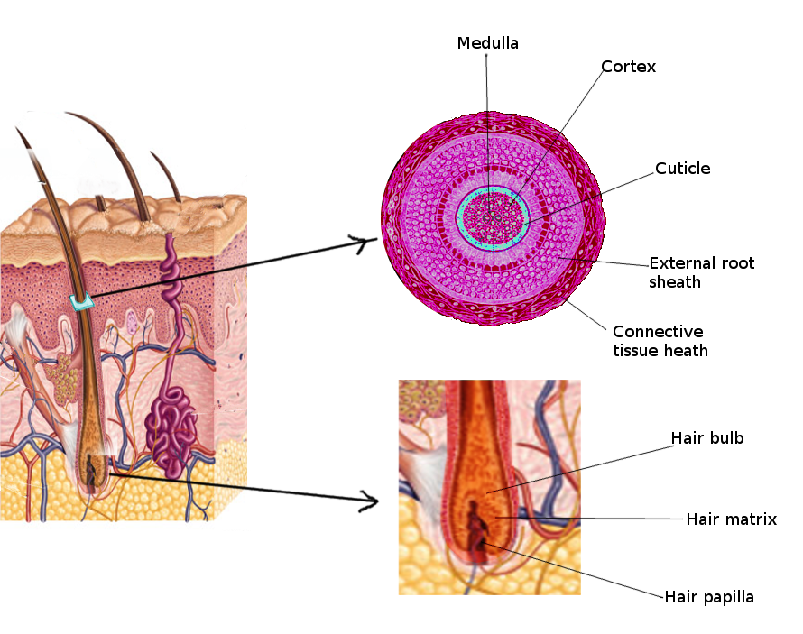
Hair Anatomy.
Contributed by S Bhimji, MD
References
McCord CD, Miotto GC. Dynamic diagnosis of "fishmouthing" syndrome, an overlooked complication of blepharoplasty. Aesthetic surgery journal. 2013 May:33(4):497-504. doi: 10.1177/1090820X13479970. Epub 2013 Apr 1 [PubMed PMID: 23547249]
Level 2 (mid-level) evidenceElder MJ. Anatomy and physiology of eyelash follicles: relevance to lash ablation procedures. Ophthalmic plastic and reconstructive surgery. 1997 Mar:13(1):21-5 [PubMed PMID: 9076779]
Byun TH, Kim JT, Park HW, Kim WK. Timetable for upper eyelid development in staged human embryos and fetuses. Anatomical record (Hoboken, N.J. : 2007). 2011 May:294(5):789-96. doi: 10.1002/ar.21366. Epub 2011 Mar 17 [PubMed PMID: 21416630]
Munger BL, Halata Z. The sensorineural apparatus of the human eyelid. The American journal of anatomy. 1984 Jun:170(2):181-204 [PubMed PMID: 6465049]
Procianoy F, Mendonça TB, Bins CA, Lang MP. Characterization of Normal Mediolateral Angular Direction of Lower Eyelid Eyelashes in Different Age Groups. Ophthalmic plastic and reconstructive surgery. 2015 Jul-Aug:31(4):332-3. doi: 10.1097/IOP.0000000000000473. Epub [PubMed PMID: 25902388]
Glaser DA, Jones D, Carruthers J, Campo A, Moench S, Tardie G, Largent J, Caulkins C. Epidemiologic analysis of change in eyelash characteristics with increasing age in a population of healthy women. Dermatologic surgery : official publication for American Society for Dermatologic Surgery [et al.]. 2014 Nov:40(11):1208-13. doi: 10.1097/DSS.0000000000000170. Epub [PubMed PMID: 25347452]
Thibaut S, De Becker E, Caisey L, Baras D, Karatas S, Jammayrac O, Pisella PJ, Bernard BA. Human eyelash characterization. The British journal of dermatology. 2010 Feb 1:162(2):304-10. doi: 10.1111/j.1365-2133.2009.09487.x. Epub 2009 Sep 1 [PubMed PMID: 19804590]
Level 3 (low-level) evidence