 Tools
Tools
 Adenomyosis
Introduction
Adenomyosis is a gynecologic condition characterized by ectopic endometrial tissue located within the uterine myometrium. German pathologist Carl von Rokitansky first described the condition in 1860 and originally referred to it as cystosarcoma adenoides uterinum. The clinical presentation of adenomyosis varies widely; however, dysmenorrhea and heavy menstrual bleeding represent the most frequently reported symptoms.
Historically, the diagnosis of adenomyosis relied on histopathologic examination, most commonly established after hysterectomy specimens. Advances in imaging have enabled noninvasive diagnosis, with transvaginal ultrasound and magnetic resonance imaging (MRI) now serving as key modalities for identifying characteristic uterine changes. Current management depends on reproductive goals and symptom severity. Hysterectomy remains the definitive treatment for individuals who have completed childbearing. For those seeking fertility preservation or wishing to avoid major surgery, multiple medical therapies and minimally invasive treatment options are available.[1]
Etiology
Register For Free And Read The Full Article
 Search engine and full access to all medical articles
- 10 free questions in your specialty
- Free CME/CE Activities
- Free daily question in your email
- Save favorite articles to your dashboard
- Emails offering discounts
Learn more about a Subscription to StatPearls Point-of-Care
Etiology
Although the histopathologic features of adenomyosis are well described, its etiology remains incompletely understood. Several mechanisms have been proposed. The most widely accepted theory suggests that adenomyosis results from the disruption of the interface between the endometrium basalis and the underlying myometrium. This disruption permits endometrial glands and stroma to invaginate into the myometrium, leading to cyclic proliferation, small-vessel angiogenesis, and adjacent smooth muscle hypertrophy and hyperplasia. The increased prevalence of adenomyosis following uterine instrumentation, eg, dilation and curettage or cesarean delivery, supports this mechanism.
A second theory proposes an embryologic origin in which pluripotent Müllerian stem cells undergo aberrant differentiation, giving rise to ectopic endometrial tissue. Evidence for this mechanism includes altered expression of specific genetic markers and case reports of endometrial tissue in individuals with Rokitansky–Küster–Hauser syndrome (Müllerian agenesis). Additionally, less widely supported theories of the underlying etiologies of adenomyosis include altered lymphatic drainage pathways and the potential contribution of displaced bone marrow–derived stem cells to the development of ectopic endometrial tissue.[2]
Adenomyosis Risk Factors
Risk factors for adenomyosis include conditions associated with increased estrogen exposure, eg, early menarche, short menstrual cycles, elevated body mass index, increased parity, oral contraceptive use, and tamoxifen therapy, as well as prior uterine surgery, including dilation and curettage, cesarean delivery, and myomectomy.[3]
Epidemiology
Accurate estimates of adenomyosis prevalence remain challenging due to historical underreporting and underdiagnosis. Reported rates of adenomyosis range widely from 5% to 70%, although more recent data suggest a prevalence of approximately 20% to 35%. Adenomyosis has traditionally been described as a condition affecting premenopausal, multiparous individuals in their 30s and 40s; however, this characterization largely reflects sampling bias from hysterectomy-based studies. Advances in transvaginal ultrasound and MRI have improved the accuracy of noninvasive diagnosis, providing greater insight into the broader population affected.
A rare variant of adenomyosis, juvenile cystic adenomyosis, is characterized by hemorrhagic myometrial cysts and typically presents in individuals younger than 30. Symptoms of juvenile cystic adenomyosis are often refractory to medical therapy and may require surgical management, including myomectomy or, less commonly, hysterectomy.
Pathophysiology
Ectopic endometrial tissue within the myometrium contributes to the clinical manifestations of adenomyosis through multiple interrelated mechanisms. Normal endometrial tissue produces prostaglandins that regulate uterine contractions during menstruation. When endometrial glands and stroma become embedded within the myometrium, these ectopic foci produce increased prostaglandin levels, resulting in exaggerated uterine contractility and dysmenorrhea. Estrogen-dependent proliferation of both eutopic and ectopic endometrial tissue further amplifies symptom severity and serves as a central target for medical therapy.
Heavy menstrual bleeding develops through a multifactorial process involving increased endometrial surface area, heightened vascularity, abnormal myometrial contractility, and elevated levels of signaling mediators, including prostaglandins, eicosanoids, and estrogen. These combined physiologic alterations contribute to the severity and persistence of bleeding symptoms associated with adenomyosis.[4][1]
Histopathology
The histologic diagnosis of adenomyosis is defined by the presence of endometrial glands and stroma within the smooth muscle of the myometrium. Criteria for the depth of myometrial invasion vary across definitions, with some approaches using an absolute measurement, typically ranging from 2.5 to 8 mm. In contrast, others apply a proportional threshold based on overall myometrial thickness. Multiple histologic grading systems have also been proposed to further characterize disease extent and burden.
Despite these frameworks, histopathologic diagnosis remains incompletely standardized and highly dependent on the quality, depth, and representativeness of tissue sampling.[5] Variability in specimen acquisition and interpretation continues to influence diagnostic consistency across clinical settings.
History and Physical
Clinical Features of Adenomyosis
Patients with adenomyosis may present with a broad spectrum of symptoms, although dysmenorrhea and heavy menstrual bleeding represent the most frequently reported clinical manifestations. Some individuals experience chronic pelvic pain, dyspareunia, or symptoms that closely resemble other gynecologic disorders, eg, endometriosis or uterine fibroids. A subset of patients remains asymptomatic, with adenomyosis identified incidentally during imaging performed for unrelated indications.
Physical examination findings tend to be nonspecific. Bimanual examination may reveal a uniformly enlarged, globular, or tender uterus, and in some cases, the uterus may feel boggy or softened. However, many patients have a completely normal pelvic examination, and reliance on physical findings alone is insufficient for diagnostic accuracy.
Given the substantial overlap of symptoms with other causes of abnormal uterine bleeding and pelvic pain, comprehensive history taking and physical examination remain essential components of evaluation. These steps guide the appropriate selection of imaging studies and further diagnostic evaluation.[6][7]
Evaluation
Imaging Studies
The evaluation of suspected adenomyosis begins with a thorough history and physical examination; however, clinical findings alone are insufficient for diagnosis due to significant overlap with other causes of abnormal uterine bleeding and pelvic pain. As a result, imaging plays a central role in the diagnostic workup. A comprehensive evaluation integrates clinical presentation with imaging findings to differentiate adenomyosis from other gynecologic conditions and guide appropriate management (see Table. Imaging Studies for Adenomyosis).[8][9]
Transvaginal ultrasound
Transvaginal ultrasound is typically the preferred initial imaging modality because of its accessibility, low cost, and ability to identify characteristic features, eg, a heterogeneous myometrium, myometrial cysts, asymmetrical myometrial thickening, or a poorly defined endometrial–myometrial junction. A 3-dimensional ultrasound may further improve visualization of the junctional zone.
Magnetic resonance imaging
MRI offers higher diagnostic accuracy and is particularly useful when ultrasound findings are inconclusive or when coexisting pathology, eg, leiomyomas or endometriosis, complicates interpretation. MRI features suggestive of adenomyosis include thickening of the junctional zone, high-signal myometrial foci on T2-weighted images, and diffuse or focal myometrial infiltration (see Image. Adenomyosis Imaging Findings).
Laboratory Studies
Laboratory testing is generally nonspecific. Hemoglobin levels may be assessed in patients with heavy menstrual bleeding, and pregnancy testing is appropriate when indicated, but no serum biomarkers reliably diagnose adenomyosis.
Table. Imaging Studies for Adenomyosis
| Role | First-line imaging modality | Second-line; highest diagnostic accuracy |
| Key Strengths | Widely available, low-cost, real-time assessment | Superior soft-tissue contrast; best for complex or mixed pathology |
| Typical Findings |
Heterogeneous myometrium |
Treatment / Management
Management of adenomyosis is guided primarily by the patient’s desire for future fertility and individualized symptom burden. Hysterectomy remains the definitive cure, while medical and minimally invasive approaches focus on reducing heavy menstrual bleeding and dysmenorrhea while preserving uterine function.
Medical Therapies
Nonhormonal medical therapies include nonsteroidal anti-inflammatory drugs (NSAIDs), which provide symptom relief by inhibiting cyclooxygenase and reducing prostaglandin production, thereby decreasing menstrual cramping and dysmenorrhea. Hormonal therapies aim to suppress estrogen-driven endometrial proliferation and include combined oral contraceptive pills, the levonorgestrel-releasing intrauterine device (LNG-IUD), danazol, and aromatase inhibitors. Although high-quality randomized controlled trial data remain limited, the LNG-IUD is generally regarded as the most effective first-line hormonal option due to its favorable adverse effect profile and strong clinical efficacy.
Minimally Invasive and Surgical Therapies
Minimally invasive and surgical options include high-intensity focused ultrasound (HIFU), which uses MRI-guided or ultrasound-guided energy delivery to target focal adenomyotic lesions and may benefit patients who fail medical therapy while preserving fertility. Uterine artery embolization (UAE) reduces uterine blood flow, leading to ischemic necrosis and decreased uterine volume. However, long-term outcomes and comparative effectiveness require further study, and fertility preservation cannot be guaranteed.[8][9][10](A1)
Endometrial ablation may be considered for patients who do not desire future fertility; however, limited treatment depth reduces effectiveness for deeper disease. Excision of deeper adenomyotic tissue while preserving the uterus is an additional surgical approach, but disruption of the endometrial–myometrial interface increases recurrence risk and may contribute to adverse pregnancy outcomes, including uterine rupture, premature rupture of membranes, preterm labor, and spontaneous abortion.
However, hysterectomy remains the only definitive cure for adenomyosis and is appropriate for patients who have completed childbearing or who experience refractory symptoms despite conservative medical and procedural management.[10][11][12][13](A1)
Differential Diagnosis
The symptoms of adenomyosis, particularly dysmenorrhea, chronic pelvic pain, and heavy menstrual bleeding, overlap significantly with several other gynecologic conditions. Accurate diagnosis, therefore, requires careful correlation of clinical presentation with imaging findings. Conditions commonly considered in the differential diagnosis of adenomyosis include:
- Uterine leiomyomas (fibroids): Fibroids may cause uterine enlargement, abnormal bleeding, and pelvic pain similar to adenomyosis. Unlike adenomyosis, leiomyomas typically appear as well-circumscribed masses on ultrasound or MRI, whereas adenomyosis produces diffuse myometrial heterogeneity or ill-defined focal thickening.
- Endometriosis: Both conditions are estrogen-responsive and associated with dysmenorrhea, dyspareunia, and chronic pelvic pain. Endometriosis is characterized by ectopic endometrial tissue outside the uterus, whereas adenomyosis involves invasion into the myometrium. Furthermore, the 2 conditions frequently coexist. Please see StatPearls' companion resource, "Endometriosis," for further information.
- Abnormal uterine bleeding due to ovulatory dysfunction (AUB-O): Anovulatory cycles can cause heavy or irregular bleeding but typically lack the uterine enlargement or imaging features seen in adenomyosis. Please see StatPearls' companion resource, "Abnormal Uterine Bleeding," for further information.
- Endometrial polyps: Polyps may cause intermenstrual or heavy bleeding. Polyps are usually focal and intracavitary, distinguishable on ultrasound or hysteroscopy.
- Adenomyoma: Adenomyomas are focal, well-circumscribed forms of adenomyosis that can mimic fibroids. MRI is often required to differentiate them.
- Chronic endometritis: May present with abnormal bleeding and pelvic discomfort, but is associated with endometrial inflammation rather than myometrial invasion.
- Malignancy (rare but important): Endometrial carcinoma or uterine sarcoma may present with abnormal bleeding or uterine enlargement. Suspicious imaging features or postmenopausal bleeding warrant further evaluation.
Therefore, a structured diagnostic approach that integrates history, physical examination, and targeted imaging helps distinguish adenomyosis from these overlapping conditions and guides appropriate management.[14][15]
Prognosis
The prognosis of adenomyosis varies widely and depends on multiple clinical factors, including the patient’s fertility goals, confidence in the diagnosis, tolerance of medical therapies, and willingness to pursue invasive procedures. Increasing recognition of adenomyosis in younger and asymptomatic individuals suggests a broader disease spectrum that remains incompletely understood. Symptom severity appears to correlate with both the number of adenomyotic foci and the depth of myometrial invasion.
A stepwise approach to management is generally recommended, beginning with medical therapy, typically NSAIDs and hormonal agents, and progressing to minimally invasive procedures, eg, endometrial ablation, excision of adenomyosis, or uterine artery embolization when symptoms persist. While these interventions may provide significant relief, hysterectomy remains the only definitive cure.[16][2]
Complications
The association between adenomyosis and infertility remains unclear. Some studies report an infertility rate of approximately 11% to 12%, but contradictory data exist, and no definitive causal relationship has been established. Numerous confounders, including coexisting gynecologic conditions, heterogeneous diagnostic criteria, and historical reliance on postoperative histopathology, limit interpretation. As imaging criteria, particularly transvaginal ultrasound and MRI, continue to be standardized and validated, future research will be better positioned to clarify the true relationship between adenomyosis and reproductive outcomes.[11][8]
Deterrence and Patient Education
Patient education plays a central role in the management of adenomyosis. Individuals should be counseled that the condition is benign but chronic, with symptoms that may fluctuate over time and overlap with other gynecologic disorders. Because the etiology is not fully understood and no proven preventive strategies exist, early recognition of symptoms and timely evaluation are essential.
Patients should be informed that dysmenorrhea and heavy menstrual bleeding are the most common manifestations and that these symptoms may significantly impact quality of life. Education regarding available medical and minimally invasive therapies, including their goals, expected benefits, and limitations, helps support shared decision-making. For those desiring fertility, counseling should emphasize that the relationship between adenomyosis and infertility remains uncertain, and that treatment choices may influence reproductive outcomes.
Clear discussion of imaging findings, the potential need for longitudinal follow-up, and the role of hysterectomy as the definitive treatment can help set realistic expectations. Encouraging patients to report worsening symptoms, changes in bleeding patterns, or inadequate response to therapy promotes ongoing engagement in care and may improve long-term outcomes.[17]
Pearls and Other Issues
Ultrasound is the preferred diagnostic imaging modality for the initial workup of adenomyosis with high sensitivity and specificity. MRI can be helpful in equivocal cases. Diagnostic imaging using either ultrasound or MRI has limitations due to numerous mimics and the uterus's variable physiologic appearance. Up to a third of women with a sonographic diagnosis of adenomyosis may be asymptomatic.
While adenomyosis is classically taught as a disease of premenopausal, multiparous women, some data show an increased prevalence in undiagnosed younger populations. Treatment options should target primary symptoms. NSAIDs for painful cramping, oral contraceptive pills or levonorgestrel IUD for heavy bleeding, and hysterectomy for women no longer desiring fertility. Minimally invasive therapies should be a consideration in refractory cases in women who wish to preserve fertility.
Adenomyosis can coexist with leiomyoma (50%), endometriosis (11%), and endometrial polyps (7%). These associations merit attention in patients who have refractory symptoms. Utilization of the PALM-COEIN mnemonic can be used to broaden the differential diagnosis for abnormal uterine bleeding.[6][18]
Enhancing Healthcare Team Outcomes
Adenomyosis is a gynecologic disorder characterized by ectopic endometrial tissue within the myometrium, leading to dysmenorrhea, heavy menstrual bleeding, and chronic pelvic pain. Pathophysiology involves disruption of the endometrial–myometrial interface with estrogen-dependent proliferation, increased prostaglandin production, and myometrial hypertrophy. Diagnosis relies on clinical suspicion, supported by transvaginal ultrasound and MRI, though findings may be variable and operator-dependent. Management is guided by symptom severity and fertility goals, ranging from NSAIDs and hormonal therapies such as levonorgestrel intrauterine devices to minimally invasive procedures and definitive hysterectomy.
Interprofessional collaboration improves diagnostic accuracy, safety, and patient-centered outcomes by integrating primary care clinicians and advanced practitioners for early recognition and management; radiologists for optimized imaging interpretation; gynecologists for treatment planning; and interventional radiologists for procedural options. Nurses support symptom monitoring, education, and adherence, while pharmacists guide the safe selection of hormonal therapy and monitor adverse effects. Coordinated communication and shared decision-making ensure timely referral, individualized care, and improved long-term outcomes.[19]
Media
(Click Image to Enlarge)
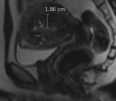
Adenomyosis Imaging Findings. T2-weighted image of adenomyosis. Imaging features of myometrial cysts include a globular contour and a thickened junctional zone measuring >12 mm.
Contributed by R Gunther, MD, MPH
References
Kho KA, Gingold JA, Shin JH. Adenomyosis Pathophysiology, Diagnostic Advances, and Therapeutic Options. Obstetrics and gynecology. 2026 Apr 2:():. doi: 10.1097/AOG.0000000000006276. Epub 2026 Apr 2 [PubMed PMID: 41926770]
Level 3 (low-level) evidenceGuo SW, Canis M, Vercellini PP. Is endometriosis a progressive disease? Trends in molecular medicine. 2026 Mar 20:():. pii: S1471-4914(26)00041-9. doi: 10.1016/j.molmed.2026.02.011. Epub 2026 Mar 20 [PubMed PMID: 41864757]
Schildroth S, Upson K, Bond J. An Epidemiologic Review of Endocrine-disrupting Chemicals and Understudied Gynecologic Health Conditions Affecting Quality of Life. Current environmental health reports. 2026 Apr 1:13(1):. pii: 15. doi: 10.1007/s40572-026-00534-w. Epub 2026 Apr 1 [PubMed PMID: 41920432]
Level 2 (mid-level) evidenceYang J, Li X, Peng Y, Sun Z, Zhang L, Duan J, Zhong N. Pathogenesis of Adenomyosis: An Integrated Review of Cellular Origins, Molecular Mechanisms, and Intersecting Diseases. Journal of cellular and molecular medicine. 2026 Apr:30(7):e71129. doi: 10.1111/jcmm.71129. Epub [PubMed PMID: 41968335]
Berlanda N, Bandini V, Croci GA, Dridi D, Nobili MV, Cipriani S, Vercellini P. Histological and immunohistochemical characteristics of superficial peritoneal endometriotic lesions of clinical responders and non-responders to dienogest treatment. Human reproduction (Oxford, England). 2026 Jun 1:41(6):882-891. doi: 10.1093/humrep/deag059. Epub [PubMed PMID: 41999626]
Piriyev E, Mennicken C, Schiermeier S, Römer T. Symptom Burden in Adenomyosis: A Comparative Analysis With Peritoneal and Deep Infiltrating Endometriosis. In vivo (Athens, Greece). 2026 May-Jun:40(3):1614-1620. doi: 10.21873/invivo.14311. Epub [PubMed PMID: 42049408]
Level 2 (mid-level) evidenceXu Z, Guo W, Shen Y. Association Between Impaired Dendritic Cell Maturation and Uterine Adenomyosis Pain. The journal of obstetrics and gynaecology research. 2026 Apr:52(4):e70283. doi: 10.1111/jog.70283. Epub [PubMed PMID: 41968803]
Koroneos Z, Cao M, Mooney S, Tyson K, Holdsworth-Carson S. Comorbidity of endometriosis and adenomyosis: A systematic review examining the impact of co-morbid disease on fertility and pregnancy outcomes. European journal of obstetrics, gynecology, and reproductive biology. 2026 Jun:322():115094. doi: 10.1016/j.ejogrb.2026.115094. Epub 2026 Apr 8 [PubMed PMID: 41996904]
Level 1 (high-level) evidenceMohamed Y, Kol SQ, Stoker J, Bipat S. Deep endometriosis: evaluation of ENZIAN and #ENZIAN classification systems by MRI compared with surgery-a meta-analysis. European radiology. 2026 Jun:36(6):4489-4502. doi: 10.1007/s00330-025-12154-3. Epub 2025 Nov 29 [PubMed PMID: 41317188]
Level 1 (high-level) evidenceChêne G, Chauleur C. [New thermal ablation modalities for uterine fibroids and adenomyosis: Radiofrequency, microwaves and high-intensity focused ultrasounds]. Gynecologie, obstetrique, fertilite & senologie. 2026 May:54(5):241-242. doi: 10.1016/j.gofs.2026.04.001. Epub [PubMed PMID: 42062011]
De Cicco Nardone C, Sangiovanni MC, Sangiovanni GM, Plotti F, Montera R, Luvero D, Angioli R, Terranova C. Intravaginal progesterone as a fertility-sparing treatment for symptomatic adenomyosis: preliminary results from a monocentric study. Archives of gynecology and obstetrics. 2026 Apr 28:313(1):. doi: 10.1007/s00404-026-08361-y. Epub 2026 Apr 28 [PubMed PMID: 42050103]
Ding K, Shi QL, Sang CM, Zhao L, Zhao SP. [Investigation of pharmacological regimens combined with focused ultrasound ablation surgery for alleviating dysmenorrhea in extrinsic adenomyosis]. Zhonghua fu chan ke za zhi. 2026 Apr 25:61(4):277-288. doi: 10.3760/cma.j.cn112141-20250915-00433. Epub [PubMed PMID: 42045779]
Sun R, Xu H, Ma R, Xu J, Liu Y, Mao D. Clinical and endocrine effects of pharmacological therapy in endometriosis: a systematic review and meta-analysis. Frontiers in endocrinology. 2026:17():1792793. doi: 10.3389/fendo.2026.1792793. Epub 2026 Apr 1 [PubMed PMID: 41993993]
Level 1 (high-level) evidenceEgashira H, Ishida H, Takashima A. Adenocarcinoma Arising in Adenomyosis: A Narrative Review of Disease Concept, Molecular Pathogenesis, and Clinical Challenges. Cureus. 2026 Feb:18(2):e104162. doi: 10.7759/cureus.104162. Epub 2026 Feb 24 [PubMed PMID: 41909345]
Level 3 (low-level) evidenceAli A, Youssef A, Saman AE, Taleb HA. Management of uterine leiomyoma and adenomyosis: role of hysteroscopy in diagnosis and norethindrone in the treatment. Scientific reports. 2025 Dec 3:15(1):43019. doi: 10.1038/s41598-025-28540-2. Epub 2025 Dec 3 [PubMed PMID: 41339448]
Otgontuya A, Jeng CJ, Chuang LT, Shen J. Improving the symptoms of dysmenorrhea and menorrhagia of women with adenomyosis and myoma in short- and long-term treatment effectiveness of microwave ablation: A systematic review and meta-analysis. Taiwanese journal of obstetrics & gynecology. 2026 Mar:65(2):188-197. doi: 10.1016/j.tjog.2025.05.023. Epub [PubMed PMID: 41813372]
Level 1 (high-level) evidence. Adenomyosis. American family physician. 2022 Jan 1:105(1):Online [PubMed PMID: 35029958]
Deniz R, Baykus Y, Tigli A, Uzuner MB, Sener N, Degirmenci YE, Akinci GE, Gurkan E, Karakoc O, Aydin MA, Emre E, Aydin S. Evaluation of pre-operative endometrial biopsy accuracy and etiological distribution in abnormal uterine bleeding using the PALM-COEIN classification. Frontiers in medicine. 2026:13():1784420. doi: 10.3389/fmed.2026.1784420. Epub 2026 Feb 24 [PubMed PMID: 41816678]
Burla L, Kalaitzopoulos DR, Samartzis N, Khazali S, Bokor A, Renner SP, Hudelist G, Constantin AS, Schäfer SD, Nassif J, Naem A, Keckstein J, Krentel H, EEL MDT meeting Consensus Group. Recommendations for the implementation and conduct of multidisciplinary team meetings for those providing endometriosis and adenomyosis care - a Delphi consensus of the European Endometriosis League (EEL). Facts, views & vision in ObGyn. 2024 Sep:16(3):337-350. doi: 10.52054/FVVO.16.3.038. Epub [PubMed PMID: 39357865]
Level 3 (low-level) evidence