 Tools
Tools
 Atrioventricular Reciprocating Tachycardia
Introduction
Atrioventricular reciprocating tachycardia (AVRT) is a type of supraventricular tachycardia (SVT) that requires specific electrophysiologic and electroanatomic characteristics. AVRT uses a circuit that consists of at least 2 different pathways with different electrical properties, including conduction velocity, refractory periods, and directionality. The anatomical substrate to sustain an AVRT can vary because AVRT may involve different configurations as follows:
- Two accessory pathways (APs) that enable communication between the atria and the ventricles.
- A single accessory pathway, in addition to the regular conduction system through the atrioventricular node, completes the circuit.
- Multiple accessory pathways through which the SVT can be perpetuated.[1]
AVRT commonly starts with an ectopic atrial or ventricular beat that travels through one of the circuit's limbs due to the distinct electrical properties of the tissues involved. These properties allow the initiation and persistence of an SVT at a heart rate ranging from 150 to 250 bpm.[2][3]
AVRT is the most common type of arrhythmia associated with Wolff-Parkinson-White (WPW) syndrome. In this condition, antegrade conduction at rest occurs through an accessory pathway (or accessory pathways) that manifests on the surface electrocardiogram (ECG). The ECG shows a shortened PR interval followed by a delta wave or slurring of the initial portion of the QRS complex, which results in a widened QRS complex. AVRT can use an antegrade and non-decremental accessory pathway in these patients, and retrograde conduction can occur through the atrioventricular node or another accessory pathway. These patients can also develop accelerated conduction of atrial arrhythmias through the accessory pathway or accessory pathways that bypass the atrioventricular node. These preexcited atrial arrhythmias, especially atrial fibrillation, can be lethal due to rapid conduction into the ventricles.[4]
Permanent junctional reciprocating tachycardia is a rare type of AVRT, which uses the atrioventricular node as its antegrade limb and a decrementally conducting accessory pathway as its retrograde limb. This type of tachycardia typically has a slower heart rate, between 130 and 150 bpm, and is commonly refractory to medical management.[5][6] Other unusual forms of AVRT use atrioventricular or nodoventricular accessory pathways, known as Mahaim pathways. These are decrementally conducting anomalous connections between the right atrium or the atrioventricular node and the right ventricle.[7][8]
Wolff-Parkinson-White (WPW) syndrome is a congenital cardiac preexcitation syndrome that arises from abnormal cardiac electrical conduction through an accessory pathway that can result in symptomatic and life-threatening arrhythmias. The hallmark electrocardiographic (ECG) finding of WPW pattern or preexcitation consists of a short PR interval and prolonged QRS with an initial slurring upstroke (“delta” wave) in the presence of sinus rhythm. The term WPW syndrome is reserved for an ECG pattern consistent with the above-described findings along with the coexistence of a tachyarrhythmia and clinical symptoms of tachycardia such as palpitations, episodic lightheadedness, presyncope, syncope, or even cardiac arrest.
The normal heart consists of two electrically insulated units, the atria and the ventricles. These units are connected by a conduction system that allows for normal cardiac synchrony and function. The cardiac electrical potential originates from the sinoatrial node of the right atrium and propagates through the atria to the atrioventricular (AV) node. The action potential is delayed in the AV node and is then quickly transmitted through the His-Purkinje system to the ventricular myocytes allowing for rapid ventricular depolarization and synchronized contraction. Patients with WPW syndrome have an accessory pathway that violates the electrical isolation of the atria and ventricles, which can allow electrical impulses to bypass the AV node. In some settings, this pathway can result in the transmission of abnormal electrical impulses leading to malignant tachyarrhythmias. The ECG findings of the WPW pattern are caused by the fusion of ventricular preexcitation through the accessory pathway and normal electrical conduction. Most patients with WPW pattern will never develop arrhythmia and will remain asymptomatic. Some accessory pathways will not manifest the described typical ECG findings, and as a result, some patients can develop a tachyarrhythmia with no prior ECG evidence that the pathway exists. These are referred to as concealed bypass tracts.
In the early 1900s, Frank Wilson and Alfred Wedd are thought to have first described ECG patterns that would later be recognized as a WPW pattern. In 1930, Louis Wolff, Sir John Parkinson, and Dr. Paul Dudley White published a case series consisting of 11 patients who experienced paroxysmal tachycardia associated with an underlying ECG pattern of sinus rhythm with short PR and bundle branch block/wide QRS. This phenomenon was subsequently named as Wolff-Parkinson-White (WPW) syndrome. The electrocardiographic features of preexcitation were first correlated with anatomic evidence of anomalous conducting tissue or bypass tracts in 1943.
Etiology
Register For Free And Read The Full Article
 Search engine and full access to all medical articles
- 10 free questions in your specialty
- Free CME/CE Activities
- Free daily question in your email
- Save favorite articles to your dashboard
- Emails offering discounts
Learn more about a Subscription to StatPearls Point-of-Care
Etiology
Typically, the fibrous annuli of the tricuspid and mitral valves provide electrical insulation between the atria and ventricles, except at the level of the atrioventricular node. The accessory pathways required to sustain an AVRT consist of myocardial muscle bundles that connect the atria to the ventricles, thus bypassing the insulation provided by the fibrous annuli of tricuspid and mitral valves. Accessory pathways can conduct impulses in an anterograde, retrograde, or bidirectional manner.[2] Patients with Ebstein anomaly of the tricuspid valve commonly have more than one myocardial bundle or accessory pathway, which makes treatment of the AVRT challenging.[1]
The electrophysiologic properties of the accessory pathways typically resemble those of the Purkinje fibers, exhibiting rapid conduction, lack of decremental conduction, and often resistance to adenosine.[2] However, some accessory pathways can exhibit decremental conduction. These are known as Mahaim accessory pathways and are characterized by their sensitivity to adenosine.[7][9]
WPW pattern arises from the fusion of ventricular preexcitation through the accessory pathway and normal electrical conduction through the AV node. This accessory pathway is thought to arise from chamber myocardium during improper early atrial and ventricular folding in cardiac embryogenesis. As a result, electrically conductive myocardial bundles violate the normal electrical insulation of the atrium and ventricle, forming the accessory pathway. This pathway usually has non-decremental or non-delayed conduction, which is in contrast to the properties of the normal AV node. The electrical conducting characteristics of the accessory pathway can vary and depend upon factors such as the speed of conduction, direction of conduction, and refractory period. These characteristics, along with location and number of pathways, will determine how the pathway may be involved in the initiation or transmission of an arrhythmia leading to WPW syndrome
Epidemiology
AVRT is the fourth most common type of SVT in patients older than 20, after atrial fibrillation, atrial flutter, and atrioventricular nodal reentrant tachycardia (AVNRT). However, it is the most common type of SVT in the pediatric population.[2] In children, the highest incidence of SVT occurs during infancy, typically ranging from 1 month to 1 year (12 months), with spontaneous resolution in 90% of patients by 12 months. Of these, approximately 30% will have a recurrence during early childhood, between the ages of 6 and 9.[5] Multiple accessory pathways can be found in 4% to 10% of patients, especially those with Ebstein anomaly. Assessing the presence of other accessory pathways during an electrophysiologic study is crucial. Detecting and eliminating any additional accessory pathways with radiofrequency ablation or cryoablation will prevent AVRT recurrence.[10]
The natural history of asymptomatic WPW patients has been speculated from the available data on symptomatic WPW patients and from those who have been incidentally discovered to have a WPW ECG pattern. In large-scale population-based studies involving pediatric and adult populations, the general prevalence of WPW has been estimated between 1 to 3 per 1000 individuals (0.1 to 0.3 %). Identification of the truly asymptomatic patients with WPW pattern is difficult, as these individuals by definition are those who have no clinical symptoms. A general estimate by experts suggests that about 65% of adolescents and 40% of individuals over 30 years with a WPW pattern on a resting ECG are asymptomatic. The incidence of patients with the WPW pattern progressing to arrhythmia is thought to be around 1% to 2% per year, and WPW syndrome prevalence peaks from age 20 to 24.
Familial studies have shown a slightly higher incidence of WPW, about 0.55% among first-degree relatives of an index patient with WPW. A familial form of WPW syndrome has been observed with a missense mutation in the PRAKAG2 gene leading to an increase in prevalence to 3.4% in first-degree relatives, and the condition is associated with congenital structural heart disease including Ebstein anomaly and hypertrophic cardiomyopathy
Pathophysiology
AVRT can be orthodromic, antidromic, or junctional reciprocating tachycardia.[11][12][2] These AVRTs have certain electrophysiologic features that can differentiate one from the other.
Orthodromic Atrioventricular Reentry Tachycardia
This form comprises 80% to 87% of AVRTs and can be initiated with atrial premature beats or ventricular premature beats. An atrial premature beat that initiates the AVRT is typically blocked in the accessory pathway. Subsequently, it conducts to the ventricles in an antegrade manner through the atrioventricular node and the His-Purkinje system. However, before reaching the His-Prkinje system, the electrical impulse travels retrogradely through the accessory pathway, establishing a reentry circuit that uses the accessory pathway and the atrioventricular node.[3] A ventricular premature beat initiating the AVRT conducts to the atria through the accessory pathway in a retrograde manner. The impulse travels back to the ventricles through the atrioventricular node to complete the circuit. In both cases, the impulse will travel through the atrioventricular node and the His-Purkinje system in an antegrade fashion and through the accessory pathway (or accessory pathways) in a retrograde manner.[13]
Antidromic Atrioventricular Reentry Tachycardia
This mechanism occurs in 5% to 10% of AVRTs. As with orthodromic AVRT, the tachyarrhythmia is initiated by either an atrial premature beat or a ventricular premature beat. When initiated by an atrial premature beat, the impulse is blocked in the atrioventricular node. Subsequently, it conducts to the ventricles through a manifest accessory pathway in an anterograde manner, preexciting the ventricles. The impulse travels back to the atria through the His-Purkinje system and atrioventricular node or another accessory pathway in a retrograde manner. If a ventricular premature beat initiates the reentrant tachycardia, the impulse conducts from the ventricles to the atria in a retrograde manner either through the His-Purkinje system and the atrioventricular node or through an accessory pathway. Subsequently, it returns to the ventricles in an antegrade manner through another accessory pathway, also displaying preexcitation. In both cases, the impulse will travel through the His-Purkinje system and the atrioventricular node or an accessory pathway in a retrograde manner and through another accessory pathway in an antegrade manner, which produces preexcitation with a wide QRS.[13]
Junctional Reciprocating Tachycardia
Junctional reciprocating tachycardia is a type of AVRT that occurs predominantly in infants and children, accounting for 1% of the SVTs in this age group. If left untreated, it can evolve into an incessant tachycardia, resulting in tachycardia-induced cardiomyopathy. During junctional reciprocating tachycardia, the atrioventricular node acts as the antegrade limb, whereas an accessory pathway with physiologic characteristics similar to the AVN, such as slow and decremental conduction, functions as the retrograde limb. The similar conduction characteristics shared between this type of accessory pathway and the atrioventricular node enable the formation of a stable, and thus persistent, reentrant circuit, resulting in a narrow-QRS tachycardia with heart rates ranging between 200 and 300 bpm in infancy and around 250 bpm in early childhood. If present during adulthood, heart rates can be slower, typically in the range of 120 bpm. Inverted P waves can typically be observed in inferior leads II, III, and aVF. This type of AVRT does not require a premature atrial contraction or a premature ventricular contraction for initiation. Typically, it exhibits a temporary response to vagal maneuvers or adenosine, resulting in slowing before termination. However, recurrence often follows shortly afterward.[12]
WPW ECG pattern is caused by abnormal electrical conduction through an accessory pathway that bypasses the normal cardiac conduction system. This accessory pathway allows cardiac electrical activity to bypass the atrioventricular node conduction delay, and arrive early at the ventricle, leading to premature ventricular depolarization. This preexcitation also bypasses the fast conducting His-Purkinje system and results in early but slowly propagated ventricular depolarization, which gives rise to the ECG pattern of a short PR interval with a “slurred” start to the QRS complex termed a delta wave. The remainder of a normal QRS obliterates this delta wave as the normal cardiac conduction catches up following AV node delay and fast conduction through the His-Purkinje system.
There are two ways in which an accessory pathway can lead to WPW syndrome. The pathway can either initiate and maintain an arrhythmia or allow conduction of an arrhythmia generated elsewhere. The first type occurs when a circuit is formed between the normal conduction system of the heart and the accessory pathway (or two or more accessory pathways), allowing for atrioventricular reentrant tachycardia (AVRT). An incorrectly timed extra electrical impulse can lead to a recurring cycle between the atria, AV node, ventricles, and the accessory pathway. Orthodromic AVRT occurs when conduction progresses from the atria with antegrade conduction through the AV node to the ventricle and retrograde conduction through the accessory pathway. This will usually result in a narrow complex QRS as the His-Purkinje system is used unless aberrant conduction is present. Antidromic AVRT is the opposite with antegrade conduction passing from the atria through the accessory pathway to the ventricle and retrograde conduction back up the AV node and is usually associated with a wide complex QRS.
The other way an accessory pathway can lead to arrhythmia is by allowing conduction of an arrhythmia that is generated elsewhere to propagate to a portion of the heart that would normally be electrically insulated from this arrhythmia. The accessory pathway is typically comprised of myocardial tissue and usually has non-decremental or non-delayed conduction allowing immediate ventricular activation. This non-decremental conduction property predisposes patients with WPW syndrome to sudden cardiac death. This occurs due to rapid ventricular rates in conditions with rapid atrial depolarization, such as atrial fibrillation (AF) or atrial flutter. These fast ventricular rates can degenerate into ventricular fibrillation (VF) and cardiac arrest
History and Physical
Similar to other SVTs, the clinical presentation of AVRT depends on the associated comorbidities. The most commonly reported symptom is palpitations. However, dizziness, chest pain, and syncope can occur.[13]
Patients with a WPW pattern who have never developed an arrhythmia will be asymptomatic, and therefore, their history and physical exam will be mostly unremarkable. Prior ECG may have diagnosed the pattern, and the patient may be aware of their condition, but some accessory pathway conduction is transient or concealed, which may lead to prior normal or intermittently normal ECGs. Patients with WPW pattern may have a family history of WPW pattern or syndrome.
Patients with WPW pattern who develop a tachyarrhythmia will often experience symptoms associated with the arrhythmia including palpitations, chest pain, dyspnea, dizziness, lightheadedness, presyncope, syncope, collapse, and/or sudden death. The history will be notable for these symptoms, which may be episodic and resolved, or ongoing at presentation if the arrhythmia persists. Physical exam should be focused on the patient's cardiovascular, pulmonary perfusion status, and neurological exam and may be completely normal if the arrhythmia has resolved. A persisting arrhythmia will usually be symptomatic, and vitals signs will be notable for tachycardia. Blood pressure may range from normal or elevated to hypotension depending on the severity of the tachyarrhythmia, comorbidities, and the patient's ability to compensate for the arrhythmia. Respiratory rate will vary based on the patient's level of distress and the ability to maintain perfusing blood pressure. The physical exam will again vary depending on the severity of the arrhythmia. The cardiac exam will demonstrate a regular or irregular tachycardia. The remainder of the physical exam may be normal or show signs of discomfort, distress, hypoperfusion, cardiogenic shock, and unresponsiveness depending on the severity of the arrhythmia
Evaluation
Electrocardiogram
An experienced electrophysiologist can use a 12-lead ECG during SVT to differentiate between different mechanisms, such as an orthodromic AVRT using a concealed accessory pathway as the retrograde limb of the tachycardia, AVNRT, antidromic AVRT using the accessory pathway as the antegrade limb of the tachycardia (presenting as wide QRS tachycardia), and ventricular tachycardia. Artificial intelligence shows promising results in differentiating SVT mechanisms, offering comparable performance to experienced electrophysiologists.[14] See Image. AVRT ECG strip.
Narrow QRS Tachycardia: Differential diagnosis between Orthodromic AVRT and AVNRT
In patients without intraventricular conduction delay or a bundle branch block, an orthodromic AVRT will exhibit narrow QRS complexes, with a heart rate ranging from 150 to 250 bpm. When the duration of the RP interval is relatively long (>70 msec), retrograde P waves can be observed following the QRS complex because the activation of the atria is reversed. Clinically, carotid palpitation can be an indicator of AVNRT.[15]
Typical or slow-fast AVNRT usually has a short RP interval (<70 msec) with the retrograde P wave buried within the preceding QRS complex. Therefore, the P waves are not visible on the surface ECG. Occasionally, a pseudo r' deflection in V1 or QRS alternans will favor the diagnosis of AVNRT over AVRT. Orthodromic AVRT and atypical AVNRT, such as fast-slow and slow-slow AVNRT, may show retrograde P waves or pseudo repolarization changes after the QRS complexes during the tachycardia, which may be visible on the surface ECG. If a bundle branch block occurs ipsilaterally to the accessory pathway during the tachycardia with lengthening of the R-R interval (that is, lengthening of the tachycardia cycle length), orthodromic AVRT is the probable diagnosis.[16] This phenomenon is described in Coumel's law, which states that when a wide QRS tachycardia abruptly becomes a narrow QRS tachycardia with a simultaneous increase in heart rate, orthodromic AVRT using an accessory pathway ipsilateral to the bundle branch block is the probable diagnosis.[17]
Wide QRS Tachycardia: Differential diagnosis between Antidromic AVRT, AVNRT with a bundle branch block, and ventricular tachycardia
In patients with WPW syndrome, the resting ECG shows a delta wave with a short PR interval, resulting from the early depolarization of the ventricles through the accessory pathway.[18] During antidromic AVRT, the QRS complexes are wide as the impulse travels to the ventricles through an accessory pathway that bypasses the atrioventricular node. This type of AVRT might be difficult to differentiate from ventricular tachycardia (VT). Wide QRS tachycardia can also be observed in the setting of AVNRT with a bundle branch block, as described above. In this case, the heart rate during SVT typically falls within the range of 150 to 250 bpm, and retrograde P waves may be seen following the QRS complexes. Generally, ventricular tachycardia is more likely when the duration of the QRS complex is greater than 140 msec.[19][20]
Electrophysiologic Study
An electrophysiologic study involves inserting intracardiac catheters with electrodes to track and differentiate the arrhythmias. In addition, an electrophysiologic study allows radiofrequency ablation or cryoablation of an accessory pathway or tachycardia focus, which can cure the arrhythmia. Traditionally, catheter ablations have used fluoroscopy, which exposes the patient to radiation that is not insignificant, as such procedures can last several hours. However, several centers are achieving a decrease and even elimination of fluoroscopy by using intracardiac electrograms, electroanatomic mapping, and intracardiac echocardiography.[21]
During an electrophysiologic study, AVRT can be diagnosed when a bundle branch block occurs simultaneously with an increase in the VA interval (time interval between the earliest QRS deflection on the surface ECG and the earliest atrial deflection in the His bundle intracardiac electrogram) greater than 20 msec. As with the standard surface ECG, this intracardiac physiologic phenomenon helps narrow the localization of the accessory pathway during an electrophysiologic study. A sudden bundle branch block with prolongation in the VA interval occurs on the side where the functional block is occurring, that is, on the side of the accessory pathway. Another technique used during an electrophysiologic study involves producing a His-synchronous ventricular premature beat delivered either during His depolarization or within 40 msec of His depolarization. This technique is performed to determine whether the ventricular premature beat resulted in an advancement of atrial depolarization. If the subsequent atrial signal is advanced by the ventricular premature beat (that is, arrived earlier than expected), it indicates the presence of an accessory pathway. When the SVT terminates without conducting to the atrium, this shows not only the presence of an accessory pathway but also a pathway necessary for the arrhythmia circuit and not just a possible bystander accessory pathway. Differential ventricular pacing performed at the base and the apex of the right ventricle can also help detect an accessory pathway. In the absence of an accessory pathway, the VA interval is shorter when pacing is performed from the apex compared to the base of the ventricle, as it retrogradely conducts through the His-Purkinje system and atrioventricular node to the right atrium. However, in the presence of an accessory pathway, ipsilateral stimulation (to the accessory pathway) renders the same VA interval, whether pacing occurs from the apex or the base of the ventricle.[22]
Asymptomatic patients with WPW pattern do not require any immediate treatment. It may be beneficial for them to undergo evaluation by a cardiologist or electrophysiologist to try to determine the risk of the patient developing a tachyarrhythmia. Patients deemed to be at high risk may benefit from preventative antiarrhythmic medications or prophylactic accessory pathway ablation depending on their level of risk, the type and characteristics of the pathway, their cardiac comorbidities, and other medical conditions. In these cases, the risk of developing a dangerous arrhythmia must be weighed against the benefits and risks of medications and invasive interventions.
In general, patients with asymptomatic WPW pattern are considered at low-risk of a cardiac arrest. Those patients who have had a cardiac arrest usually almost always experience preceding tachycardia related symptoms. Thus, most asymptomatic patients may be managed by reassurance and close watchful clinical monitoring. Patients may be advised to notify their clinician urgently in case of rapid palpitations or syncope. Alternatively, an additional risk stratification strategy may be utilized. Risk stratification of asymptomatic WPW pattern may be performed either invasively or by non-invasive means. Neither risk-stratification scheme is 100% perfect due to some false positives or false negatives. Non-invasive evaluation is usually a preferred initial modality. Patients can undergo exercise treadmill testing, ambulatory ECG monitoring, or sodium channel blocker challenge. The presence of an abrupt and clear loss of preexcitation at faster sinus rates on electrocardiogram suggests a weak or low-risk pathway. These pathways thus are unlikely to result in life-threatening ventricular rates during AF. They can usually be managed with watchful monitoring alone without subjecting them to an invasive EP study. On the other hand, if preexcitation persists at faster heart rates during exercise testing, or is persistent during the entire ambulatory monitoring period, then it may suggest that an invasive evaluation may be further needed. However, it does not necessarily mean that the pathway is "high-risk."
The 2015 ACC/AHA/HRS guidelines indicate that in asymptomatic patients with WPW pattern, EP study is reasonable, and ablation is reasonable for accessory pathways found to be either at high risk or in patients with high-risk occupations. The measurement of the Shortest Pre-Excited R-R Interval (SPERRI) is used to determine accessory pathway properties on invasive study. A SPERRI of 220 to 250 ms and especially less than 220 ms is more commonly seen in patients with WPW who have experienced cardiac arrest. Thus, SPERRI less than 250 ms may be considered as an indication for consideration of ablation. All of these options should involve a careful discussion of the risks/benefits with the patient and their family.
Treatment of patients who have demonstrated WPW syndrome can be broken down into two categories. Patients presenting with an acute tachyarrhythmia and patients with known WPW pattern and prior symptomatic episodes but currently without an arrhythmia or symptoms. This second group of patients is similar to patients with known WPW pattern only as they do not require immediate treatment. They differ because they have proven that their accessory pathway is capable of initiating and maintaining or conducting an arrhythmia. This places them at a higher risk of recurrent arrhythmias, and they should be evaluated and treated. The treatment of choice for symptomatic patients is an accessory pathway catheter ablation. This is commonly done by radiofrequency current ablation, but cryoablation can also be utilized. Catheter ablation has become the first-line treatment for symptomatic patients due to its high success rate and low-risk profile. The 2015 ACC/AHA/HRS guidelines utilize this as first-line therapy for symptomatic patients. Other treatment options include surgical ablation, which has a better success rate than catheter ablation and overall low mortality rate but is generally only attempted after failed catheter ablation due to the increased invasiveness of the procedure.[23] Medical therapy is available for patients who are not a candidate for catheter or surgical ablation or who do not wish to pursue these therapies. In patients without structural or ischemic heart disease, flecainide, and propafenone are deemed reasonable by the 2015 ACC/AHA/HRS guidelines, while dofetilide or sotalol are reasonable options in patients with structural heart disease. AV nodal blocking agents, including beta-blockers, verapamil, diltiazem, or digoxin, may be reasonable only in the setting or orthodromic AVRT or WPW pattern on ECG. Amiodarone may be considered should other medical therapies be ineffective or contraindicated in patients with AVRT or pre-excited AF.
A patient with known WPW pattern on baseline ECG or prior episode of WPW syndrome-related tachyarrhythmia who presents with an acute tachyarrhythmia will require acute medical management. The patient who presents with an acute tachyarrhythmia can be managed following the "2010 American Heart Association guidelines for Cardiopulmonary Resuscitation and Emergency Cardiovascular Care" The first step in this algorithm is determining if the patient has a pulse. If no pulse is identified, CPR should be initiated, and the patient should be managed using the Advanced Cardiac Life Support (ACLS) Cardiac Arrest Algorithm. If a pulse is Identified, then management can proceed using the ACLS Tachycardia Algorithm.
According to the ACLS tachycardia algorithm, patients with persisting tachyarrhythmia who are deemed hemodynamically unstable as determined by identification of hypotension, acutely altered mental status, signs of shock, ischemic chest discomfort, or acute heart failure should undergo synchronized cardioversion or defibrillation. A trial of adenosine may be considered for a regular narrow complex tachycardia.
Pharmacological treatment of a hemodynamically stable acute tachyarrhythmia suspected to involve an accessory pathway must be based on the type of arrhythmia and accessory pathway present as certain pharmacological treatments may be detrimental or even fatal. Pathways that are not involved in the initiation and maintenance of an arrhythmia leading to AVRT will conduct an arrhythmia generated elsewhere in the heart. In the setting of an accessory pathway capable of rapid antegrade conduction, rapid atrial arrhythmias can be conducted to the ventricle inducing rapid ventricular rates that may deteriorate into ventricular fibrillation and cardiac arrest. AV nodal blockade can induce this scenario if given in the setting of a bystander accessory pathway and rapid atrial rhythm, as seen in atrial fibrillation, atrial flutter, or other atrial tachycardias. These arrhythmias will be wide complex tachycardia and may be regular or irregular, depending on the underlying arrhythmia. Without electrophysiologic study, antidromic AVRT will be difficult to diagnose definitively and should, therefore, be managed similarly with avoidance of nodal blocking agents. The pharmacologic treatment of choice for antidromic AVRT is procainamide. In the setting of preexcitation and atrial fibrillation, AV nodal blockade is contraindicated. Procainamide and ibutilide are agents of choice in atrial fibrillation with preexcitation on ECG. Amiodarone has been used in WPW pattern with atrial fibrillation, but some evidence suggests that it is less effective and has a higher risk of precipitating ventricular fibrillation. If there remains any doubt about the diagnosis of a wide complex tachycardia, it is recommended that it be managed as suspected ventricular tachycardia.
Patients with WPW pattern on ECG who present with hemodynamically stability, orthodromic AVRT will demonstrate a regular narrow complex tachycardia. They can be managed similarly to other regular narrow complex supraventricular tachycardias. Vagal maneuvers followed by a trial of adenosine are the first-line therapy. The 2015 ACC/AHA/HRS guidelines recommend beta-blockers or calcium channel blockers as the second-line agents with electric cardioversion being reserved for refractory arrhythmias. If there is any doubt about the diagnosis of orthodromic AVRT or in the setting of aberrant conduction leading to a wide complex appearance, caution should be used with nodal blocking agents. Managing as an undifferentiated wide complex tachycardia may be prudent
Treatment / Management
The management of AVRT seeks the termination of the acute arrhythmia and the prevention of its recurrence. If the patient is hemodynamically unstable, has altered mental status, or is suspected to have ischemic chest pain, immediate electrical cardioversion is recommended.[24]
Acute Termination of Orthodromic AVRT
If the patient is hemodynamically stable, vagal maneuvers can be attempted first. A Valsalva maneuver involves instructing the patient to bear down against a closed glottis for 10 to 30 seconds. Carotid massage can be performed by applying steady pressure over the left and right carotid sinuses, one at a time. If vagal maneuvers are unsuccessful, the subsequent intervention aimed at terminating tachycardia is the intravenous administration of adenosine at a dose of 6 mg (0.1 mg/kg for children with a maximum dose of 6 mg). Adenosine has been reported to be 91% effective in terminating paroxysmal SVT with minimal and brief adverse effects. Adenosine can be increased to 12 mg if the initial dose is ineffective. Verapamil can also be effective in terminating AVRT when administered intravenously at a dosage of 5 mg every 2 to 3 minutes, with a maximum dose of 15 mg. Verapamil should be used with caution in patients with heart failure and reduced ejection fraction. The intravenous administration of procainamide or beta-blockers can also be attempted. Procainamide can be administered intravenously at 20 to 50 mg per minute until the arrhythmia stops, hypotension occurs, or the QRS complex widens by more than 50% (with a maximum dose of 17 mg/kg). Metoprolol can be administered intravenously at a dose of 5 mg through a slow push over 2 minutes and may be repeated every 10 minutes, up to a total dosage of 15 mg.[25](A1)
Acute Termination of Antidromic AVRT
In practice, confirming the diagnosis of antidromic AVRT, which presents as a wide QRS complex tachycardia, can be challenging in the acute setting. If antidromic AVRT is confirmed, atrioventricular node blockade agents should be avoided, and intravenous procainamide administration can be considered an alternative option.[25](A1)
Treatment to Prevent Recurrence of the Arrhythmia
In symptomatic individuals and select asymptomatic individuals, particularly younger patients, radiofrequency ablation of the accessory pathway is the preferred treatment to prevent arrhythmia recurrence. For individuals experiencing arrhythmia recurrence after radiofrequency ablation and refusing subsequent procedures, for those who refuse to undergo RFA, in cases involving very young and very small patients where radiofrequency ablation can be challenging, or when arrhythmia is expected to subside with age, antiarrhythmic medications are considered as an option.[19] Prophylactic antiarrhythmic medications are often successfully used as a single medication to prevent SVT. Beta-blockers such as propranolol are commonly used as the first line of therapy, especially in infants, due to their effectiveness in preventing arrhythmia recurrence.[26] Sodium channel blockers such as propafenone can also be used with caution due to QRS prolongation. Amiodarone is a broad-spectrum antiarrhythmic medication primarily blocking potassium channels, with additional blocking effects on sodium and calcium channels, and alpha- and beta-adrenergic receptors. While amiodarone is typically quite effective, its long-term use may lead to adverse effects and toxicity, including thyroid toxicity, interstitial lung disease, hepatotoxicity, and others. Therefore, amiodarone is used when other antiarrhythmic medications have failed to control the arrhythmia. Antiarrhythmic therapy can be discontinued in infants after 12 months, as approximately 70% are expected to become free of tachycardia by that age. However, infants diagnosed with SVT in utero appear to face a significantly higher risk of persistent tachycardia. In infants, radiofrequency ablation is limited to those with refractory SVT. In children weighing greater than 15 kg with persistent SVT, radiofrequency ablation has shown favorable results with a lower risk of complications.[27]
Asymptomatic patients with WPW pattern do not require any immediate treatment. It may be beneficial for them to undergo evaluation by a cardiologist or electrophysiologist to try to determine the risk of the patient developing a tachyarrhythmia. Patients deemed to be at high risk may benefit from preventative antiarrhythmic medications or prophylactic accessory pathway ablation depending on their level of risk, the type and characteristics of the pathway, their cardiac comorbidities, and other medical conditions. In these cases, the risk of developing a dangerous arrhythmia must be weighed against the benefits and risks of medications and invasive interventions.
In general, patients with asymptomatic WPW pattern are considered at low-risk of a cardiac arrest. Those patients who have had a cardiac arrest usually almost always experience preceding tachycardia related symptoms. Thus, most asymptomatic patients may be managed by reassurance and close watchful clinical monitoring. Patients may be advised to notify their clinician urgently in case of rapid palpitations or syncope. Alternatively, an additional risk stratification strategy may be utilized. Risk stratification of asymptomatic WPW pattern may be performed either invasively or by non-invasive means. Neither risk-stratification scheme is 100% perfect due to some false positives or false negatives. Non-invasive evaluation is usually a preferred initial modality. Patients can undergo exercise treadmill testing, ambulatory ECG monitoring, or sodium channel blocker challenge. The presence of an abrupt and clear loss of preexcitation at faster sinus rates on electrocardiogram suggests a weak or low-risk pathway. These pathways thus are unlikely to result in life-threatening ventricular rates during AF. They can usually be managed with watchful monitoring alone without subjecting them to an invasive EP study. On the other hand, if preexcitation persists at faster heart rates during exercise testing, or is persistent during the entire ambulatory monitoring period, then it may suggest that an invasive evaluation may be further needed. However, it does not necessarily mean that the pathway is "high-risk."
The 2015 ACC/AHA/HRS guidelines indicate that in asymptomatic patients with WPW pattern, EP study is reasonable, and ablation is reasonable for accessory pathways found to be either at high risk or in patients with high-risk occupations. The measurement of the Shortest Pre-Excited R-R Interval (SPERRI) is used to determine accessory pathway properties on invasive study. A SPERRI of 220 to 250 ms and especially less than 220 ms is more commonly seen in patients with WPW who have experienced cardiac arrest. Thus, SPERRI less than 250 ms may be considered as an indication for consideration of ablation. All of these options should involve a careful discussion of the risks/benefits with the patient and their family.
Treatment of patients who have demonstrated WPW syndrome can be broken down into two categories. Patients presenting with an acute tachyarrhythmia and patients with known WPW pattern and prior symptomatic episodes but currently without an arrhythmia or symptoms. This second group of patients is similar to patients with known WPW pattern only as they do not require immediate treatment. They differ because they have proven that their accessory pathway is capable of initiating and maintaining or conducting an arrhythmia. This places them at a higher risk of recurrent arrhythmias, and they should be evaluated and treated. The treatment of choice for symptomatic patients is an accessory pathway catheter ablation. This is commonly done by radiofrequency current ablation, but cryoablation can also be utilized. Catheter ablation has become the first-line treatment for symptomatic patients due to its high success rate and low-risk profile. The 2015 ACC/AHA/HRS guidelines utilize this as first-line therapy for symptomatic patients. Other treatment options include surgical ablation, which has a better success rate than catheter ablation and overall low mortality rate but is generally only attempted after failed catheter ablation due to the increased invasiveness of the procedure. Medical therapy is available for patients who are not a candidate for catheter or surgical ablation or who do not wish to pursue these therapies. In patients without structural or ischemic heart disease, flecainide, and propafenone are deemed reasonable by the 2015 ACC/AHA/HRS guidelines, while dofetilide or sotalol are reasonable options in patients with structural heart disease. AV nodal blocking agents, including beta-blockers, verapamil, diltiazem, or digoxin, may be reasonable only in the setting or orthodromic AVRT or WPW pattern on ECG. Amiodarone may be considered should other medical therapies be ineffective or contraindicated in patients with AVRT or pre-excited AF.
A patient with known WPW pattern on baseline ECG or prior episode of WPW syndrome-related tachyarrhythmia who presents with an acute tachyarrhythmia will require acute medical management. The patient who presents with an acute tachyarrhythmia can be managed following the "2010 American Heart Association guidelines for Cardiopulmonary Resuscitation and Emergency Cardiovascular Care" The first step in this algorithm is determining if the patient has a pulse. If no pulse is identified, CPR should be initiated, and the patient should be managed using the Advanced Cardiac Life Support (ACLS) Cardiac Arrest Algorithm. If a pulse is Identified, then management can proceed using the ACLS Tachycardia Algorithm.
According to the ACLS tachycardia algorithm, patients with persisting tachyarrhythmia who are deemed hemodynamically unstable as determined by identification of hypotension, acutely altered mental status, signs of shock, ischemic chest discomfort, or acute heart failure should undergo synchronized cardioversion or defibrillation. A trial of adenosine may be considered for a regular narrow complex tachycardia.
Pharmacological treatment of a hemodynamically stable acute tachyarrhythmia suspected to involve an accessory pathway must be based on the type of arrhythmia and accessory pathway present as certain pharmacological treatments may be detrimental or even fatal. Pathways that are not involved in the initiation and maintenance of an arrhythmia leading to AVRT will conduct an arrhythmia generated elsewhere in the heart. In the setting of an accessory pathway capable of rapid antegrade conduction, rapid atrial arrhythmias can be conducted to the ventricle inducing rapid ventricular rates that may deteriorate into ventricular fibrillation and cardiac arrest. AV nodal blockade can induce this scenario if given in the setting of a bystander accessory pathway and rapid atrial rhythm, as seen in atrial fibrillation, atrial flutter, or other atrial tachycardias. These arrhythmias will be wide complex tachycardia and may be regular or irregular, depending on the underlying arrhythmia. Without electrophysiologic study, antidromic AVRT will be difficult to diagnose definitively and should, therefore, be managed similarly with avoidance of nodal blocking agents. The pharmacologic treatment of choice for antidromic AVRT is procainamide. In the setting of preexcitation and atrial fibrillation, AV nodal blockade is contraindicated. Procainamide and ibutilide are agents of choice in atrial fibrillation with preexcitation on ECG. Amiodarone has been used in WPW pattern with atrial fibrillation, but some evidence suggests that it is less effective and has a higher risk of precipitating ventricular fibrillation. If there remains any doubt about the diagnosis of a wide complex tachycardia, it is recommended that it be managed as suspected ventricular tachycardia.
Patients with WPW pattern on ECG who present with hemodynamically stability, orthodromic AVRT will demonstrate a regular narrow complex tachycardia. They can be managed similarly to other regular narrow complex supraventricular tachycardias. Vagal maneuvers followed by a trial of adenosine are the first-line therapy. The 2015 ACC/AHA/HRS guidelines recommend beta-blockers or calcium channel blockers as the second-line agents with electric cardioversion being reserved for refractory arrhythmias. If there is any doubt about the diagnosis of orthodromic AVRT or in the setting of aberrant conduction leading to a wide complex appearance, caution should be used with nodal blocking agents. Managing as an undifferentiated wide complex tachycardia may be prudent
Differential Diagnosis
The differential diagnosis of an orthodromic AVRT includes all regular, narrow, complex tachycardias such as sinus tachycardia, atrial flutter, and AVNRT. The differential diagnosis of an antidromic AVRT includes regular, wide QRS tachycardias such as ventricular tachycardia.[3][28]
The differential diagnosis for WPW pattern and syndrome is broad and can be broken down by the symptoms, ECG pattern, or with the type of dysrhythmia that the patient presents. Since the WPW pattern is defined by its ECG findings of a short PR interval, widened QRS complex with a slurred delta wave, the differential diagnosis can include any condition that can cause similar ECG findings. The differential diagnosis includes myocardial infarction, bundle branch block, congenital or acquired structural heart abnormalities, hypertrophy, premature junctional or ventricular complexes, ventricular bigeminy, accelerated idioventricular rhythms, electrical alternans, pacemaker, or metabolic/electrolyte abnormalities.
For patients with a history of or symptoms concerning for an episode of a tachyarrhythmia who have WPW pattern on a resting ECG, the differential diagnosis includes other causes or manifestations of these arrhythmias. An accessory pathway that participates in initiating and maintaining an arrhythmia can cause a regular narrow complex tachycardia from orthodromic AVRT or a regular wide complex tachycardia from antidromic AVRT. The arrhythmia may be irregular if there is variable conduction, recurring episodes of initiation and termination of AVRT, or the accessory pathway is propagating an arrhythmia generated elsewhere. Therefore, the differential must include causes of both wide and narrow, regular, or irregular complex tachycardias. These categories are broken down into groups below.
Regular narrow complex tachycardia: Sinus tachycardia, atrial tachycardia, atrial flutter (with regular AV block), AVNRT, AVRT, junctional tachycardia.
Irregular narrow complex tachycardia: Atrial fibrillation, atrial flutter (with variable AV block), multifocal atrial tachycardia, sinus tachycardia with ectopic complexes, and any cause of a regular narrow complex tachycardia with variable conduction or ectopic complexes.
Regular wide complex tachycardia: Ventricular tachycardia, accelerated idioventricular rhythm, paced rhythm, artifact, and any SVT associated with aberrant ventricular conduction or accessory pathway or metabolic/electrolyte abnormalities.
Irregular wide complex tachycardia: Torsades de pointes, nonsustained ventricular tachycardia, any cause of an irregular narrow complex tachycardia associated with abnormally aberrant conduction, metabolic/electrolyte abnormalities, any regular wide complex tachycardia associated with variable conduction or frequent ectopic complexes.
Prognosis
Patients with AVRT generally have a favorable prognosis as the arrhythmia can typically be terminated using interventions such as vagal maneuvers, adenosine, or through treatments such as radiofrequency ablation or cryoablation. When AVRT occurs in a patient with WPW syndrome with an antegrade-conducting accessory pathway, the occurrence of atrial fibrillation can lead to sudden cardiac death if atrial fibrillation is conducted rapidly into the ventricles. The prevalence of ventricular fibrillation in these patients can be up to 0.3%, and the occurrence of life-threatening events can be 70 times higher compared to the general population.[29]
WPW pattern is a rare condition, and most patients with preexcitation on ECG will never have symptoms, associated arrhythmias, or the most feared complication of sudden cardiac death. Two population studies put the rate of sudden cardiac death between 0.0002 to 0.0015 per patient-years for patients with WPW pattern. Some risk factors place a patient at higher risk for sudden cardiac death, including male gender, age less than 35 years, history of atrial fibrillation or AVRT, multiple accessory pathways, septal location of the accessory pathway, the ability for rapid anterograde conduction of the accessory pathway. Despite the low prevalence of WPW pattern or the low incidence of serious complications, it remains a dangerous medical condition. The prognosis for patients with WPW pattern has improved significantly as antiarrhythmic medications, and ablation techniques were developed over the last 80 years. For patients who have WPW syndrome, high-risk factors, or strong preference, radiofrequency catheter ablation can be curative and has high success rates with low rates of complications
Complications
Complications from AVRT can include dyspnea, chest pain, pre-syncope, or syncope.[5] Although rare, sudden cardiac death can occur as the initial presentation of an antidromic AVRT in a patient with atrial fibrillation who has an accessory pathway or accessory pathways with a short RR interval, indicating rapid conduction through the accessory pathway.[30]
Complications from radiofrequency ablation can occur in 2% to 4% of cases. Information gathered from a national registry involving 3357 patients reported cardiac tamponade, acute myocardial infarction, femoral artery pseudoaneurysms, atrioventricular block, pneumothorax, and pericarditis. Complications have been reduced with the advent of cryoablation and modern electrophysiologic mapping systems.[18]
The feared complication of WPW syndrome is sudden cardiac death (SCD). Population studies suggest that SCD is most often a result of ventricular fibrillation leading to cardiac arrest or with atrial fibrillation or circus movement tachycardia. The mechanism for deterioration to ventricular fibrillation leading to SCD is an accessory pathway that is capable of rapid antegrade conduction that allows rapid transmission of atrial impulses to the ventricle. This can be exacerbated or initiated by the use of AV nodal blocking agents, and care should be taken to avoid these medications in the setting of WPW pattern on resting ECG with rapid atrial arrhythmias; atrial fibrillation and flutter are the most dangerous given their excessively rapid rates. Recurrent or prolonged tachyarrhythmias can predispose to heart failure. Hemodynamic instability during a tachyarrhythmia can initiate or exacerbate comorbid medical conditions. Patients that experience syncope with acute arrhythmia are at risk for traumatic injuries
Consultations
A shared decision-making conversation should be held with patients who have WPW pattern on their ECG but no history concerning for or documented arrhythmia. Cardiology or electrophysiology referral is reasonable to discuss electrophysiologic testing for risk stratification. Catheter ablation is the recommended first-line therapy for patients with preexcitation on ECG who have a history of involved arrhythmias. Electrophysiology consultation or referral should be obtained.
Deterrence and Patient Education
Patient education plays a significant role in management. Patients should be cognizant of the signs and symptoms of a tachyarrhythmia, the risks and benefits of RFA, and the long-term effects of antiarrhythmic drugs when appropriate.
The dysrhythmias causing electrical abnormalities associated with WPW syndrome are a result of a congenital abnormality forming an accessory pathway. There is nothing that can be done to prevent WPW pattern. After WPW syndrome has manifested with the presentation of a tachyarrhythmia, an electrophysiologic study can be performed to map and assess risks of the accessory pathway, and catheter radiofrequency ablation of the pathway can be curative. For patients that this is not an option or preference, antiarrhythmic medications can be a reasonable alternative option
Pearls and Other Issues
SVT in Sports
Athletes with frequent SVTs should be evaluated for an underlying cardiac or thyroid disease and the use of stimulants or performance-enhancing drugs. If SVT, such as AVRT, occurs during exercise with very rapid heart rates, it may cause hemodynamic impairment; thus, radiofrequency ablation is recommended as a treatment option. For athletes with sporadic SVT that is not associated with exercise and is well-tolerated, the sport does not pose a high risk of loss of consciousness during the SVT (in contrast to activities such as diving, piloting, and horse riding). In cases where radiofrequency ablation has been unsuccessful, these athletes may still be eligible to compete. Certain antiarrhythmic drugs are discouraged in athletes. Sodium channel blockers have a limited ability to prevent SVT recurrence during exercise. Beta-blockers reduce athletic performance and are banned by the World Anti-Doping Agency in certain sports requiring precision and stillness, such as golf, shooting, and archery.[19][31]
WPW syndrome in Athletes and Risk for AVRT and Sudden Cardiac Death
Although many young athletes with WPW syndrome remain asymptomatic, AVRT remains a possibility. Managing ventricular preexcitation in affected athletes is challenging due to the small but ever-present risk of sudden cardiac death, which appears to be higher in individuals younger than 40, especially males.[4] The current guidelines recommend performing an electrophysiologic study to risk-stratify the accessory pathway, irrespective of symptoms, especially in competitive athletes (and individuals with high-risk occupations such as airline pilots). Radiofrequency ablation or cryoablation is recommended if the accessory pathway is considered as high risk based on factors such as a shortest preexcited R-R interval or SPERRI of ≤250 msec, an effective refractory period or ERP of ≤250 msec, the presence of multiple accessory pathways, and the presence of an inducible accessory pathway-mediated tachycardia. Asymptomatic individuals stratified as having a low-risk accessory pathway can be allowed to participate in competitive sports, as AVRT over a concealed accessory pathway is not listed as a cause of sudden cardiac death during exercise in those with normal cardiac anatomy and function.[19] It appears that anteroseptal accessory pathways are more likely to be linked with the development of atrial fibrillation.[32]
SVT and Driving
Although the contribution of arrhythmia to motor vehicle accidents is unknown, it is believed to be small. The European Society of Cardiology (ESC) has established specific guidelines that prohibit driving for individuals who have experienced syncope during SVT until the SVT has been adequately treated. Driving can be continued in the absence of syncope or troublesome symptoms such as palpitations with dizziness. However, if the patient has WPW syndrome, driving is allowed only after an electrophysiologist evaluates their condition.[19]
Artificial Intelligence
Machine learning, or artificial intelligence, is now used to identify SVT mechanisms that may appear visually imperceptible. Adequate algorithms have successfully differentiated AVRT from AVNRT and atrial tachycardia with sensitivities similar to those of experienced cardiac electrophysiologists. Interestingly, with time, artificial intelligence can improve its diagnostic accuracy as it feeds from further ECG data.[33]
Fetal SVT
Fetal tachyarrhythmias occur in less than 0.1% of pregnancies, with orthodromic AVRT and atrial flutter being the most common types. When persistent, they can result in fetal cardiac failure, followed by hydrops and demise. Preterm delivery can be lifesaving in viable fetuses with hydrops. However, extreme prematurity still places these patients at a high morbidity and mortality risk. Thus, maternal antiarrhythmic therapy is considered the first line of treatment, which appears to be effective and well-tolerated in most cases. Due to the lack of large controlled studies, the choice of antiarrhythmic drugs varies based on institutional protocols and physician preferences. However, it is not uncommon to administer digoxin intravenously or orally to the mother in the absence of fetal hydrops. If digoxin fails to control fetal tachyarrhythmia after 3 days of treatment, it can be replaced by sotalol. Sotalol dosage can be increased; however, if it proves ineffective, flecainide can be added. An echocardiographic assessment of fetal heart rate and rhythm is used to evaluate treatment effectiveness.[34] Some practitioners might use beta-blockers. In this case, metoprolol or propranolol is favored over atenolol due to its reported association with small infants for gestational age. If calcium channel blockers are used, verapamil is recommended over diltiazem due to its reported teratogenicity in animal studies.[19]
Tachycardia-Induced Cardiomyopathy
The diagnosis and treatment of tachycardia-induced cardiomyopathy are crucial, as it is a reversible cause of cardiac failure. If left unrecognized, it can progress to the extent of requiring a cardiac transplant or could lead to death. The incidence of tachycardia-induced cardiomyopathy is unknown, but it has been reported in various age groups, ranging from fetuses to older adults. Any chronic arrhythmia can cause tachycardia-induced cardiomyopathy, such as incessant AVRT due to a septal accessory pathway, permanent junctional reciprocating tachycardia, rapid AF, idiopathic ventricular tachycardia, and focal atrial tachycardia. Cardiac function typically improves after 3 months of sinus rhythm restoration through radiofrequency ablation of the tachycardia focus and using a beta-blocker and angiotensin-converting enzyme inhibitor or an angiotensin II receptor blocker.[19]
Patients with atrial fibrillation and rapid ventricular response are often treated with amiodarone or procainamide. Procainamide and cardioversion are accepted treatments for conversion of tachycardia associated with Wolff Parkinson White syndrome (WPW). In acute AF associated with WPW syndrome, the use of IV amiodarone may potentially lead to ventricular fibrillation in some reports and thus should be avoided.
AV node blockers should be avoided in atrial fibrillation and atrial flutter with Wolff Parkinson White syndrome (WPW). In particular, avoid adenosine, diltiazem, verapamil, and other calcium channel blockers and beta-blockers. They can exacerbate the syndrome by blocking the heart's normal electrical pathway and facilitating antegrade conduction via the accessory pathway.
An acutely presenting wide complex tachycardia should be assumed to be ventricular tachycardia if doubt remains about the etiology.
Enhancing Healthcare Team Outcomes
Recognizing an AVRT and seeking the appropriate specialist are crucial steps in managing this condition. A cardiologist, especially specialized in electrophysiology, is best suited to diagnose patients and assess whether radiofrequency ablation of an accessory pathway is warranted. This evaluation involves weighing the benefits and risks of undergoing such a procedure, which can provide a definitive cure compared to long-term antiarrhythmic medication. Effective communication between the specialist and the primary care provider is crucial for a comprehensive approach to patient care. This collaboration should consider factors such as the patient's age, any underlying heart disease, comorbidities, and the individual's social support.
Wolff-Parkinson-White syndrome is a rare but dangerous condition. A high index of clinical suspicion and close attention to concerning symptoms may be crucial in making a diagnosis. Once a diagnosis or sufficient concern is established, an interprofessional approach will be necessary for further evaluation and management. This approach, paired with education and shared decision making with patients and their families, will help guide treatment plans.
It is often difficult to develop and carry out well structured and rigorous studies in rare medical conditions. Wolff-Parkinson-White syndrome is no exception, and most of the evidence is drawn from case series and population studies. The pathophysiologic basis is well understood, and surgical or catheter ablation has been shown to be successful and low risk. In high-risk patients, ablation is the most definitive treatment, but more future studies would help delineate medical management and ablation thresholds in some low-risk patients.
Media
(Click Image to Enlarge)
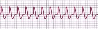
AVRT ECG strip Contributed by Joseph Heaton, MD
References
Zachariah JP, Walsh EP, Triedman JK, Berul CI, Cecchin F, Alexander ME, Bevilacqua LM. Multiple accessory pathways in the young: the impact of structural heart disease. American heart journal. 2013 Jan:165(1):87-92. doi: 10.1016/j.ahj.2012.10.025. Epub 2012 Nov 20 [PubMed PMID: 23237138]
Level 2 (mid-level) evidenceLink MS. Clinical practice. Evaluation and initial treatment of supraventricular tachycardia. The New England journal of medicine. 2012 Oct 11:367(15):1438-48. doi: 10.1056/NEJMcp1111259. Epub [PubMed PMID: 23050527]
Colucci RA, Silver MJ, Shubrook J. Common types of supraventricular tachycardia: diagnosis and management. American family physician. 2010 Oct 15:82(8):942-52 [PubMed PMID: 20949888]
Leung LWM, Gallagher MM. Review paper on WPW and athletes: Let sleeping dogs lie? Clinical cardiology. 2020 Aug:43(8):897-905. doi: 10.1002/clc.23399. Epub 2020 Jun 27 [PubMed PMID: 32592213]
Salerno JC, Seslar SP. Supraventricular tachycardia. Archives of pediatrics & adolescent medicine. 2009 Mar:163(3):268-74. doi: 10.1001/archpediatrics.2008.547. Epub [PubMed PMID: 19255396]
Rodríguez-Mañero M, Fernández-López XA, González-Melchor L, García-Seara J, Martínez-Sande JL, González-Juanatey JR. Permanent junctional reciprocating tachycardia in a patient with an atypically located accessory pathway in the left lateral mitral annulus. Revista portuguesa de cardiologia : orgao oficial da Sociedade Portuguesa de Cardiologia = Portuguese journal of cardiology : an official journal of the Portuguese Society of Cardiology. 2016 Jan:35(1):59.e1-5. doi: 10.1016/j.repc.2015.07.012. Epub 2015 Dec 31 [PubMed PMID: 26749575]
Katritsis DG, Wellens HJ, Josephson ME. Mahaim Accessory Pathways. Arrhythmia & electrophysiology review. 2017 Apr:6(1):29-32. doi: 10.15420/aer.2016:35:1. Epub [PubMed PMID: 28507744]
Kanzaki Y, Morishima I, Furui K, Yamauchi R. The spatial and temporal visualization of the entire atriofascicular fiber conduction during antidromic reciprocating tachycardia. HeartRhythm case reports. 2021 Mar:7(3):150-152. doi: 10.1016/j.hrcr.2020.11.023. Epub 2020 Dec 3 [PubMed PMID: 33786309]
Level 3 (low-level) evidenceLerman BB, Markowitz SM, Cheung JW, Liu CF, Thomas G, Ip JE. Supraventricular Tachycardia: Mechanistic Insights Deduced From Adenosine. Circulation. Arrhythmia and electrophysiology. 2018 Dec:11(12):e006953. doi: 10.1161/CIRCEP.118.006953. Epub [PubMed PMID: 30562103]
Gonzalez JE, Zipse MM, Nguyen DT, Sauer WH. Antidromic Atrioventricular Reciprocating Tachycardia Using a Concealed Retrograde Conducting Left Lateral Accessory Pathway. Cardiac electrophysiology clinics. 2016 Mar:8(1):37-43. doi: 10.1016/j.ccep.2015.10.001. Epub 2016 Jan 11 [PubMed PMID: 26920167]
Ko JK, Deal BJ, Strasburger JF, Benson DW Jr. Supraventricular tachycardia mechanisms and their age distribution in pediatric patients. The American journal of cardiology. 1992 Apr 15:69(12):1028-32 [PubMed PMID: 1561973]
Kylat RI, Samson RA. Permanent junctional reciprocating tachycardia in infants and Children. Journal of arrhythmia. 2019 Jun:35(3):494-498. doi: 10.1002/joa3.12193. Epub 2019 May 15 [PubMed PMID: 31293698]
Page RL, Joglar JA, Caldwell MA, Calkins H, Conti JB, Deal BJ, Estes NA 3rd, Field ME, Goldberger ZD, Hammill SC, Indik JH, Lindsay BD, Olshansky B, Russo AM, Shen WK, Tracy CM, Al-Khatib SM, Evidence Review Committee Chair‡. 2015 ACC/AHA/HRS Guideline for the Management of Adult Patients With Supraventricular Tachycardia: Executive Summary: A Report of the American College of Cardiology/American Heart Association Task Force on Clinical Practice Guidelines and the Heart Rhythm Society. Circulation. 2016 Apr 5:133(14):e471-505. doi: 10.1161/CIR.0000000000000310. Epub 2015 Sep 23 [PubMed PMID: 26399662]
Level 1 (high-level) evidenceHiguchi S, Li R, Gerstenfeld EP, Liem LB, Im SI, Kalantarian S, Ansari M, Abreau S, Barrios J, Scheinman MM, Tison GH. Identification of supraventricular tachycardia mechanisms with surface electrocardiograms using a convolutional neural network. Heart rhythm O2. 2023 Aug:4(8):491-499. doi: 10.1016/j.hroo.2023.07.004. Epub 2023 Jul 13 [PubMed PMID: 37645266]
González-Torrecilla E, Almendral J, Arenal A, Atienza F, Atea LF, del Castillo S, Fernández-Avilés F. Combined evaluation of bedside clinical variables and the electrocardiogram for the differential diagnosis of paroxysmal atrioventricular reciprocating tachycardias in patients without pre-excitation. Journal of the American College of Cardiology. 2009 Jun 23:53(25):2353-8. doi: 10.1016/j.jacc.2009.02.059. Epub [PubMed PMID: 19539146]
González-Torrecilla E, Arenal A, Atienza F, Datino T, Atea LF, Calvo D, Pachón M, Miracle A, Fernández-Avilés F. EGC diagnosis of paroxysmal supraventricular tachycardias in patients without preexcitation. Annals of noninvasive electrocardiology : the official journal of the International Society for Holter and Noninvasive Electrocardiology, Inc. 2011 Jan:16(1):85-95. doi: 10.1111/j.1542-474X.2010.00399.x. Epub [PubMed PMID: 21251139]
Sasmita BR, Luo S, Huang B. Wide and Narrow QRS Complex Tachycardia With Cycle Length Alternans: What Is the Mechanism? Circulation. 2021 Nov 30:144(22):1824-1826. doi: 10.1161/CIRCULATIONAHA.121.057578. Epub 2021 Nov 29 [PubMed PMID: 34843393]
Pediatric and Congenital Electrophysiology Society (PACES), Heart Rhythm Society (HRS), American College of Cardiology Foundation (ACCF), American Heart Association (AHA), American Academy of Pediatrics (AAP), Canadian Heart Rhythm Society (CHRS), Cohen MI, Triedman JK, Cannon BC, Davis AM, Drago F, Janousek J, Klein GJ, Law IH, Morady FJ, Paul T, Perry JC, Sanatani S, Tanel RE. PACES/HRS expert consensus statement on the management of the asymptomatic young patient with a Wolff-Parkinson-White (WPW, ventricular preexcitation) electrocardiographic pattern: developed in partnership between the Pediatric and Congenital Electrophysiology Society (PACES) and the Heart Rhythm Society (HRS). Endorsed by the governing bodies of PACES, HRS, the American College of Cardiology Foundation (ACCF), the American Heart Association (AHA), the American Academy of Pediatrics (AAP), and the Canadian Heart Rhythm Society (CHRS). Heart rhythm. 2012 Jun:9(6):1006-24. doi: 10.1016/j.hrthm.2012.03.050. Epub 2012 May 10 [PubMed PMID: 22579340]
Level 3 (low-level) evidenceBrugada J, Katritsis DG, Arbelo E, Arribas F, Bax JJ, Blomström-Lundqvist C, Calkins H, Corrado D, Deftereos SG, Diller GP, Gomez-Doblas JJ, Gorenek B, Grace A, Ho SY, Kaski JC, Kuck KH, Lambiase PD, Sacher F, Sarquella-Brugada G, Suwalski P, Zaza A, ESC Scientific Document Group. 2019 ESC Guidelines for the management of patients with supraventricular tachycardiaThe Task Force for the management of patients with supraventricular tachycardia of the European Society of Cardiology (ESC). European heart journal. 2020 Feb 1:41(5):655-720. doi: 10.1093/eurheartj/ehz467. Epub [PubMed PMID: 31504425]
Brugada P, Brugada J, Mont L, Smeets J, Andries EW. A new approach to the differential diagnosis of a regular tachycardia with a wide QRS complex. Circulation. 1991 May:83(5):1649-59 [PubMed PMID: 2022022]
Razminia M, Willoughby MC, Demo H, Keshmiri H, Wang T, D'Silva OJ, Zheutlin TA, Jibawi H, Okhumale P, Kehoe RF. Fluoroless Catheter Ablation of Cardiac Arrhythmias: A 5-Year Experience. Pacing and clinical electrophysiology : PACE. 2017 Apr:40(4):425-433. doi: 10.1111/pace.13038. Epub 2017 Mar 3 [PubMed PMID: 28160298]
Abisse S, Adelstein E, Jain S, Saba S. Choice and Utility of Pacing Maneuver in Establishing the Mechanism of Supraventricular Tachycardia: A Single Center Experience. Cardiology research. 2012 Feb:3(1):28-33. doi: 10.4021/cr135w. Epub 2012 Jan 20 [PubMed PMID: 28357021]
Holman WL, Kay GN, Plumb VJ, Epstein AE. Operative results after unsuccessful radiofrequency ablation for Wolff-Parkinson-White syndrome. The American journal of cardiology. 1992 Dec 1:70(18):1490-1 [PubMed PMID: 1442625]
Neumar RW, Otto CW, Link MS, Kronick SL, Shuster M, Callaway CW, Kudenchuk PJ, Ornato JP, McNally B, Silvers SM, Passman RS, White RD, Hess EP, Tang W, Davis D, Sinz E, Morrison LJ. Part 8: adult advanced cardiovascular life support: 2010 American Heart Association Guidelines for Cardiopulmonary Resuscitation and Emergency Cardiovascular Care. Circulation. 2010 Nov 2:122(18 Suppl 3):S729-67. doi: 10.1161/CIRCULATIONAHA.110.970988. Epub [PubMed PMID: 20956224]
Page RL, Joglar JA, Caldwell MA, Calkins H, Conti JB, Deal BJ, Estes NA 3rd, Field ME, Goldberger ZD, Hammill SC, Indik JH, Lindsay BD, Olshansky B, Russo AM, Shen WK, Tracy CM, Al-Khatib SM, Evidence Review Committee Chair‡. 2015 ACC/AHA/HRS Guideline for the Management of Adult Patients With Supraventricular Tachycardia: A Report of the American College of Cardiology/American Heart Association Task Force on Clinical Practice Guidelines and the Heart Rhythm Society. Circulation. 2016 Apr 5:133(14):e506-74. doi: 10.1161/CIR.0000000000000311. Epub 2015 Sep 23 [PubMed PMID: 26399663]
Level 1 (high-level) evidenceMecklin M, Linnanmäki A, Hiippala A, Leino T, Arola A, Leskinen M, Ruotsalainen H, Happonen JM, Poutanen T. Multicenter cohort study on duration of antiarrhythmic medication for supraventricular tachycardia in infants. European journal of pediatrics. 2023 Mar:182(3):1089-1097. doi: 10.1007/s00431-022-04757-5. Epub 2022 Dec 28 [PubMed PMID: 36576576]
Bruder D, Weber R, Gass M, Balmer C, Cavigelli-Brunner A. Antiarrhythmic Medication in Neonates and Infants with Supraventricular Tachycardia. Pediatric cardiology. 2022 Aug:43(6):1311-1318. doi: 10.1007/s00246-022-02853-9. Epub 2022 Mar 8 [PubMed PMID: 35258638]
Helton MR. Diagnosis and Management of Common Types of Supraventricular Tachycardia. American family physician. 2015 Nov 1:92(9):793-800 [PubMed PMID: 26554472]
Janson CM, Millenson ME, Okunowo O, Dai D, Christmyer Z, Tan RB, Ramesh Iyer V, Shah MJ, O'Byrne ML. Incidence of life-threatening events in children with Wolff-Parkinson-White syndrome: Analysis of a large claims database. Heart rhythm. 2022 Apr:19(4):642-647. doi: 10.1016/j.hrthm.2021.12.009. Epub 2021 Dec 10 [PubMed PMID: 34902591]
Skanes AC, Obeyesekere M, Klein GJ. Electrophysiology testing and catheter ablation are helpful when evaluating asymptomatic patients with Wolff-Parkinson-White pattern: the con perspective. Cardiac electrophysiology clinics. 2015 Sep:7(3):377-83. doi: 10.1016/j.ccep.2015.05.002. Epub 2015 Jul 7 [PubMed PMID: 26304516]
Level 3 (low-level) evidenceCáceres C, Del Pilar Garcia Morgado M, Bozo FC, Piletsky S, Moczko E. Rapid Selective Detection and Quantification of β-Blockers Used in Doping Based on Molecularly Imprinted Nanoparticles (NanoMIPs). Polymers. 2022 Dec 11:14(24):. doi: 10.3390/polym14245420. Epub 2022 Dec 11 [PubMed PMID: 36559787]
Borregaard R, Lukac P, Gerdes C, Møller D, Mortensen PT, Pedersen L, Nielsen JC, Jensen HK. Radiofrequency ablation of accessory pathways in patients with the Wolff-Parkinson-White syndrome: the long-term mortality and risk of atrial fibrillation. Europace : European pacing, arrhythmias, and cardiac electrophysiology : journal of the working groups on cardiac pacing, arrhythmias, and cardiac cellular electrophysiology of the European Society of Cardiology. 2015 Jan:17(1):117-22. doi: 10.1093/europace/euu176. Epub 2014 Jul 10 [PubMed PMID: 25013013]
Level 2 (mid-level) evidenceKsiążczyk TM, Pietrzak R, Werner B. Management of Young Athletes with Asymptomatic Preexcitation-A Review of the Literature. Diagnostics (Basel, Switzerland). 2020 Oct 15:10(10):. doi: 10.3390/diagnostics10100824. Epub 2020 Oct 15 [PubMed PMID: 33076240]
Miyoshi T, Maeno Y, Hamasaki T, Inamura N, Yasukochi S, Kawataki M, Horigome H, Yoda H, Taketazu M, Nii M, Hagiwara A, Kato H, Shimizu W, Shiraishi I, Sakaguchi H, Ueda K, Katsuragi S, Yamamoto H, Sago H, Ikeda T, Japan Fetal Arrhythmia Group. Antenatal Therapy for Fetal Supraventricular Tachyarrhythmias: Multicenter Trial. Journal of the American College of Cardiology. 2019 Aug 20:74(7):874-885. doi: 10.1016/j.jacc.2019.06.024. Epub [PubMed PMID: 31416531]
Level 1 (high-level) evidence