 Tools
Tools
 Anatomy, Arteries
Introduction
Arteries constitute a major component of the circulatory system, with veins and the heart as the other main components. Arteries are tubelike structures that transport fluid (ie, blood in the circulatory system and lymph in the lymphatic system) to and from every organ in the body. Arteries primarily transport oxygen, nutrients, and hormones throughout the body. Arteries deliver fresh oxygen to the body after it is loaded onto the Fe2+ at the center of hemoglobin. Oxygen binds to hemoglobin and is transported by the arteries to tissues that lack oxygen. Through a shift in oxygen affinity, it is then unloaded into specific tissues via capillaries with high surface area.[1] Far from being static structures, arteries adapt in response to signals from the central nervous system and to external stimuli, including pressure, temperature, and chemical agents. Vascular nerves innervate arteries, enabling them to respond to stimuli. As catecholamines are released into the bloodstream, nerves signal to arteries to constrict or dilate, thereby altering blood pressure.[2]
Arteries are composed of smooth muscle, which allows constriction and dilation via the parasympathetic nervous system.[3] Arteries differ from veins in that they typically carry oxygenated blood away from the heart and into the rest of the body. However, this is not always the case, as the pulmonary artery transports unoxygenated blood from the heart to the lungs for gas exchange in the alveoli.[4] Additionally, arteries play an important role in maintaining adequate blood flow to the uterus during pregnancy, thereby supporting fetal growth. Arteries play a crucial role in maintaining homeostasis in the body. Additionally, arteries become clogged by a thickening of the plaque, a process known as atherosclerosis.[5]
As individuals age, health issues often manifest as arterial stiffening or thickening. This issue may develop due to a variety of issues, ranging from advanced age, poor diet, sedentary lifestyle, and/or genetic factors such as hypercholesterolemia. As problems arise in the structure of the arteries, it begins leading to more strain on the heart, which develops congestive heart failure, and which is often fatal. More commonly, arteries continue to develop plaque, which eventually leads to an obstruction of blood flow to vital organs, including the heart. Coronary arteries are crucial for supplying the heart with blood; however, like other arteries, they are prone to atherosclerosis in untreated high-risk individuals. Depending on the location of the obstruction in the coronary arteries, blood flow to a particular segment or, in some cases, to the entire heart may be arrested. Ischemia of the cardiac muscle may be evidenced by ST elevations on EKG monitoring strips and by a rise in troponin, a significant marker of cardiac injury. If the ischemia persists, the individual may experience myocardial infarction, otherwise known as a heart attack. The arteries are vital to maintaining a healthy cardiovascular system and, consequently, a healthy lifestyle.
Structure and Function
Register For Free And Read The Full Article
 Search engine and full access to all medical articles
- 10 free questions in your specialty
- Free CME/CE Activities
- Free daily question in your email
- Save favorite articles to your dashboard
- Emails offering discounts
Learn more about a Subscription to StatPearls Point-of-Care
Structure and Function
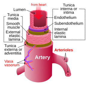
The arteries throughout the body are composed of 3 different layers (see Image. Artery Layers). The innermost layer of the artery, known as the intima, consists of a single layer of endothelial cells overlying a smooth muscle layer; the remainder comprises smooth muscle and elastin. The tunica intima forms a tube through which oxygen-rich blood flows to the appropriate perfusion site. See Image. Structure of an Artery Wall. In this way, there is no leakage from the artery, and nutrient-rich blood can reach the appropriate area before it delivers oxygen and other nutrients. The second layer is known as the media or the middle layer. This media layer consists of smooth muscle that can dilate or constrict, thereby adjusting the pressures experienced by the arterial walls during systolic pumping. As the muscle contracts, the walls experience increased pressure from the left ventricle; similarly, as the vessels dilate, the observed pressure decreases.[6] The last layer is the outermost, known as the adventitia.
The adventitia is crucial for connecting arteries to other tissues in the body, including the vascular nerves that control the smooth muscle in the arteries. In this way, arteries do not move freely throughout the body; instead, they are anchored in place to maintain a consistent and effective cardiovascular system. Usually, arteries are under the greatest pressure because they receive blood from the left ventricle, the heart's most powerful chamber. However, the pulmonary artery is different for 2 reasons. Not only does it move unoxygenated blood from the heart to the lungs, but it also operates at a lower pressure because the right ventricle exerts less force than the left. The pulmonary artery delivers blood to the lungs, whereas the remaining arteries deliver blood to specific areas of the body. Blood from the left ventricle is pumped through the aorta, the largest artery. The aorta then splits into 4 sections: the ascending aorta, the aortic arch, which curves over the heart, the descending thoracic aorta, and the abdominal aorta. The ascending aorta arises from the heart and further branches into the carotid artery, which supplies the brain with blood.[7]
The aortic arch is similar to the ascending aorta and carries blood up to the back and neck. The descending aorta brings blood from the heart to smaller vessels in the chest or ribs. Finally, the abdominal aorta supplies blood to the iliac arteries, which provide blood flow to many organs in the abdominal region.[8]
Embryology
During development, arteries continually remodel to maintain homeostasis in changing environments. The heart and arteries develop from the mesoderm germ layer as it is composed of smooth muscle. After the mesoderm differentiates into segments of the circulatory system through specific growth factor signaling, arteries begin to form, and circulation, powered by the heart, initiates. At the beginning of development, the arteries form from the pharyngeal arches. Each arch gives rise to a specific arterial segment during the 9-month pregnancy.
During pregnancy, the fetus shares a circulatory system with the mother, resulting in unique blood flow patterns. The uterine arteries carry blood from the mother to the placenta, where it perfuses the placenta and then flows to the fetus, supplying it with oxygen. Fetal hearts also have a unique blood flow as blood does not become oxygenated in its lungs but instead by the mother. A fetus has a unique connection between the aorta and the pulmonary artery called the ductus arteriosus.[9] This connection allows blood to flow past the fetal lungs, which are not functional, while the fetus is in the amniotic sac.
Muscles
In all of these examples, arteries are comprised of smooth muscle. They are nonstriated muscles that surround the vessels, providing support and integrity to the entire artery. This smooth muscle responds to various signals and innervation to constrict or dilate, thereby maintaining a stable blood pressure, including via epinephrine and angiotensin II. The sympathetic nervous system can release high levels of epinephrine, which activates alpha-adrenergic receptors, thereby causing arterial constriction. This increase in pressure can aid in perfusion during both trauma and hormonal imbalance.[10]
Additionally, the kidney can indirectly affect blood pressure through its release of renin. Renin catalyzes the formation of angiotensin II, a potent vasoconstrictor.[11] Angiotensin II interacts directly with smooth muscle in arterial walls, causing vasoconstriction when blood pressure falls below normal. Additionally, when blood pressure decreases due to bleeding or peripheral arterial dilation, as in shock, the arteries interact with the renin-angiotensin-aldosterone system to compensate for the decrease.
As arteries age, they harden and become less compliant, creating a systemic imbalance that can lead to secondary hypertension through reduced renal glomerular filtration rate. Therefore, through the endocrine system, hormones are released into the bloodstream, which then signal the vascular nerves to alter their tone to maintain homeostasis.
Surgical Considerations
The different surgical considerations with regard to arterial anatomy are:
- Aneurysms - May need surgical ligation, stenting, or clipping based on the location
- Arteriovenous fistula - May need separation of the vessels
- Ligation of an arterial bleeder - May need a trans-fixation suture for major arteries
- Thrombo-embolism - May need open/interventional radiology-assisted evacuation
Clinical Significance
There are many additional ways in which arteries help maintain balance, including through vasodilators such as histamine and serotonin. This, however, can become dangerous if balance is not maintained. An angiogram, or the insertion of a catheter and contrast agent into an artery, can be used to monitor blood flow and identify bleeding or blockages. However, environmental factors also influence arterial structure. For example, during anaphylaxis shock, massive amounts of histamine are released into the body, causing the arteries to dilate to an unsafe amount. This reduces pressure for the perfusion of nutrient-rich blood. In an attempt to compensate for this loss, the body enters a state of compensated shock, as indicated by increased heart rate. This does not work forever, and without dilation, the patient soon goes into a state of shock. In addition to being balanced, many medical issues may arise, further complicating the situation.
One of the most common medical conditions affecting arteries is atherosclerosis, in which arteries accumulate plaque, impairing normal cardiovascular function. This accumulation of plaque can occur anywhere, allowing a blockage to develop. A myocardial infarction occurs if the blood supply to the heart is cut off.[12] Similarly, if blood flow to the brain is blocked, this is known as a stroke. A lack of blood supply to any area of the body can cause permanent severe damage or even death. Ischemia of any vital organ is severe and may present in a wide variety of forms. For example, if the blood supply to the retina is cut off, one may develop amaurosis fugax or blindness.[13] Trans fats and high cholesterol have been shown to adversely affect the arteries, leading to atherosclerosis. Therefore, it is crucial to maintain a healthy lifestyle through diet and exercise. Individuals who maintain a healthy lifestyle yet have elevated LDL levels should also be screened for genetic causes, such as familial hypercholesterolemia.
Individuals with a history of stroke or transient ischemic attack (TIA) should be evaluated with a carotid ultrasound to assess the level of stenosis. Vascular surgery may be performed to further dilate the arterial lumen, depending on the degree of stenosis or obstruction in these major vessels. This process is often achieved by balloon angioplasty and stenting of the artery via subclavian artery access. This procedure is minimally invasive and referred to as TCAR. If TCAR is unsuccessful, an endarterectomy may be performed to open the artery and remove any atherosclerotic plaque buildup directly. The cardiovascular system is essential for keeping all other systems in the body. Arteries, in particular, are vital in supplying nutrients, including oxygen, to the rest of the body. It is crucial to recognize the role of arteries in the cardiovascular system to prevent future medical complications.
Media
(Click Image to Enlarge)

(Click Image to Enlarge)
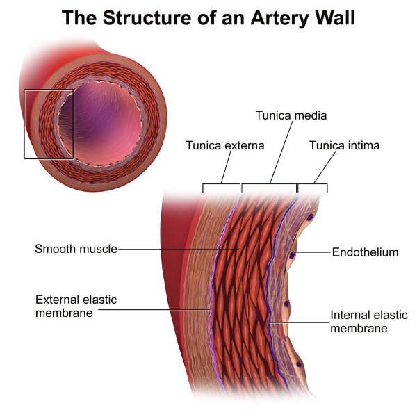
Structure of an Artery Wall.
Blausen.com staff. Medical Gallery of Blausen Medical 2014. WikiJournal of Medicine. doi: 10.15347/wjm/2014.010.
ISSN 2002-4436. [CC BY 3.0 (https://creativecommons.org/licenses/by/3.0)] via Wikimedia Commons.
References
El Shahawy MS, Shady ZM, Gaafar A. The Efficacy of Argon Plasma Coagulation versus Carvedilol for Treatment of Portal Hypertensive Gastropathy. Digestion. 2020:101(6):651-658. doi: 10.1159/000502814. Epub 2019 Sep 27 [PubMed PMID: 31563912]
Sjöberg RL, Wu WY, Dahlin AM, Tsavachidis S, Gliogene Group, Bondy ML, Melin B. Role of monoamine-oxidase-A-gene variation in the development of glioblastoma in males: a case control study. Journal of neuro-oncology. 2019 Nov:145(2):287-294. doi: 10.1007/s11060-019-03294-w. Epub 2019 Sep 25 [PubMed PMID: 31556016]
Level 2 (mid-level) evidenceWentzel A, Malan L, von Känel R, Smith W, Malan NT. Heart rate variability, the dynamic nature of the retinal microvasculature and cardiac stress: providing insight into the brain-retina-heart link: the SABPA study. Eye (London, England). 2020 May:34(5):835-846. doi: 10.1038/s41433-019-0515-y. Epub 2019 Jul 5 [PubMed PMID: 31278382]
Richardson M. The physiology of mucus and sputum production in the respiratory system. Nursing times. 2003 Jun 10-16:99(23):63-4 [PubMed PMID: 12838653]
Yeh HC, Lin YT, Ting IW, Huang HC, Chiang HY, Chung CW, Chang SN, Kuo CC. Variability of red blood cell size predicts all-cause mortality, but not progression to dialysis, in patients with chronic kidney disease: A 13-year pre-ESRD registry-based cohort. Clinica chimica acta; international journal of clinical chemistry. 2019 Oct:497():163-171. doi: 10.1016/j.cca.2019.07.035. Epub 2019 Jul 30 [PubMed PMID: 31374189]
Valbusa F, Angheben A, Mantovani A, Zerbato V, Chiampan A, Bonapace S, Rodari P, Agnoletti D, Arcaro G, Fava C, Bisoffi Z, Targher G. Increased aortic stiffness in adults with chronic indeterminate Chagas disease. PloS one. 2019:14(8):e0220689. doi: 10.1371/journal.pone.0220689. Epub 2019 Aug 2 [PubMed PMID: 31374101]
Furuta A, Morimoto H, Mukai S, Futagami D, Okubo S. Valve-sparing partial root repair for aortic dissection limited to the right coronary sinus of Valsalva. Journal of cardiac surgery. 2019 Oct:34(10):1133-1136. doi: 10.1111/jocs.14203. Epub 2019 Aug 2 [PubMed PMID: 31374594]
Yan GW, Li HW, Yang GQ, Bhetuwal A, Liu JP, Li Y, Fu QS, Zhao LW, Chen H, Hu N, Wu L, Yan J, Wang W, Shuang JY, Ge J. Iatrogenic arteriovenous fistula of the iliac artery after lumbar discectomy surgery: a systematic review of the last 18 years. Quantitative imaging in medicine and surgery. 2019 Jun:9(6):1163-1175. doi: 10.21037/qims.2019.05.12. Epub [PubMed PMID: 31367570]
Level 1 (high-level) evidencePeter ID, Oladele DM, Kefas GJ, Kayode OV, Iseko II. Challenges with Managing Delayed Presentation of Persistent Truncus Arteriosus with Torrential Pulmonary Blood Flow in a Resource-Limited Setting. Journal of cardiovascular echography. 2019 Apr-Jun:29(2):75-77. doi: 10.4103/jcecho.jcecho_69_18. Epub [PubMed PMID: 31392125]
Stannov SU, Brasen JC, Salomonsson M, Holstein-Rathlou NH, Sorensen CM. Interactions between renal vascular resistance and endothelium-derived hyperpolarization in hypertensive rats in vivo. Physiological reports. 2019 Aug:7(15):e14168. doi: 10.14814/phy2.14168. Epub [PubMed PMID: 31368238]
Lu Q, Davel AP, McGraw AP, Rao SP, Newfell BG, Jaffe IZ. PKCδ Mediates Mineralocorticoid Receptor Activation by Angiotensin II to Modulate Smooth Muscle Cell Function. Endocrinology. 2019 Sep 1:160(9):2101-2114. doi: 10.1210/en.2019-00258. Epub [PubMed PMID: 31373631]
Chu AA, Li W, Zhu YQ, Meng XX, Liu GY. Effect of coronary collateral circulation on the prognosis of elderly patients with acute ST-segment elevation myocardial infarction treated with underwent primary percutaneous coronary intervention. Medicine. 2019 Aug:98(31):e16502. doi: 10.1097/MD.0000000000016502. Epub [PubMed PMID: 31374011]
Inan S, Yavas G, Inan UU. Homocysteinemia as a cause for amaurosis fugax in a patient without an apparent embolic source. Romanian journal of ophthalmology. 2019 Apr-Jun:63(2):188-192 [PubMed PMID: 31334400]