 Tools
Tools
 Carotid Artery Stenting
Introduction
Traditionally, carotid endarterectomy (CEA) has been the primary treatment for high-grade asymptomatic and symptomatic carotid artery stenosis. A carotid endarterectomy involves exposure of the carotid artery and removal of plaque, most typically from the carotid bulb and the proximal internal carotid artery, via a neck incision. However, in vascular surgery, as in many other surgical specialties, minimally invasive techniques have evolved over the years. These techniques offer the advantages of smaller incisions, reduced postoperative pain, reduced risk of postoperative wound complications, and a shorter hospital length of stay. Carotid artery stenting (CAS) is one such technique that can be performed via a transfemoral or transcarotid approach.[1][2][3]
Anatomy and Physiology
Register For Free And Read The Full Article
 Search engine and full access to all medical articles
- 10 free questions in your specialty
- Free CME/CE Activities
- Free daily question in your email
- Save favorite articles to your dashboard
- Emails offering discounts
Learn more about a Subscription to StatPearls Point-of-Care
Anatomy and Physiology
The common carotid artery (CCA) arises from the aortic arch (left) or the brachiocephalic trunk (right) and divides into the internal and external carotid arteries (ICA and ECA). The internal carotid artery provides blood to the brain, while the external carotid artery provides blood to the face, scalp, and neck. The internal and external carotid arteries form collaterals at several locations. This helps maintain blood flow through collateral circulation if either artery becomes occluded. The aortic arch can be divided into 3 anatomic types.
To simplify, an imaginary horizontal line is drawn through the top of the aortic arch, and imaginary parallel lines marking the origins of the great vessels coming off the arch are drawn for comparison. For a type 1 arch, all great vessels originate close to the imaginary horizontal line drawn through the top of the aortic arch. If the origins of all great vessels fall within the second parallel line, this is considered a type 2 aortic arch. For a type 3 aortic arch, the origins of all great vessels fall within the third parallel line. Angulation, as it relates to the type of aortic arch, has important implications for CAS.
Indications
CAS is a reasonable alternative to CEA in select patients with high-grade asymptomatic (more than 70%) or symptomatic CAS. Indications for CAS include high surgical patient risk, such as severe pulmonary disease, recent myocardial infarction, unstable angina, or severe congestive heart failure; a history of prior neck radiation that is anticipated to make open surgical dissection difficult; a history of damage to contralateral vocal cords; the presence of a tracheostomy, contralateral carotid occlusion; and previous CEA with recurrent stenosis.[4][5][6][7]
Contraindications
The main contraindication to CAS via a transfemoral approach is unfavorable aortic arch anatomy. This can include a heavily calcified aortic arch or a type 3 aortic arch. A relative contraindication is a history of a severe allergic reaction to intravenous contrast dye. However, patients can be premedicated to mitigate some of this risk. For a transcarotid approach, a CCA length of less than 5 cm from the clavicle is considered inadequate.
Equipment
CAS requires meticulous preparation and specialized equipment to ensure procedural precision, neuroprotection, and patient safety. Core components include an ultrasound unit for vascular access and imaging guidance, a fluoroscopic imaging system (such as a C-arm) for real-time visualization, lead shielding for radiation protection, sterile drapes, heparinized saline, and systemic heparin for anticoagulation. An activated clotting time (ACT) machine should be available to verify adequate anticoagulation, maintaining an ACT greater than 250 seconds after heparin administration.
Transfemoral Approach
The transfemoral approach remains the traditional route for CAS and requires equipment to facilitate percutaneous access and precise stent deployment. Necessary instruments include:
- Micropuncture kit for percutaneous access of the common femoral artery
- Guidewires: 0.035-inch and 0.014-inch
- 6 French short and long sheaths
- Angled catheters for selective vessel engagement and contrast injection
- Angioplasty balloons for pre- and postdilation of the lesion
- An embolic protection device or flow reversal system to minimize cerebral embolization
Transcarotid Approach
The transcarotid artery revascularization (TCAR) approach involves direct access to the common carotid artery, reducing the risk of aortic arch manipulation and embolic events. Equipment required includes:
- Surgical tray with standard vascular instruments
- Vascular clamps to facilitate surgical exposure of the common carotid artery at the base of the neck
- Micropuncture kit for direct arterial puncture
- Guidewires, sheaths, and angioplasty balloons, similar to the transfemoral technique
- Flow reversal system for neuroprotection against potential embolization
Additional Considerations
Regardless of approach, heparinized saline, strict sterile technique, and continuous ACT monitoring are essential to procedural safety. Real-time imaging and embolic protection strategies significantly reduce periprocedural stroke risk. The coordinated use of this equipment ensures precise stent placement, optimal revascularization, and improved neurologic outcomes.
Personnel
CAS is a complex, high-stakes procedure that requires a multidisciplinary team with advanced technical expertise and coordinated communication to optimize outcomes and patient safety.
The core procedural team typically includes:
- Interventionalist (vascular surgeon, interventional radiologist, or interventional cardiologist) experienced in endovascular techniques and carotid anatomy
- Assistant or scrub nurse/technologist, trained in catheter and wire handling, balloon and stent preparation, and maintenance of a sterile field
- Circulating nurse, responsible for patient monitoring, medication administration (eg, heparin, antiplatelets), and coordination between team members
- Radiologic technologist, responsible for fluoroscopic operation, imaging optimization, and radiation safety compliance
- Anesthesiologist or nurse anesthetist, providing sedation, hemodynamic management, and airway support as needed
- Neurophysiologist (when available), for intraoperative neurologic monitoring and early detection of cerebral ischemia
For TCAR, an additional surgical assistant or vascular-trained scrub is essential to assist with carotid exposure, vascular control, and the setup of the flow reversal system. Optimal outcomes depend on team coordination, preprocedural briefing, and adherence to established protocols for anticoagulation, imaging, and neuroprotection. Simulation training and morbidity and mortality reviews enhance procedural efficiency and patient safety across carotid intervention institutions.
Preparation
Comprehensive preprocedural preparation is critical for optimizing patient outcomes and minimizing complications during CAS. Preparation begins with a detailed medical history, neurologic assessment, and review of comorbidities. Imaging, via Duplex ultrasonography, computed tomography angiography (CTA), or magnetic resonance angiography (MRA), is used to confirm stenosis, assess vessel tortuosity, and evaluate aortic arch anatomy. Patients should receive dual antiplatelet therapy (aspirin and clopidogrel) at least 3 to 5 days before the procedure or a loading dose if urgent intervention is needed. All patients should be asked explicitly about aspirin and clopidogrel use in the preoperative holding area to confirm compliance.
For transfemoral CAS, most physicians perform the procedure under local anesthesia with or without sedation to allow continuous neurologic monitoring. For the transcarotid approach, either local anesthesia with sedation or general anesthesia may be employed; if general anesthesia is chosen, intraoperative electroencephalogram (EEG) or brain mapping is recommended to detect ischemic changes early. Regardless of the approach, patients must be closely monitored for hemodynamic instability—CAS can induce bradycardia or hypotension due to carotid sinus stimulation. Anesthesia personnel should have vasoactive medications readily available, and an arterial line is recommended for continuous invasive hemodynamic monitoring.
The procedure should be performed in a hybrid operating room or interventional angiography suite equipped with high-resolution fluoroscopy, hemodynamic monitoring, and radiation protection (lead aprons, thyroid shields, dosimeters). The operative field must be shaved, prepped with chlorhexidine, and draped using full sterile technique. Heparinized saline and heparin are prepared in advance, and the ACT should be monitored to maintain levels above 250 seconds.
For patient positioning, those undergoing a transfemoral approach should be placed supine with the neck turned to the contralateral side, and both groins prepped and draped sterilely. For transcarotid CAS, the patient should also be supine, with a shoulder roll placed under the scapulae and the neck rotated away from the operative side. The neck and groin should be prepped, draped sterilely, and arms tucked. Equipment—including guidewires, sheaths, stents, angioplasty balloons, and embolic protection or flow reversal devices—should be verified and ready for use before access is obtained. Effective team communication, adherence to sterile technique, and vigilant neurologic and hemodynamic monitoring are essential to procedural safety and success.
Technique or Treatment
Transfemoral Approach
The transfemoral approach remains the most commonly employed method for CAS, offering excellent maneuverability and familiarity for most interventionalists (see Image. Transfemoral Carotid Artery Stenting, Right Common Femoral Artery via Micropuncture). However, it can be limited by aortic arch tortuosity, type 3 arch configuration, severe atherosclerosis, or calcifications, which may increase procedural risk and embolic potential. Under ultrasound guidance, percutaneous access to the common femoral artery is obtained using a micropuncture kit and the Seldinger technique. A femoral angiogram should be performed to confirm appropriate entry into the common femoral artery and assess suitability for a vascular closure device at the procedure’s conclusion.
A 6 French short sheath is then advanced over a 0.035-inch guidewire. Using fluoroscopy, the wire is navigated carefully into the aortic arch. With an angled catheter in a left anterior oblique projection, the CCA is selectively cannulated, and the anatomy is mapped. The guidewire is advanced into the ECA, followed by advancement of the catheter itself. Over a stiffer wire, a shuttle sheath or guide catheter is advanced into the CCA for stability.
Dedicated carotid arteriography is performed in multiple projections to identify the stenotic lesion and assess collateral circulation. A 0.014-inch microwire is advanced across the lesion, after which an embolic protection device—typically a distal filter or proximal flow-reversal system—is deployed to minimize risk of cerebral embolization. Predilation with a low-profile angioplasty balloon is performed cautiously, with close attention to hemodynamic changes (bradycardia or hypotension due to carotid baroreceptor stimulation). After predilation, an appropriately sized self-expanding stent is deployed across the lesion. If residual stenosis >30% persists postdeployment, postdilation balloon angioplasty may be performed.
Upon completion, carotid and cerebral angiography confirm stent patency, adequate flow restoration, and absence of distal emboli. All devices are removed, and the arteriotomy site in the femoral artery is closed using a vascular closure device. The patient is monitored for groin complications and hemodynamic stability post-procedure.
Transcarotid Approach
The transcarotid approach, often called TCAR, provides a direct route to the carotid artery and uses flow-reversal neuroprotection, which has been shown to reduce embolic events compared to the transfemoral route. This technique begins with surgical exposure of the CCA at the base of the neck, typically guided by duplex ultrasound and anatomic landmarks to place the incision.
After prepping and draping, dissection proceeds through the subcutaneous tissues using electrocautery, followed by division of the platysma. The CCA is circumferentially dissected and controlled with vessel loops or umbilical tape, with additional proximal and distal exposure to facilitate vascular control. The artery is accessed directly using a micropuncture kit, and an initial angiogram confirms anatomy and lesion characteristics. A guidewire is placed, and the arterial sheath component of the flow-reversal system is introduced.
The common femoral vein is then accessed under ultrasound guidance, and a 0.035-inch guidewire is used to insert the venous sheath of the flow-reversal system. The arterial and venous sheaths are connected, establishing temporary flow reversal that diverts potential emboli away from the cerebral circulation. Carotid angiography is performed in multiple projections, and the lesion is traversed with a guidewire. The proximal CCA is clamped to maintain reverse flow during intervention. Predilation is performed, followed by deployment of a self-expanding stent. Any residual stenosis that is 30% or greater is managed with balloon angioplasty.
Following a completion angiogram confirming satisfactory results, the flow-reversal system is disconnected, and sheaths and wires are removed. The arteriotomy is closed with Prolene suture after clamping the distal CCA, and the neck incision is closed in layers. The femoral venous sheath is removed, and manual pressure is applied until hemostasis is achieved. Both transfemoral and transcarotid techniques require meticulous hemodynamic monitoring, neuroprotection, and interdisciplinary coordination between interventionalists, anesthesiologists, and nursing staff. Choice of approach depends on patient anatomy, comorbidities, and operator expertise, with transcarotid methods increasingly favored in patients with complex arch anatomy or high embolic risk.
Complications
Complications following CAS vary depending on the access route and patient factors, but can involve both local vascular injury and serious cerebrovascular events. For the transfemoral approach, complications are often related to femoral artery access, including bleeding, arterial perforation, dissection, thrombosis, pseudoaneurysm formation, and distal embolization. Access-site hematomas and retroperitoneal hemorrhage are also potential risks, particularly in anticoagulated patients. In both transfemoral and transcarotid approaches, procedural complications may include carotid artery dissection, thromboembolism, incomplete stent deployment, and stent fracture or migration in rare cases.
Neurologic complications represent the most serious category, with stroke and transient ischemic attacks resulting from embolization of atheromatous debris during wire manipulation, balloon inflation, or stent placement. Hyperperfusion syndrome, characterized by severe headache, seizures, or intracerebral hemorrhage, may develop postrevascularization due to impaired cerebrovascular autoregulation. Careful blood pressure control is essential in the postoperative period to mitigate this risk.
Hemodynamic instability, specifically bradycardia and hypotension, is typical during angioplasty or stent deployment due to carotid sinus stimulation and typically responds to atropine or vasopressors. Access-site infections, contrast-induced nephropathy, and allergic reactions to contrast agents are less frequent but essential considerations. Long-term complications include in-stent restenosis from neointimal hyperplasia (occurring in 5%–10% of cases) and stent thrombosis, underscoring the need for adherence to dual antiplatelet therapy (aspirin and clopidogrel). Prevention of complications relies on careful patient selection, meticulous procedural technique, and interprofessional coordination between surgeons, anesthesiologists, nurses, and technologists. Neuroprotection devices, close hemodynamic monitoring, and timely recognition of access or stent-related issues are critical for minimizing morbidity and optimizing patient outcomes.
Clinical Significance
Multiple studies have compared CAS with CEA in symptomatic and asymptomatic patients. The risk of myocardial ischemia appears to be higher with CEA, while the risk of a cerebrovascular accident appears to be higher with CAS. Restenosis rates appear similar between CEA and CAS at 2 years. As a procedure, CAS appears to have worse outcomes in patients aged 70 and over and patients with a high burden of white-matter lesions on brain imaging. Therefore, patient selection is important for CAS. The transcarotid approach of CAS potentially reduces the risk of embolization in a hostile aortic arch. More experience with this technique is being gained in ongoing trials. Hospitalization and long-term costs of CAS and CEA appear similar.[8][9][10]
Because randomized controlled trials have demonstrated a higher periprocedural stroke risk with transfemoral carotid artery stenting (TF-CAS) compared with CEA, current guidelines restrict its indications. Consensus supports the use of TF-CAS primarily in symptomatic patients with high-grade stenosis who are considered high medical risk for open surgery. In contrast, TF-CAS use in low- or moderate-risk patients, particularly those without prior neck radiation, prior carotid surgery, or other anatomic contraindications to CEA, remains less well supported. One report suggests a potential benefit in lower-medical-risk patients with high anatomic risk factors for CEA, such as restenosis or prior neck radiation. However, many experts continue to express concern regarding the long-term durability of TF-CAS.
Accordingly, societal guidelines remain cautious. The Society for Vascular Surgery guidelines specifically recommend that asymptomatic high-surgical-risk cases be managed with optimal medical therapy rather than carotid stenting. Notably, most existing guidelines were established before the advent of TCAR; as a result, the role of this newer technique has yet to be clearly defined.
Enhancing Healthcare Team Outcomes
Optimal outcomes in carotid artery stenting (CAS) rely on a coordinated, multidisciplinary approach that integrates the skills and strategies of clinicians, nurses, pharmacists, and allied health professionals. Interventionalists must possess expertise in endovascular techniques, imaging interpretation, and real-time management of hemodynamic instability. Advanced practitioners and nurses are essential in preprocedural assessment, patient education, and postprocedural monitoring for complications such as access-site bleeding, stroke symptoms, or hemodynamic fluctuations. Pharmacists contribute to patient safety by optimizing antiplatelet and anticoagulant therapy, monitoring for drug interactions, and ensuring appropriate perioperative medication management. Collectively, these professionals implement strategies that emphasize patient-centered decision-making, adherence to evidence-based protocols, and meticulous procedural planning to reduce risk and improve outcomes.
Effective interprofessional communication and care coordination are crucial throughout all stages of CAS—from preoperative evaluation through postoperative follow-up. Clear, structured communication ensures that all team members consistently understand diagnostic findings, procedural plans, and evolving patient conditions. Regular interdisciplinary briefings, standardized checklists, and timely electronic documentation foster seamless care transitions. By collaborating across disciplines, healthcare professionals can anticipate complications early, enhance procedural safety, and deliver comprehensive care that improves stroke prevention, functional recovery, and patient satisfaction while reinforcing a culture of teamwork and continuous quality improvement.
Media
(Click Image to Enlarge)
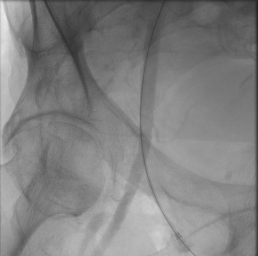
Transfemoral Carotid Artery Stenting, Right Common Femoral Artery via Micropuncture. This image shows the micropuncture system accessing the right common femoral artery during transfemoral carotid artery stenting. This angiogram shows adequate entry into the artery below the most superior aspect of the femoral head, a useful fluoroscopic landmark; there is no dissection in the artery.
Contributed by T Saleem, MD
References
Texakalidis P, Chaitidis N, Giannopoulos S, Giannopoulos S, Machinis T, Jabbour P, Rivet D, Reavey-Cantwell J, Rangel-Castilla L. Carotid Revascularization in Older Adults: A Systematic Review and Meta-Analysis. World neurosurgery. 2019 Jun:126():656-663.e1. doi: 10.1016/j.wneu.2019.02.030. Epub 2019 Feb 22 [PubMed PMID: 30797928]
Level 1 (high-level) evidenceGaba K, Bulbulia R. Identifying asymptomatic patients at high-risk for stroke. The Journal of cardiovascular surgery. 2019 Jun:60(3):332-344. doi: 10.23736/S0021-9509.19.10912-3. Epub 2019 Feb 20 [PubMed PMID: 30785251]
Mukherjee D, Roffi M. Minimizing Distal Embolization During Carotid Artery Stenting. JACC. Cardiovascular interventions. 2019 Feb 25:12(4):404-405. doi: 10.1016/j.jcin.2018.12.024. Epub [PubMed PMID: 30784648]
Rasheed AS, White RS, Tangel V, Storch BM, Pryor KO. Carotid Revascularization Procedures and Perioperative Outcomes: A Multistate Analysis, 2007-2014. Journal of cardiothoracic and vascular anesthesia. 2019 Jul:33(7):1963-1972. doi: 10.1053/j.jvca.2019.01.022. Epub 2019 Jan 9 [PubMed PMID: 30773439]
Lan Y, Lyu J, Ma X, Ma L, Lou X. Longitudinal assessment of cerebral blood flow changes following carotid artery stenting and endarterectomy. La Radiologia medica. 2019 Jul:124(7):636-642. doi: 10.1007/s11547-018-00986-7. Epub 2019 Feb 15 [PubMed PMID: 30771219]
Chung JW, Kim SJ, Hwang J, Lee MJ, Lee J, Lee KY, Park MS, Sung SM, Kim KH, Jeon P, Bang OY. Comparison of Clopidogrel and Ticlopidine/Ginkgo Biloba in Patients With Clopidogrel Resistance and Carotid Stenting. Frontiers in neurology. 2019:10():44. doi: 10.3389/fneur.2019.00044. Epub 2019 Jan 30 [PubMed PMID: 30761076]
Lim S, Javorski MJ, Nassoiy SP, Park Y, Halandras PM, Bechara CF, Aulivola B, Crisostomo PR. Long-Term Hemodynamic Effects After Carotid Artery Revascularization. Vascular and endovascular surgery. 2019 May:53(4):297-302. doi: 10.1177/1538574419828088. Epub 2019 Feb 11 [PubMed PMID: 30744510]
Xin WQ, Li MQ, Li K, Li QF, Zhao Y, Wang WH, Gao YK, Wang HY, Yang XY. Systematic and Comprehensive Comparison of Incidence of Restenosis Between Carotid Endarterectomy and Carotid Artery Stenting in Patients with Atherosclerotic Carotid Stenosis. World neurosurgery. 2019 May:125():74-86. doi: 10.1016/j.wneu.2019.01.118. Epub 2019 Jan 31 [PubMed PMID: 30710719]
Level 1 (high-level) evidenceHintze AJ, Greenleaf EK, Schilling AL, Hollenbeak CS. Thirty-day Readmission Rates for Carotid Endarterectomy Versus Carotid Artery Stenting. The Journal of surgical research. 2019 Mar:235():270-279. doi: 10.1016/j.jss.2018.10.011. Epub 2018 Nov 1 [PubMed PMID: 30691806]
Clavel P, Hebert S, Saleme S, Mounayer C, Rouchaud A, Marin B. Cumulative incidence of restenosis in the endovascular treatment of extracranial carotid artery stenosis: a meta-analysis. Journal of neurointerventional surgery. 2019 Sep:11(9):916-923. doi: 10.1136/neurintsurg-2018-014534. Epub 2019 Jan 31 [PubMed PMID: 30705056]
Level 1 (high-level) evidence