 Tools
Tools
 Embryology, Optic Cup
Introduction
Eye development is a complex, multifactorial process organized in both temporal and spatial patterns during embryogenesis. PAX6 is the master control gene, modulated by various signaling pathways governing optic vesicle induction, patterning, and ocular structural specification. Cellular differentiation involves neuroectoderm formation of the optic cup, surface ectoderm-derived lens and anterior segment structures, and neural crest contribution to corneal, trabecular, scleral, and optic nerve connective tissues. The optic cup plays a central role in ocular development, contributing to the formation of all major structures of the globe, with the exception of the lens. Conditions such as congenital glaucoma, retinal detachment, and coloboma are associated with defects of the optic cup or its associated structures and may result in significant visual impairment. Understanding ocular embryogenesis provides a basis for elucidating the pathogenesis of congenital ocular diseases.
Development
Register For Free And Read The Full Article
 Search engine and full access to all medical articles
- 10 free questions in your specialty
- Free CME/CE Activities
- Free daily question in your email
- Save favorite articles to your dashboard
- Emails offering discounts
Learn more about a Subscription to StatPearls Point-of-Care
Development
Development of the primitive eye begins at approximately week 3 of gestation with the emergence of the optic sulcus from periocular mesenchyme. The optic sulci are bilateral invaginations on both sides of the anterior neural tube at the level of the diencephalon. Progressive deepening of the sulci leads to the formation of optic pits, followed by transformation into optic vesicles as extension toward the surface ectoderm occurs. Surface ectoderm and optic vesicles both secrete extracellular matrix and establish adhesion between the 2 structures. Continued outward expansion of the optic vesicles shifts the proximal attachment to the forebrain into the optic stalk. Migration of neural crest cells is facilitated by the optic vesicles, with a contribution to subsequent ocular development.
At approximately week 5 of gestation, a portion of the optic vesicle undergoes auto-invagination, forming a 2-layered optic cup. Asymmetric invagination results in the formation of a groove along the inferior margin. This structure is designated the optic fissure, permitting entry of early vasculature and periocular mesenchyme into the developing eye. Closure of the optic fissure occurs by the end of week 6.[1][2][3][4]
The 2 layers of the optic cup constitute a continuous tissue that bends at a hinge point termed the "optic cup lip," which delineates the border between the outer pigmented layer and the inner neural layer.[5] The anterior portion of the optic cup gives rise to the pars caeca retinae.[6] Subsequent development of this structure leads to the formation of the ciliary body, iris, and pupillary muscles.[7] The posterior portion of the optic cup forms the retina.[8] The outer layer of the optic cup differentiates into the retinal pigmented epithelium. The inner layer of the optic cup differentiates into the outer nuclear layer, containing rods and cones, the inner nuclear layer, containing bipolar cells, and the ganglion cell layer, containing ganglion cells. Axonal growth into the optic stalk begins during week 7 of gestation, and completion of optic nerve development occurs by week 8.
Cellular
Neuroectodermal cells comprise the optic cup.[9] Direct derivatives of the optic cup are described in the preceding section. However, embryologic origins of additional ocular structures are relevant. Surface ectoderm contributes to the development of the lens, conjunctiva, eyelids, and corneal epithelium. The optic cup lip forms the adult iris margin. Neural crest cells give rise to the corneal endothelium, trabecular meshwork, scleral fibroblasts, connective fascia of extraocular muscles, and meninges of the optic nerve.
Molecular Level
Multiple genes interact during the development of the primitive eye, consistent with the embryogenesis of other organ systems. In ocular development, PAX6 functions as the primary regulatory gene while WNT (wingless-related integration site) and FGF (fibroblast growth factor) signaling provide supportive roles during optic vesicle development.[10][11][12] Sonic hedgehog (Shh) signaling is another key regulator of ocular embryogenesis and mediates suppression of PAX6 expression.[13] Inhibition of Shh signaling results in dysregulated PAX6 expression and cyclopia. Conversely, increased Shh signaling results in loss of ocular structures. Retinoic acid also plays an essential role in ocular development, primarily through paracrine regulation of mesenchymal tissue surrounding the optic cup. Retinoic acid deficiency leads to anterior segment malformations and may result in blindness.
However, the interplay between genes and molecular signaling factors is highly complex and extends beyond the scope of this article. Additional genes involved in ocular development include, but are not limited to, PITX2, PITX3, FOXC1, FOXE3, LMX1B, GPR48, TFAP2A, and TFAP2B.[14][15]
Function
The optic cup gives rise to the entire globe and its associated internal structures, with the exception of the lens (see Image. Development of the Embryonic Eye). Retinal development includes the formation of 10 layers containing rods, cones, bipolar cells, amacrine cells, and horizontal cells, along with the organized synaptic interactions among these neuronal populations that process photic input into visual perception.
Clinical Significance
Defects in optic cup embryogenesis carry broad clinical significance because the optic cup gives rise to multiple ocular structures. A classic example is coloboma, a developmental defect characterized by the absence of tissue within a part of an ocular structure. Coloboma is thought to result from the failed closure of the optic fissure. The condition may involve most ocular structures and is most commonly associated with defects of the iris, cornea, retina, optic nerve, or choroid. Association with microphthalmia is well documented, with coloboma accounting for up to 2% of blindness in adults and up to 11% of blindness in children.[16] The mechanism underlying co-occurrence with microphthalmia is unclear but may relate to globe ectasia secondary to coloboma formation.[17][18]
Axenfeld-Rieger syndrome (ARS) is a rare genetic condition that may result from defects affecting the optic cup.[19][20] ARS is an autosomal dominant disorder with multiple effects on the anterior segment and may lead to glaucoma.[21] Numerous abnormalities may be present in ARS, with manifestations occurring either in combination or in isolation. Findings are generally classified into 3 categories: iris, corneal, and chamber angle abnormalities.
Iris defects include hypoplasia, corectopia, and polycoria.[22] Posterior embryotoxon is the most common corneal sign of ARS but is not present in all affected patients. Still, detection of posterior embryotoxon warrants evaluation for ARS. In the chamber angle, iris strands may bridge from the iridocorneal angle to the trabecular meshwork. A major mutation associated with ARS involves the transcription factor PITX2. A specific G-protein-coupled receptor, GPR48, expressed extensively in the optic cup, is thought to regulate PITX2.[23] Therefore, GPR48 mutations may result in ARS and contribute to glaucoma in affected populations.
Defects in optic cup development contribute to a spectrum of rare ocular diseases. Complex interactions between embryologic tissue types involved in ocular formation, including neuroectodermal, neural crest, and surface ectoderm derivatives, influence structural development. Conditions affecting neural crest– or surface ectoderm–derived structures may be modified by abnormal neuroectodermal signaling. A comprehensive discussion of rare diseases influenced by neuroectodermal signaling without primary neuroectodermal structural defects is beyond the scope of this article.
Morning glory syndrome is another rare anomaly potentially related to optic cup dysgenesis.[24] Clinical presentation may include retinal detachment, a glial tuft at the optic nerve head, and microphthalmos.[25]
Retinoic acid is essential for proper optic cup development. Accordingly, deficiency or mutations affecting retinoic acid signaling result in abnormal ocular development, ranging from relatively mild disorders such as fundus albipunctatus to severe conditions such as Matthew-Wood syndrome or Leber congenital amaurosis.[26]
In summary, the optic cup is a key embryologic structure responsible for the formation of multiple ocular structures within the globe. Optic cup–derived signaling also contributes to the development of additional intraocular and extraocular structures. Dysgenesis results in a broad spectrum of disorders, including retinal detachment, glaucoma, and anterior segment dysgenesis.
Media
(Click Image to Enlarge)
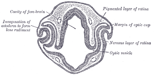
Development of the Embryonic Eye. This transverse section of a 48-hour chick embryo head shows the early formation of the optic cup and lens. Key structures include the invaginating ectoderm forming the lens rudiment and the differentiation of the retinal layers.
Henry Vandyke Carter, Public Domain, via Wikimedia Commons
References
Tawfik HA, Dutton JJ. Embryologic and Fetal Development of the Human Orbit. Ophthalmic plastic and reconstructive surgery. 2018 Sep/Oct:34(5):405-421. doi: 10.1097/IOP.0000000000001172. Epub [PubMed PMID: 30134385]
Langenberg T, Kahana A, Wszalek JA, Halloran MC. The eye organizes neural crest cell migration. Developmental dynamics : an official publication of the American Association of Anatomists. 2008 Jun:237(6):1645-52. doi: 10.1002/dvdy.21577. Epub [PubMed PMID: 18498099]
Level 3 (low-level) evidenceChow RL, Lang RA. Early eye development in vertebrates. Annual review of cell and developmental biology. 2001:17():255-96 [PubMed PMID: 11687490]
Level 3 (low-level) evidenceEckert P, Knickmeyer MD, Schütz L, Wittbrodt J, Heermann S. Morphogenesis and axis specification occur in parallel during optic cup and optic fissure formation, differentially modulated by BMP and Wnt. Open biology. 2019 Feb 28:9(2):180179. doi: 10.1098/rsob.180179. Epub [PubMed PMID: 30958096]
Wang X, Xiong K, Lu L, Gu D, Wang S, Chen J, Xiao H, Zhou G. Developmental origin of the posterior pigmented epithelium of iris. Cell biochemistry and biophysics. 2015 Mar:71(2):1067-76. doi: 10.1007/s12013-014-0310-0. Epub [PubMed PMID: 25344647]
Level 3 (low-level) evidenceIwatsuki H, Sasaki K, Suda M, Itano C. Vimentin intermediate filament protein as differentiation marker of optic vesicle epithelium in the chick embryo. Acta histochemica. 1999 Nov:101(4):369-82 [PubMed PMID: 10611926]
Level 3 (low-level) evidenceVenters SJ, Cuenca PD, Hyer J. Retinal and anterior eye compartments derive from a common progenitor pool in the avian optic cup. Molecular vision. 2011:17():3347-63 [PubMed PMID: 22219630]
Level 3 (low-level) evidenceAavani T, Tachibana N, Wallace V, Biernaskie J, Schuurmans C. Temporal profiling of photoreceptor lineage gene expression during murine retinal development. Gene expression patterns : GEP. 2017 Jan:23-24():32-44. doi: 10.1016/j.gep.2017.03.001. Epub 2017 Mar 10 [PubMed PMID: 28288836]
Onwochei BC, Simon JW, Bateman JB, Couture KC, Mir E. Ocular colobomata. Survey of ophthalmology. 2000 Nov-Dec:45(3):175-94 [PubMed PMID: 11094243]
Level 3 (low-level) evidenceHung SS, Tsai PS, Po CW, Hou PS. Pax6 isoforms shape eye development: Insights from developmental stages and organoid models. Differentiation; research in biological diversity. 2024 May-Jun:137():100781. doi: 10.1016/j.diff.2024.100781. Epub 2024 Apr 10 [PubMed PMID: 38631141]
Shah R, Amador C, Chun ST, Ghiam S, Saghizadeh M, Kramerov AA, Ljubimov AV. Non-canonical Wnt signaling in the eye. Progress in retinal and eye research. 2023 Jul:95():101149. doi: 10.1016/j.preteyeres.2022.101149. Epub 2022 Nov 25 [PubMed PMID: 36443219]
Makrides N, Wang Q, Tao C, Schwartz S, Zhang X. Jack of all trades, master of each: the diversity of fibroblast growth factor signalling in eye development. Open biology. 2022 Jan:12(1):210265. doi: 10.1098/rsob.210265. Epub 2022 Jan 12 [PubMed PMID: 35016551]
Azuma N, Tadokoro K, Yamada M, Nakafuku M, Nishina H. Sonic Hedgehog Determines Early Retinal Development and Adjusts Eyeball Architecture. International journal of molecular sciences. 2025 Jan 9:26(2):. doi: 10.3390/ijms26020496. Epub 2025 Jan 9 [PubMed PMID: 39859210]
Fritzsch B, Martin PR. Vision and retina evolution: How to develop a retina. IBRO neuroscience reports. 2022 Jun:12():240-248. doi: 10.1016/j.ibneur.2022.03.008. Epub 2022 Apr 1 [PubMed PMID: 35449767]
Qu Z, Swaroop A, Del Debbio CB. Genetic and epigenetic control of photoreceptor development. Current topics in developmental biology. 2025:165():449-491. doi: 10.1016/bs.ctdb.2025.03.007. Epub 2025 Apr 10 [PubMed PMID: 40973235]
MACDONALD AE. CAUSES OF BLINDNESS IN CANADA: AN ANALYSIS OF 24,605 CASES REGISTERED WITH THE CANADIAN NATIONAL INSTITUTE FOR THE BLIND. Canadian Medical Association journal. 1965 Feb 6:92(6):264-79 [PubMed PMID: 14270210]
Level 3 (low-level) evidenceHarding P, Gore S, Malka S, Rajkumar J, Oluonye N, Moosajee M. Real-world clinical and molecular management of 50 prospective patients with microphthalmia, anophthalmia and/or ocular coloboma. The British journal of ophthalmology. 2023 Nov 22:107(12):1925-1935. doi: 10.1136/bjo-2022-321991. Epub 2023 Nov 22 [PubMed PMID: 36192130]
Russo M, Palmeri S, Zucconi A, Vagge A, Arioni C. Management of anophthalmia, microphthalmia and coloboma in the newborn, shared care between neonatologist and ophthalmologist: a literature review. Italian journal of pediatrics. 2025 Mar 5:51(1):65. doi: 10.1186/s13052-025-01882-3. Epub 2025 Mar 5 [PubMed PMID: 40038803]
Muzyka L, Winterhalter E, LoPresti MA, Scoville J, Bohnsack BL, Lam SK. Axenfeld-Rieger syndrome: A systematic review examining genetic, neurological, and neurovascular associations to inform screening. Heliyon. 2023 Jul:9(7):e18225. doi: 10.1016/j.heliyon.2023.e18225. Epub 2023 Jul 13 [PubMed PMID: 37539177]
Level 1 (high-level) evidenceReis LM, Maheshwari M, Capasso J, Atilla H, Dudakova L, Thompson S, Zitano L, Lay-Son G, Lowry RB, Black J, Lee J, Shue A, Kremlikova Pourova R, Vaneckova M, Skalicka P, Jedlickova J, Trkova M, Williams B, Richard G, Bachman K, Seeley AH, Costakos D, Glaser TM, Levin AV, Liskova P, Murray JC, Semina EV. Axenfeld-Rieger syndrome: more than meets the eye. Journal of medical genetics. 2023 Apr:60(4):368-379. doi: 10.1136/jmg-2022-108646. Epub 2022 Jul 26 [PubMed PMID: 35882526]
Seifi M, Walter MA. Axenfeld-Rieger syndrome. Clinical genetics. 2018 Jun:93(6):1123-1130. doi: 10.1111/cge.13148. Epub 2018 Jan 25 [PubMed PMID: 28972279]
Tümer Z, Bach-Holm D. Axenfeld-Rieger syndrome and spectrum of PITX2 and FOXC1 mutations. European journal of human genetics : EJHG. 2009 Dec:17(12):1527-39. doi: 10.1038/ejhg.2009.93. Epub 2009 Jun 10 [PubMed PMID: 19513095]
Level 3 (low-level) evidenceAkula M, Park JW, West-Mays JA. Relationship between neural crest cell specification and rare ocular diseases. Journal of neuroscience research. 2019 Jan:97(1):7-15. doi: 10.1002/jnr.24245. Epub 2018 Apr 16 [PubMed PMID: 29660784]
Cennamo G, de Crecchio G, Iaccarino G, Forte R, Cennamo G. Evaluation of morning glory syndrome with spectral optical coherence tomography and echography. Ophthalmology. 2010 Jun:117(6):1269-73. doi: 10.1016/j.ophtha.2009.10.045. Epub 2010 Feb 16 [PubMed PMID: 20163868]
Amador-Patarroyo MJ, Pérez-Rueda MA, Tellez CH. Congenital anomalies of the optic nerve. Saudi journal of ophthalmology : official journal of the Saudi Ophthalmological Society. 2015 Jan-Mar:29(1):32-8. doi: 10.1016/j.sjopt.2014.09.011. Epub 2014 Sep 28 [PubMed PMID: 25859137]
Cvekl A, Wang WL. Retinoic acid signaling in mammalian eye development. Experimental eye research. 2009 Sep:89(3):280-91. doi: 10.1016/j.exer.2009.04.012. Epub 2009 May 7 [PubMed PMID: 19427305]
Level 3 (low-level) evidence