 Tools
Tools
 Anatomy, Head and Neck, Eye Corrugator Muscle
Introduction
The corrugator (or corrugator supercilii) muscles are a pair of muscles located at the medial end of the eyebrow. They are recognized as significant muscles in the glabellar and central forehead regions. The corrugator supercilii muscles (CSM) play a major role in elevating the medial segment of the eyebrow, depressing the lateral portion of the eyebrow, and producing vertical frown lines.[1]
Structure and Function
Register For Free And Read The Full Article
 Search engine and full access to all medical articles
- 10 free questions in your specialty
- Free CME/CE Activities
- Free daily question in your email
- Save favorite articles to your dashboard
- Emails offering discounts
Learn more about a Subscription to StatPearls Point-of-Care
Structure and Function
The corrugator supercilii muscles exist as a pair of small pyramidal muscles that lie deep to the frontal portion of the occipitofrontalis and the orbicularis oculi muscles. They arise from the medial supraorbital ridge of the frontal bone.[2] From the medial end of the eyebrow, the corrugator supercilii muscles travel laterally and above the orbital rim, becoming more superficial.
They are then inserted into the dermis of the medial aspect of the eyebrow, above the supraorbital margin.[1] The paired corrugator supercilii muscles have two heads: a transverse head and an oblique head. The transverse head arises from the superomedial part of the orbital rim and pulls the brows medially, while the smaller oblique head runs parallel to the depressor supercilii and depresses the medial brow.[3]
These actions lead to the vertical wrinkling of the supranasal skin of the forehead. Along with the glabellar muscles and the procerus, the corrugator supercilii also function as accessory protractors of the upper eyelid, helping reduce glare in bright sunlight.[1] Additionally, the corrugator supercilii muscles serve as mimetic muscles that contract when one is angered or perplexed and form a major component of the superficial musculoaponeurotic system (SMAS) of the upper portion of the face.[1] A previous study demonstrated that participants who categorized surprised facial expressions as negative exhibited greater corrugator supercilii muscle activity than those who viewed them as positive. Therefore, corrugator supercilii muscle activity can help evaluate differences in positivity-negativity bias among different participants.[4]
Blood Supply and Lymphatics
The corrugator supercilii muscles receive their blood supply from the supraorbital and supratrochlear arteries, branches of the ophthalmic artery, which is a branch of the internal carotid artery.[5] On the right side of the body, the common carotid arteries arise from the brachiocephalic trunk, and on the left, they arise from the aortic arch. In the neck, at the upper margin of the thyroid cartilage, each common carotid artery runs within the carotid sheath and bifurcates into the internal and external carotid arteries.
In the temporal bone, the internal carotid artery enters the skull via the carotid canal and branches into the ophthalmic artery, which passes through the optic canal into the orbit. Within the orbit, the ophthalmic artery branches into the supraorbital, supratrochlear, lacrimal, central retinal, ethmoidal, ciliary, and inferior and superior muscular arteries. The supraorbital and supratrochlear arteries, branches of the ophthalmic artery, supply the corrugator supercilii muscle. The supratrochlear artery, which initially courses beneath the frontalis and corrugator supercilii muscles, gradually becomes more superficial. The supratrochlear neuromuscular bundle passes through the corrugator supercilii muscle and provides sensory innervation and vascular supply to the forehead.[5]
Nerves
The corrugator supercilii muscles have a dual nerve supply from branches of the temporal, zygomatic, and buccal branches of the facial nerve. The motor nerve supply of the corrugator supercilii muscles originates from the frontal branch of the temporal division of the facial nerve, while the zygomatic branch appears to innervate the oblique head. After receiving innervation from the zygomatic branch, the buccal branch forms the angular nerve, which supplies the corrugator supercilii and procerus muscles.[6]
The supratrochlear nerve (STN) and supraorbital nerve (SON), which are some terminal branches of the frontal nerve, are also closely associated with the corrugator supercilii muscles. After coursing anteromedially over the orbit, the supratrochlear and supraorbital nerves pass through the corrugator supercilii muscle and enter the occipitofrontalis muscle to innervate the glabella, the lower forehead, and the upper portion of the eyelid. The supratrochlear nerve also exits lateral to the origin of the corrugator supercilii muscles, enters the corrugator supercilii muscles where it splits into 3 or 4 small branches, and then penetrates the frontalis muscle.[7]
In a study by Lee et al, among Asian individuals, the supratrochlear nerve was found to emerge separately, coming from the supraorbital nerve at the medial aspect of the orbit, and either pass through the corrugator supercilii muscles as a single branch or bifurcate into 2 divisions before entering the corrugator supercilii muscles in 69% of cases. In the other cases, the supratrochlear nerve was found to originate from the orbit at the same location as the supratrochlear nerve and then pass through the corrugator supercilii muscles. The supratrochlear nerve was also found to always travel within the fibers of the corrugator supercilii muscles, not on a surface more superficial or deeper than the corrugator supercilii muscles. These results differ from those of a previous study that demonstrated that the supratrochlear nerve penetrated the corrugator supercilii muscle in 84% of cases among White individuals.[7][8] A complete understanding of the relationships among the supratrochlear nerve, the supraorbital nerve, and the corrugator supercilii muscle, along with their anatomic locations, allows for a more precise and tailored approach to corrugator supercilii muscle resection for forehead rejuvenation and the surgical treatment of migraine headaches.
Muscles
The corrugator supercilii muscles consist of 2 heads. The transverse head arises from the superomedial margin of the orbital rim and inserts into the dermis at the middle third of the brow, where it interdigitates with the orbicularis oculi and frontalis muscles. The smaller oblique head runs parallel to the depressor supercilii after inserting into the medial brow.[3] The corrugator supercilii muscles work with other muscles of the glabellar complex, including the procerus, depressor supercilii, and the orbicularis oculi muscles, to maintain a balanced brow position and shape.[1]
Physiologic Variants
The lateral margin of the corrugator supercilii muscles may differ among different individuals. In some cases, it may extend to the lateral third of the brow. According to a review of the anatomy of the corrugator supercilii muscles, among the 30 articles included in the analysis, the corrugator supercilii muscles were found to arise mainly from the medial supraorbital rim and the medial frontal bone.[9]
Forty-one percent of the articles mentioned the corrugator supercilii muscles traveled only through the frontalis muscle. The same number of articles also indicated the corrugator supercilii muscles could travel through the frontalis and orbicularis oculi, while only 18% of the articles evaluated mentioned the corrugator supercilii muscles passed through the orbicularis oculi.[9] Fifty-nine percent of the articles reported that the corrugator supercilii muscles course superolateral to the eyebrow, while 41% reported that they traverse laterally. The corrugator supercilii muscles are mainly inserted into the middle of the eyebrow but could also insert into the glabella.[9]
The temporal division of the facial nerve is the primary source of innervation to the corrugator supercilii muscles, but innervation could also come from the zygomatic division (17%) or the buccal and zygomatic branches (17%). In most cases, the corrugator supercilii muscles were found to pull the eyebrows downward and toward the midline of the face. However, they may also pull the medial portion of the eyebrow upward and depress the lateral part of the eyebrow. These anatomical variations require consideration during surgical procedures involving the corrugator supercilii muscles.[9]
Surgical Considerations
Complete comprehension of the anatomy of the corrugator supercilii muscles and their associated nerves and blood supply is essential for corrugator supercilii muscle resections during forehead rejuvenation and supraorbital nerve/supratrochlear nerve decompression in migraine headache treatment. With regards to forehead rejuvenation, cosmetic physicians may inject neurotoxins into the glabellar complex to maintain an aesthetically pleasing brow position and shape. Precise toxin injection is vital for retaining an aesthetically pleasing brow appearance. When treating the corrugator supercilii muscles near their origin, a deeper injection is necessary to fully grasp the depressor supercilii and corrugator supercilii muscles.
When treating the corrugator supercilii muscles more laterally, however, the injection should be more superficial because they lie at a shallower level. The glabellar complex needs 15 to 20 units of onabotulinum or incobotulinum, or 38 to 50 units of abobotulinum, at the procerus (one injection site), and at the corrugator supercilii muscles (two injection sites).[10] Details regarding the physiological variations of the corrugator supercilii muscles also deserve consideration to reduce the risk of undesirable postoperative outcomes, such as forehead irregularities, asymmetries, and unequal muscle reactivation. Further, discrepancies in the locations of the corrugator supercilii muscles and the supratrochlear nerve among patients from different cultural groups warrant investigation, as previous studies have demonstrated that the positions of the corrugator supercilii muscles and the supratrochlear nerve between people of White race and Asians differ.[7][8] Therefore, it is inappropriate to apply the same surgical techniques to both patient groups.[7]
Clinical Significance
As a significant component of the glabellar complex, the corrugator supercilii muscle plays an essential role in maintaining brow position. Complete resection of the corrugator supercilii muscles has been recommended for forehead rejuvenation. Incomplete corrugator supercilii muscle resection can have harmful consequences, such as forehead asymmetries, skin dimpling, and recurrence of glabellar furrows and frown lines. One previous study suggested the amount of corrugator supercilii muscle resection can differ depending on the surgical technique used, with as much as one-third of the transverse corrugator supercilii muscle head remaining after the corrugator supercilii muscles are removed using transpalpebral methods.[3] Therefore, familiarity with the anatomy of the corrugator supercilii muscles with regard to fixed bony landmarks is essential for a more systematic approach to precise resection of the corrugator supercilii muscles.
Complete resection of the corrugator supercilii muscles has also been a recommended approach for the treatment of migraine headaches. There have been suggestions that migraine headaches are associated with peripheral nerve trigger points and the supraorbital nerve/supratrochlear nerve.[3] Chemodenervation of the corrugator supercilii muscles with botulinum toxin type A has been shown to improve migraine headaches by decompressing the supraorbital and supratrochlear nerves and relaxing the surrounding musculature. Patients who have complete corrugator supercilii muscle myectomy have also been shown to have long-term success.[3]
Patients with thyroid eye disease and upper eyelid retraction often have overactivity of the accessory muscles involved in eyelid closure and of the glabellar muscles. They may have a staring facial expression with proptosis, upper lid retraction, and a glabellar frown.[1] The frown may have vertical glabellar furrowing and an altered eyebrow contour, with medial brow ptosis and a deep horizontal line over the nasal bridge. These facial lines mainly originate from a chronic contraction of the corrugator supercilii muscles and the procerus pulling on the skin. Botulinum A chemodenervation has been shown to flatten the glabellar region and enhance the medial eyebrow contour in patients with thyroid eye disease.[1]
Among patients with abnormal Parkinson disease, the “procerus sign” is a clinical sign that is often observed, particularly in patients with progressive supranuclear palsy (PSP). PSP may be misdiagnosed as the more well-known Parkinson disease. The procerus sign involves having vertical wrinkles on the forehead and a staring, astonished look with eyelid retraction and infrequent blinking.[11] It is often believed the procerus sign is secondary to dystonia of the procerus muscle. However, the term may be misleading, as the corrugator supercilii muscles appear to be the primary muscles responsible for the frown or vertical creases.[11] Nevertheless, the generation of vertical creases on the forehead is a complex phenomenon, and the procerus sign should be prominent in early PSP.
Media
(Click Image to Enlarge)
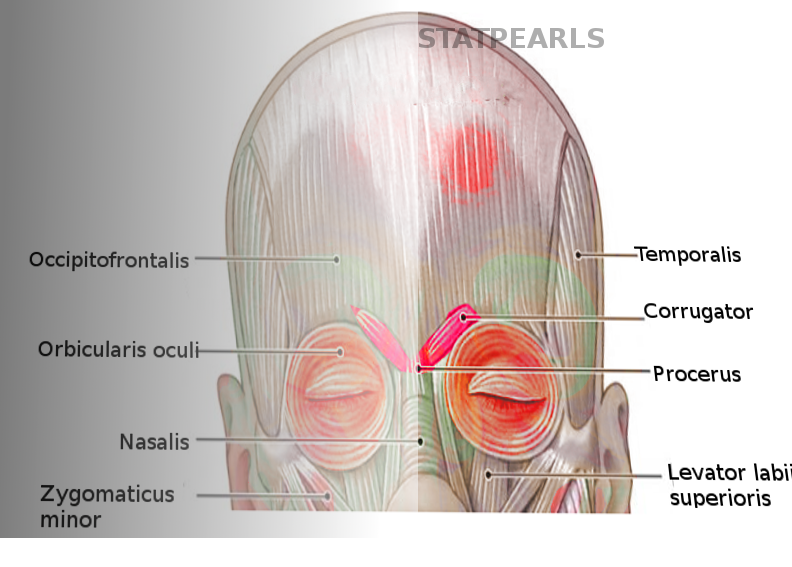
Corrugator Muscle.
Contributed by S Bhimji, MD
References
Olver JM. Botulinum toxin A treatment of overactive corrugator supercilii in thyroid eye disease. The British journal of ophthalmology. 1998 May:82(5):528-33 [PubMed PMID: 9713061]
Isse NG, Elahi MM. The corrugator supercilii muscle revisited. Aesthetic surgery journal. 2001 May:21(3):209-15. doi: 10.1067/maj.2001.116055. Epub [PubMed PMID: 19331895]
Janis JE, Ghavami A, Lemmon JA, Leedy JE, Guyuron B. Anatomy of the corrugator supercilii muscle: part I. Corrugator topography. Plastic and reconstructive surgery. 2007 Nov:120(6):1647-1653. doi: 10.1097/01.prs.0000282725.61640.e1. Epub [PubMed PMID: 18040200]
Neta M, Norris CJ, Whalen PJ. Corrugator muscle responses are associated with individual differences in positivity-negativity bias. Emotion (Washington, D.C.). 2009 Oct:9(5):640-8. doi: 10.1037/a0016819. Epub [PubMed PMID: 19803586]
. Understanding the Anatomy of the Upper Face When Providing Aesthetic Injection Treatments. Plastic surgical nursing : official journal of the American Society of Plastic and Reconstructive Surgical Nurses. 2016 Jul-Sep:36(3):E1. doi: 10.1097/PSN.0000000000000153. Epub [PubMed PMID: 27606590]
Level 3 (low-level) evidenceCaminer DM, Newman MI, Boyd JB. Angular nerve: new insights on innervation of the corrugator supercilii and procerus muscles. Journal of plastic, reconstructive & aesthetic surgery : JPRAS. 2006:59(4):366-72 [PubMed PMID: 16756251]
Lee HJ, Choi KS, Won SY, Apinuntrum P, Hu KS, Kim ST, Tansatit T, Kim HJ. Topographic Relationship between the Supratrochlear Nerve and Corrugator Supercilii Muscle--Can This Anatomical Knowledge Improve the Response to Botulinum Toxin Injections in Chronic Migraine? Toxins. 2015 Jul 17:7(7):2629-38. doi: 10.3390/toxins7072629. Epub 2015 Jul 17 [PubMed PMID: 26193317]
Janis JE, Hatef DA, Hagan R, Schaub T, Liu JH, Thakar H, Bolden KM, Heller JB, Kurkjian TJ. Anatomy of the supratrochlear nerve: implications for the surgical treatment of migraine headaches. Plastic and reconstructive surgery. 2013 Apr:131(4):743-750. doi: 10.1097/PRS.0b013e3182818b0c. Epub [PubMed PMID: 23249981]
Hwang K, Lee JH, Lim HJ. Anatomy of the Corrugator Muscle. The Journal of craniofacial surgery. 2017 Mar:28(2):524-527. doi: 10.1097/SCS.0000000000003304. Epub [PubMed PMID: 28005653]
Beer JI, Sieber DA, Scheuer JF 3rd, Greco TM. Three-dimensional Facial Anatomy: Structure and Function as It Relates to Injectable Neuromodulators and Soft Tissue Fillers. Plastic and reconstructive surgery. Global open. 2016 Dec:4(12 Suppl Anatomy and Safety in Cosmetic Medicine: Cosmetic Bootcamp):e1175. doi: 10.1097/GOX.0000000000001175. Epub 2016 Dec 14 [PubMed PMID: 28018776]
Bhattacharjee S. Procerus Sign: Mechanism, Clinical Usefulness, and Controversies. Annals of Indian Academy of Neurology. 2018 Apr-Jun:21(2):164-165. doi: 10.4103/aian.AIAN_408_17. Epub [PubMed PMID: 30122846]