 Tools
Tools
 Anatomy, Head and Neck: Foramen Magnum
Introduction
The foramen magnum holds relevance in multiple scientific disciplines, including forensic and physical anthropology, comparative anatomy, and biology. Surgical considerations involving the foramen and the craniovertebral junction further highlight the clinical importance of this structure.
The foramen magnum is the largest aperture of the skull (see Image. Skull Foramina). Located in the inferiormost portion of the cranial fossa, the foramen magnum constitutes part of the occipital bone (see Image. Foramen Magnum, Superior View). Structures traversing this opening include the medulla oblongata, meninges, spinal root of the spinal accessory nerve (cranial nerve XI), vertebral arteries, anterior and posterior spinal arteries, tectorial membrane, and alar ligaments. The foramen magnum defines 2 craniometric points. The basion is situated at the median point of the anterior margin, and the opisthion is located at the corresponding posterior margin.
The foramen magnum has critical clinical significance because its anatomy influences the presentation and progression of conditions, such as cerebellar tonsil herniation, Chiari malformations, trauma-related injuries, and vertebrobasilar vascular compromise. The structure's surgical significance arises from its variable size, shape, and surrounding bony structures, which affect access and approach during procedures such as decompressions, tumor resections, and vascular repairs. Detailed knowledge of the foramen magnum's anatomy and function enables clinicians to anticipate potential complications, plan safe surgical interventions, and accurately interpret imaging and physical findings at the craniovertebral junction.
Structure and Function
Register For Free And Read The Full Article
 Search engine and full access to all medical articles
- 10 free questions in your specialty
- Free CME/CE Activities
- Free daily question in your email
- Save favorite articles to your dashboard
- Emails offering discounts
Learn more about a Subscription to StatPearls Point-of-Care
Structure and Function
The foramen magnum is the largest foramen of the skull and lies within the occipital bone of the posterior cranial fossa. The foramen magnum functions as a passage for the central nervous system through the skull, connecting the brain to the spinal cord. The occipital condyles, located on either side of the foramen magnum, form joints with the 1st cervical vertebra (atlas).
The foramen magnum's position plays an essential role in posture during orthostatism by maintaining an appropriate relationship between the skull and the cervical spine. In fossil hominins, foramen magnum position indicates the evolution of bipedal locomotion.[1]
Embryology
All skull bones develop from paraxial mesoderm and neural crest cells. During gastrulation in the 3rd week, mesenchymal cells migrate through the primitive streak to form the mesoderm. By the end of the 3rd week, paraxial mesoderm segments into somites, which differentiate into sclerotomes.[2] During the 4th week, occipital sclerotomes concentrate around the notochord under signals from the rhombencephalon.
Sclerotomes give rise to membrane and cartilage, representing the developmental origins of the occipital bone. The anterior basioccipital (basilar), the 2 lateral exoccipital (condylar), and the posterior supraoccipital (squamous) centers are the 4 primary cartilaginous elements of the occipital bone. These centers unite with a 5th membranous element, the interparietal, around the foramen magnum to form the complete occipital bone. The mendosal suture extends horizontally between the developing inferior supraoccipital and superior interparietal bones.[3]
Fetuses at 9 weeks of gestation exhibit an ossification center around the hypoglossal canal in each exoccipital part and a single median ossification center in the basioccipital cartilage. At 12 weeks of gestation, a pair of ossification centers in the supraoccipital cartilage fuse to form the supraoccipital bone. Rostral to the supraoccipital bone, a second pair of ossification centers in the membranous portion fuse to form the interparietal bone.[4] The intraparietal portion ossifies intramembranously, whereas the remaining occipital bone ossifies endochondrally, utilizing cartilage as a precursor.
The supraoccipital and interparietal bones fuse at the midline, but lateral separation persists through the mendosal suture at this stage of development. At 14 weeks, ossification of the basioccipital progresses laterally into the ventral portion of the condylars, while ventral portions simultaneously advance into dorsal portions. Fusion of the supraoccipital and interparietal bones also progresses nearly to completion during the 14th week. Full union of these segments occurs between ages 2 and 4 years.
All intramembranous ossification centers have generally fused by the 16th week of fetal development, forming a lattice of trabeculae overlaying the external surface of the occipital squama. The exoccipitals remain separated from the basioccipital and supraoccipital segments by synchondroses and do not fuse until between ages 2 and 4 years.[5]
Blood Supply and Lymphatics
The vertebral arteries and the anterior and posterior spinal arteries traverse the foramen magnum (see Image. Vertebral Artery Anatomy in the Neck Region). The vertebral artery originates from the subclavian artery and consists of 4 segments. The 1st preforaminal segment (V1) extends from the subclavian artery to the transverse foramen of C6. The 2nd foraminal segment (V2) ascends through the transverse foramina from C6 to C2.
Emergence of the artery from the C2 foramen marks the beginning of the 3rd segment (V3), also referred to as the "extradural segment." The 3rd segment continues through the transverse foramen of C1, the suboccipital triangle, and the foramen magnum. Penetration of the dura and arachnoid mater indicates the start of the 4th and final segment (V4), the intradural segment.[6] The right and left intradural segments of the vertebral artery converge to form the basilar artery at the level of the pons.[7][8]
In addition to the basilar artery, the 4th segment of the vertebral arteries gives rise to the anterior spinal artery, the posterior spinal artery, perforating branches to the medulla, and the posterior inferior cerebellar artery. The anterior spinal artery supplies the upper cervical spinal cord and the inferior medulla. The posterior spinal artery supplies the dorsal spinal cord and the conus medullaris. The lateral medulla, cerebellar tonsils, inferior vermis, and choroid plexus receive blood from the posterior inferior cerebellar arteries. Penetrating branches supply portions of the medulla, the olives, and the inferior cerebellar peduncle.[9]
Nerves
The spinal accessory nerve originates from the upper spinal cord, specifically C1 to C5 and, occasionally, C6, and enters the skull through the foramen magnum. Inside the skull, the nerve travels superolaterally along the floor of the posterior cranial fossa before entering the jugular foramen dural canal. At the jugular foramen, the spinal accessory nerve joins small fibers from the medulla and continues caudally. The spinal accessory nerve innervates the sternocleidomastoid (SCM) and trapezius muscles, providing motor function.[10]
Muscles
The SCM muscle flexes and extends the neck and rotates the head to the contralateral side. The 2 muscle heads of the SCM originate from the sternal manubrium and the upper clavicle. The heads unite into a single muscle belly that travels superiorly and laterally. The muscle inserts on the mastoid process of the temporal bone and the anterior superior nuchal line.[11]
The trapezius muscle elevates the shoulders. The trapezius originates from the skull, the medial superior nuchal ligament, and the spinous processes of vertebrae C7 to T12. The muscle inserts on the lateral 1/3 of the clavicle, the scapular spine, and the acromion process.[12]
Physiologic Variants
Several anatomical measurements characterize the foramen magnum: the transverse diameter, the anteroposterior diameter, and the foramen magnum index. The foramen magnum index is calculated by dividing the anteroposterior diameter by the transverse diameter. These measurements exhibit physiologic variance among skulls.[13][14] For example, a foramen magnum index greater than 1.2 is considered an ovoid variant. Other named shapes of the foramen magnum include rhomboid, circle, heart, pear, and hexagon.[15] Variation in foramen magnum size and shape is most often associated with sexual dimorphism and differences among ethnic groups, respectively. Terminology describing these shapes remains inconsistent across studies.[16]
Asymmetry of the foramen magnum may also occur. Protrusion of the occipital condyles into the foramen magnum represents another source of anatomical variation. The hypocondylar arch, a feature of embryologic skulls, normally regresses at birth but is rarely retained.[17]
Surgical Considerations
Anatomical variation of the foramen magnum may influence several surgical procedures, including vertebral artery and posterior inferior cerebellar artery aneurysm repair, foramen magnum meningioma resection, and foramen magnum decompression. An ovoid foramen magnum can limit adequate surgical exposure of the anterior portion. The occipital condyle and jugular tubercle represent the principal bony prominences obstructing the anterolateral portion of the foramen magnum. Extension of the occipital condyles into the foramen magnum may indicate the need for more extensive bony removal during certain procedures.[18][19]
Clinical Significance
Several pathologies are associated with the foramen magnum. Elevated intracranial pressure, ie, greater than 20 mm Hg, often results from edema due to stroke, trauma, mass effect, or infection and can lead to obtundation and death without prompt treatment. Total intracranial volume is fixed and consists of blood, cerebrospinal fluid, and brain tissue. Therefore, increased intracranial pressure may produce compensatory cerebellar tonsillar herniation through the foramen magnum. The clinical presentation of herniation includes hypertension, bradycardia, and respiratory depression. Cerebellar tonsillar herniation causes compression of the respiratory centers of the medulla and may be fatal. Foramen magnum anatomy may influence the direction and extent of tissue displacement during herniation.
Chiari I malformation is characterized by herniation of the cerebellar tonsils through the foramen magnum. Diagnostic criteria include herniation of the cerebellar tonsils at least 5 mm below the foramen magnum.[20] Some patients remain asymptomatic, but clinical severity varies widely. Mild manifestations include infrequent exertional headaches. Severe cases present with significant myelopathy and brainstem compromise. Common complications include syringomyelia and hydrocephalus (see Image. Arnold-Chiari Malformation with Syringomyelia on Magnetic Resonance Imaging). Associated findings in infants include sleep apnea and feeding difficulties.
Chiari II malformation involves inferior displacement of the vermis, cerebellar tonsils, medulla, and 4th ventricle through the foramen magnum. Clinical presentation is more severe than in Chiari I and shows a strong association with lumbar myelomeningocele and supratentorial anomalies, including corpus callosal dysgenesis, heterotopias, and sulcal abnormalities. Approximately 80% to 90% of children with Chiari II malformations develop hydrocephalus secondary to 4th ventricle obstruction requiring shunt placement. Chiari III malformation presents with encephalocele in addition to the abnormalities observed in Chiari II.[21]
A mass represents another form of compression adjacent to the foramen magnum. Meningiomas are benign central nervous system tumors arising from arachnoid cells of the dura mater and most commonly occur in the basal region of the cerebrum. Occurrence within the foramen magnum is rare and may present with posterior headache, paresthesias, and motor deficits. Symptom patterns vary because of the anatomic proximity to the cerebellar tonsils, caudal medulla, lower cranial nerves, rostral spinal cord, and upper cervical nerves.[22]
Chronic rheumatoid arthritis is another cause of pathology associated with the foramen magnum. Rheumatoid arthritis–associated facet erosion of C1 and C2, as well as laxity of ligamentous restraints, results in vertical atlantoaxial subluxation, which may lead to protrusion of the odontoid through the foramen magnum and compression of the midbrain. Other etiologies of atlantoaxial subluxation include trauma and congenital conditions such as Down syndrome. Clinical presentation varies widely, with some patients experiencing minimal symptoms and others developing significant instability and neurologic compromise.[23]
The occipital condyles form the osseous lateral boundaries of the foramen magnum. High-energy trauma mechanisms or high-impact axial loads during sporting events, including American football, mountain climbing, and other extreme sports, require careful evaluation for injury to the occipitocervical junction and the remainder of the cervical spine. Evaluation includes accurate history taking, physical examination, and appropriate imaging.
Advanced (Level I) and regional trauma centers perform computed tomography as part of the advanced imaging protocol. Recognition remains important because even displaced occipital condyle fractures may be missed on initial radiographs. Persistent neck pain in an appropriate clinical scenario may warrant transfer to a higher level of care, even in the absence of neurologic symptoms at presentation.[24]
Media
(Click Image to Enlarge)
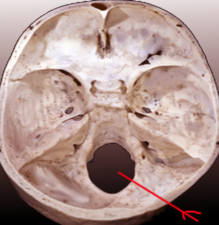
Foramen Magnum, Superior View. This image highlights the foramen magnum within the posterior cranial fossa. This opening is the largest foramen in the skull and is essential for central nervous system connectivity.
Contributed by S Bhimji, MD
(Click Image to Enlarge)
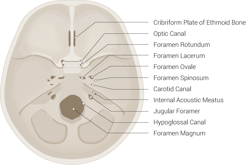
Skull Foramina. This illustration includes the cribriform plate of the ethmoid bone, optic canal, foramen rotundum, foramen lacerum, foramen ovale, foramen spinosum, carotid canal, internal acoustic meatus, jugular foramen, hypoglossal canal, and foramen magnum.
Illustrated by B Palmer
(Click Image to Enlarge)
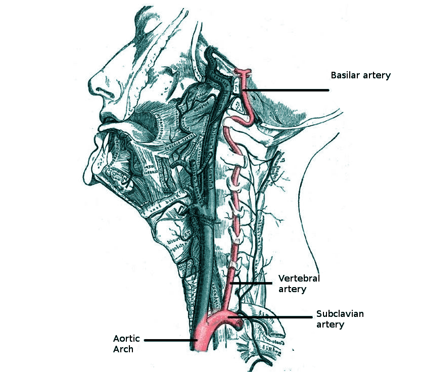
Vertebral Artery Anatomy in the Neck Region. This illustration shows the vertebral artery's course in the neck and relationships to the subclavian artery, aortic arch, cervical vertebrae, and basilar artery.
Contributed by O Chaigasame, MD
(Click Image to Enlarge)
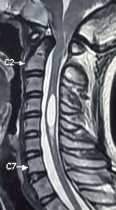
Arnold-Chiari Malformation with Syringomyelia on Magnetic Resonance Imaging. This sagittal T2-weighted image demonstrates cerebellar tonsils protruding through the foramen magnum. A large, fluid-filled syrinx is clearly visible within the cervical spinal cord between the C2 and C7 vertebrae.
Contributed by Sunil Munakomi, MD
References
Russo GA, Kirk EC. Foramen magnum position in bipedal mammals. Journal of human evolution. 2013 Nov:65(5):656-70. doi: 10.1016/j.jhevol.2013.07.007. Epub 2013 Sep 19 [PubMed PMID: 24055116]
Jin SW, Sim KB, Kim SD. Development and Growth of the Normal Cranial Vault : An Embryologic Review. Journal of Korean Neurosurgical Society. 2016 May:59(3):192-6. doi: 10.3340/jkns.2016.59.3.192. Epub 2016 May 10 [PubMed PMID: 27226848]
Bernard S, Loukas M, Rizk E, Oskouian RJ, Delashaw J, Tubbs RS. The human occipital bone: review and update on its embryology and molecular development. Child's nervous system : ChNS : official journal of the International Society for Pediatric Neurosurgery. 2015 Dec:31(12):2217-23. doi: 10.1007/s00381-015-2870-8. Epub 2015 Aug 18 [PubMed PMID: 26280629]
Matsumura G, England MA, Uchiumi T, Kodama G. The fusion of ossification centres in the cartilaginous and membranous parts of the occipital squama in human fetuses. Journal of anatomy. 1994 Oct:185 ( Pt 2)(Pt 2):295-300 [PubMed PMID: 7961136]
Shapiro R, Robinson F. Embryogenesis of the human occipital bone. AJR. American journal of roentgenology. 1976 May:126(5):1063-8 [PubMed PMID: 178231]
Peeters JB, Idriceanu T, El Hage G, Martin T, Salaud C, Champagne PO, Bojanowski MW. A comprehensive review of the vertebral artery anatomy. Neuro-Chirurgie. 2024 May:70(3):101518. doi: 10.1016/j.neuchi.2023.101518. Epub 2024 Jan 29 [PubMed PMID: 38277859]
Velasco S, Mejía JA, Vasquez A, Guarnizo A. Left vertebral artery arising from the external carotid artery: an uncommon anatomical variant. Surgical and radiologic anatomy : SRA. 2024 Nov 28:47(1):7. doi: 10.1007/s00276-024-03518-3. Epub 2024 Nov 28 [PubMed PMID: 39607500]
Zhou B, Alshareef M, Prim D, Collins M, Kempner M, Hartstone-Rose A, Eberth JF, Rachev A, Shazly T. The perivascular environment along the vertebral artery governs segment-specific structural and mechanical properties. Acta biomaterialia. 2016 Nov:45():286-295. doi: 10.1016/j.actbio.2016.09.004. Epub 2016 Sep 6 [PubMed PMID: 27612958]
Tudose RC, Rusu MC, Hostiuc S. The Vertebral Artery: A Systematic Review and a Meta-Analysis of the Current Literature. Diagnostics (Basel, Switzerland). 2023 Jun 12:13(12):. doi: 10.3390/diagnostics13122036. Epub 2023 Jun 12 [PubMed PMID: 37370931]
Level 1 (high-level) evidenceRoberts SO, Cardozo A. A detailed review of the spinal accessory nerve and its anatomical variations with cadaveric illustration. Anatomical science international. 2024 Jun:99(3):239-253. doi: 10.1007/s12565-024-00770-w. Epub 2024 May 2 [PubMed PMID: 38696101]
Silawal S, Schulze-Tanzil G. The sternocleidomastoid muscle variations: a mini literature review. Folia morphologica. 2023:82(3):507-512. doi: 10.5603/FM.a2022.0045. Epub 2022 May 24 [PubMed PMID: 35607877]
Gavid M, Mayaud A, Timochenko A, Asanau A, Prades JM. Topographical and functional anatomy of trapezius muscle innervation by spinal accessory nerve and C2 to C4 nerves of cervical plexus. Surgical and radiologic anatomy : SRA. 2016 Oct:38(8):917-22. doi: 10.1007/s00276-016-1658-1. Epub 2016 Mar 8 [PubMed PMID: 26957148]
Wolf-Vollenbröker M, Prescher A. Osseous bridging of the condylar fossa: report of a rare anatomical variation at the outer skull base. Surgical and radiologic anatomy : SRA. 2024 Aug:46(8):1231-1235. doi: 10.1007/s00276-024-03422-w. Epub 2024 Jun 26 [PubMed PMID: 38926224]
Kedar E, Ezra D, Pelleg-Kallevag R, Stein D, Peled N, May H, Hershkovitz I. Capturing the cervical spine shape: Angular measurements versus geometric morphometric methods. Clinical anatomy (New York, N.Y.). 2025 Apr:38(3):228-238. doi: 10.1002/ca.24166. Epub 2024 Apr 24 [PubMed PMID: 38655670]
Kumar R, Harode HA, Vora R, Javia M. Variations in the shape of foramen magnum at the base of human skulls among Indians in Rajasthan. Bioinformation. 2022:18(5):488-491. doi: 10.6026/97320630018488. Epub 2022 May 31 [PubMed PMID: 36945222]
Zdilla MJ, Russell ML, Bliss KN, Mangus KR, Koons AW. The size and shape of the foramen magnum in man. Journal of craniovertebral junction & spine. 2017 Jul-Sep:8(3):205-221. doi: 10.4103/jcvjs.JCVJS_62_17. Epub [PubMed PMID: 29021672]
Avci E, Dagtekin A, Ozturk AH, Kara E, Ozturk NC, Uluc K, Akture E, Baskaya MK. Anatomical variations of the foramen magnum, occipital condyle and jugular tubercle. Turkish neurosurgery. 2011:21(2):181-90. doi: 10.5137/1019-5149.JTN.3838-10.1. Epub [PubMed PMID: 21534200]
Ber R, Kay-Rivest E, Sen C. Extreme Lateral Approach to the Craniocervical Junction, Operative Technique and Approach Essentials: 2-Dimensional Operative Video. Operative neurosurgery (Hagerstown, Md.). 2023 Oct 1:25(4):e218. doi: 10.1227/ons.0000000000000739. Epub 2023 Jun 30 [PubMed PMID: 37387583]
Graffeo CS, Perry A, Carlstrom LP, Leonel L, Nguyen BT, Morris JM, Driscoll CLW, Link MJ, Peris-Celda M. Anatomical Step-by-Step Dissection of Complex Skull Base Approaches for Trainees: Surgical Anatomy of the Far Lateral Approach. Journal of neurological surgery. Part B, Skull base. 2023 Apr:84(2):170-182. doi: 10.1055/a-1760-2528. Epub 2022 Mar 8 [PubMed PMID: 36895809]
Alhosani MS, Gachechiladze S. Chiari Malformation Type I: A Review of Pathophysiology, Cerebrospinal Fluid Flow Dynamics, Diagnosis, Surgical Management, and Its Relationship to Syringomyelia. Cureus. 2026 Jan:18(1):e101226. doi: 10.7759/cureus.101226. Epub 2026 Jan 10 [PubMed PMID: 41674734]
Milhorat TH, Nishikawa M, Kula RW, Dlugacz YD. Mechanisms of cerebellar tonsil herniation in patients with Chiari malformations as guide to clinical management. Acta neurochirurgica. 2010 Jul:152(7):1117-27. doi: 10.1007/s00701-010-0636-3. Epub 2010 May 4 [PubMed PMID: 20440631]
Tsao GJ, Tsang MW, Mobley BC, Cheng WW. Foramen magnum meningioma: Dysphagia of atypical etiology. Journal of general internal medicine. 2008 Feb:23(2):206-9 [PubMed PMID: 18080720]
Level 3 (low-level) evidenceYang SY, Boniello AJ, Poorman CE, Chang AL, Wang S, Passias PG. A review of the diagnosis and treatment of atlantoaxial dislocations. Global spine journal. 2014 Aug:4(3):197-210. doi: 10.1055/s-0034-1376371. Epub 2014 May 22 [PubMed PMID: 25083363]
Bhalaik V, Fraser M. Fracture of the occipital condyle. Injury. 2001 Mar:32(2):157-8 [PubMed PMID: 11223048]
Level 3 (low-level) evidence