 Tools
Tools
 Generalized Pustular Psoriasis
Introduction
Pustular psoriasis is a rare and severe variant of psoriasis characterized by eruptions of sterile pustules that may present in distinct clinical patterns. Pathologic features of psoriasis, including keratinocyte hyperproliferation, neutrophilic infiltration, and immune dysregulation, are markedly accentuated (see Image. Pustular Psoriasis). In contrast to psoriasis vulgaris, which is primarily driven by the adaptive immune system and the interleukin 17/interleukin 23 (IL-17/IL-23) axis, pustular psoriasis appears to be characterized by a robust inflammatory response associated with hyperactivation of innate immunity, particularly involving the interleukin 36 (IL-36) axis.[1][2][3] Pustules may be widespread or localized and are characterized by a sterile, predominantly neutrophilic infiltrate. Unlike chronic plaque psoriasis, the most common variant of psoriasis vulgaris, pustular psoriasis lesions are often tender to palpation. Pustular psoriasis may be further subdivided based on clinical presentation and pustule distribution. However, all subtypes are characterized by superficial sterile pustules on an erythematous base.
Pustular psoriasis may be classified as generalized or localized based on clinical distribution and presentation. Generalized forms include the von Zumbusch subtype, which presents with diffuse pustular eruptions accompanied by systemic manifestations such as fever and arthralgias. The annular subtype is characterized by annular lesions with pustules distributed along the advancing edge. The exanthematic subtype is an acute pustular eruption that lacks systemic symptoms, such as fever or joint pain, and typically resolves within several days. Impetigo herpetiformis is pustular psoriasis occurring during pregnancy. Localized forms include acrodermatitis continua of Hallopeau (ACH), which is characterized by pustules involving the fingers, toes, and nail beds, and palmoplantar pustular psoriasis, which primarily affects the palms and soles.
Etiology
Register For Free And Read The Full Article
 Search engine and full access to all medical articles
- 10 free questions in your specialty
- Free CME/CE Activities
- Free daily question in your email
- Save favorite articles to your dashboard
- Emails offering discounts
Learn more about a Subscription to StatPearls Point-of-Care
Etiology
Pustular psoriasis occurs in genetically susceptible individuals and may be precipitated by multiple risk factors, resulting in psoriasiform changes and marked neutrophilic accumulation within the epidermis. An increased frequency of the human leukocyte antigen B27 (HLA-B27) allele has been reported among patients with pustular psoriasis compared with the general population. Most cases are idiopathic. However, several factors have been implicated in disease onset or exacerbation.
Infectious triggers, particularly bacterial and viral pathogens, are commonly reported. Staphylococcus aureus and Streptococcus epidermidis have been associated with disease exacerbation.[4] Viral infections, including influenza, have also been reported to precipitate generalized pustular psoriasis (GPP), and acute exacerbations associated with respiratory syncytial virus infection have been described.[5] Hypocalcemia may develop as a consequence of GPP or serve as a precipitating factor, particularly in the setting of hypoparathyroidism.[6]
Abrupt withdrawal of systemic corticosteroids and the use of potent topical corticosteroids under occlusion have been associated with the onset of pustular psoriasis.[7][8] Disease flares following cyclosporine withdrawal have also been reported.[9] Additional medications occasionally implicated in pustular psoriasis include terbinafine, propranolol, bupropion, lithium, phenylbutazone, salicylates, and potassium iodide. Topical therapies, such as coal tar and dithranol, may precipitate pustulation when applied inappropriately during unstable disease.[10]
Pregnancy-associated pustular psoriasis, historically termed "impetigo herpetiformis," is a distinct subtype associated with an increased risk of stillbirth and fetal anomalies.[11] A subset of patients with pustular psoriasis exhibits features of autoinflammatory disease associated with specific genetic mutations. Identified syndromes include deficiency of the IL-36 receptor antagonist (DITRA) and deficiency of the IL-1 receptor antagonist (DIRA).
Phototherapy has also been reported as a potential precipitating factor in some cases. Vaccinations, including bacillus Calmette-Guérin (BCG) and H1N1 influenza vaccines, have been associated with pustular psoriasis. Cases have also been documented in the setting of COVID-19 infection and following COVID-19 vaccination.[12][13] Psychological stress is frequently reported as a disease-exacerbating factor.
Epidemiology
Pustular psoriasis is a rare disease affecting both children and adults and demonstrates a bimodal age distribution. In adults, disease onset typically occurs between ages 40 and 50, whereas pediatric cases commonly present during infancy. Greater disease prevalence has been reported among Asian populations than among Caucasian populations.[14] Pustular variants account for approximately 1% of all clinical cases of psoriasis vulgaris.[15]
GPP is an uncommon condition with variable epidemiologic characteristics. In France, age- and sex-standardized cumulative prevalence and incidence rates have been reported at 45.2 and 7.1 cases per million, respectively.[16] In Japan, the estimated prevalence is 7.46 cases per million.[17] Pediatric GPP is uncommon but well-documented. The reported age at presentation ranges from 6 weeks to 10 years, including 1 documented case in a 6-week-old infant.[18]
Palmoplantar pustulosis and ACH have reported mean ages of onset of 43.7 and 51.8 years, respectively.[19] Annular pustular psoriasis in children has a mean age of onset of approximately 6 years.[20] Sex distribution in GPP varies geographically. In the US, the male-to-female ratio is approximately 1:1. Globally, female predominance has been reported, with female-to-male ratios of 1.5 for ACH, 1.7 for GPP, and 3.5 for palmoplantar pustulosis.[21][22] Common comorbidities include hypertension, ischemic heart disease, and hyperlipidemia.
Pathophysiology
The exact pathogenesis of pustular psoriasis remains incompletely understood. However, several mechanisms have been proposed based on cytokine expression patterns and therapeutic responses to targeted medications. A combination of genetic susceptibility and exposure to precipitating factors, such as withdrawal of systemic corticosteroids, may result in upregulation of specific cytokines and neutrophilic accumulation within the epidermis. Dermal dendritic cells also release elastase, which may contribute to pustule formation. Both pustular psoriasis and chronic plaque psoriasis demonstrate overexpression of IL-1, IL-17, IL-23, IL-36, tumor necrosis factor α (TNF-α), and interferon γ (IFN-γ). However, IL-1 and IL-36 expression appears to be more prominent in pustular psoriasis. Proposed pathogenic mechanisms are discussed below.
Deleterious germline mutations in IL36RN have been identified in both familial and sporadic cases of GPP across diverse populations, including the UK, Germany, Tunisia, Malaysia, China, and Japan. Mutation patterns vary among populations, with evidence supporting a founder effect. In European populations, the most common mutation, IL36RN p.Ser113Leu, occurs in approximately 0.03% of healthy individuals. Patients with GPP harboring IL36RN mutations are more likely to develop early-onset disease and exhibit a pronounced systemic inflammatory response.[23][24]
IL36RN encodes the IL-36 receptor antagonist, which is primarily expressed in the skin and functions as an antagonist to 3 proinflammatory IL-1 family cytokines: IL-36α, IL-36β, and IL-36γ. These cytokines activate intracellular signaling cascades, including the NF-κB and MAPK pathways, and are modulated by cytokines released by Th17 cells and TNF-α. Although IL-36 cytokines are also overexpressed in plaque psoriasis, the mechanisms of IL-36 dysregulation in GPP appear to differ.
IL-36 receptors are constitutively expressed on dermal dendrocytes, CD4+ T cells, and macrophages. Receptor activation promotes maturation of monocyte-derived dendritic cells and release of cytokines, including IL-1, IL-6, IL-23, TNF-α, and IFN-γ. These cytokines contribute to neutrophil recruitment and cutaneous inflammation. Studies have also identified IL-36 receptor antagonist deficiency in patients with pustular psoriasis. Successful treatment with a novel monoclonal antibody targeting the IL-36 receptor has been reported in affected patients.[25]
Mutations in CARD14 have been occasionally reported, particularly in individuals with coexistent plaque psoriasis. These mutations result in increased activation of NF-κB signaling and upregulation of IL-8 and IL-36γ expression. Mutations in AP1S3, more commonly observed in patients with GPP and ACH, lead to increased IL-36α expression and impaired innate immune responses through disruption of TLR3 signaling.
IL-6 signaling has gained attention for its role in the pathogenesis of pustular psoriasis. The IL-6 receptor exists in both membrane-bound and soluble forms, a feature that distinguishes it from other cytokine receptors, which are exclusively membrane-bound. The IL-6/IL-6 receptor complex, together with the ubiquitously expressed gp130 subunit, activates the JAK/STAT and RAS/MEK/ERK/MAPK signaling pathways, promoting downstream gene transcription. IL-6 exerts broad immunologic effects, including induction of acute-phase protein synthesis, maturation of B cells, differentiation of T cells, and promotion of Th17 cell development. IL-6 also stimulates neutrophil maturation from myeloid precursors, upregulates ICAM-1 and other endothelial adhesion molecules to facilitate neutrophil migration, and enhances production of proinflammatory cytokines such as IL-23 and IL-17, reinforcing the Th17-driven inflammatory loop.[26]
The pathophysiology of ACH has been a subject of prolonged debate. Case studies describing the transition from ACH to GPP, together with reported associations with IL36RN, CARD14, and AP1S3 variants, support a shared disease spectrum. ACH and GPP may represent opposite ends of a single inflammatory continuum.[27]
Histopathology
Many histopathological features of pustular psoriasis overlap with those of psoriasis vulgaris, including parakeratosis, hyperkeratosis with thickening of the stratum spinosum, elongation of rete ridges, reduction of the stratum granulosum, and thinning of the suprapapillary epidermis. In addition to the classic histologic features of psoriasis vulgaris, a prominent neutrophilic infiltrate is present in the papillary dermis and epidermis. Neutrophilic accumulation leads to disruption of keratinocyte desmosomal junctions (spongiosis), basal keratinocyte herniation into the papillary dermis, and formation of superficial microabscesses. Neutrophilic infiltration of the epidermis is more pronounced in pustular psoriasis than in other variants of psoriasis vulgaris.
The histologic findings in GPP are dominated by dense neutrophilic infiltration, which underlies the bright erythema and formation of sterile pustules. The defining feature is the accumulation of neutrophils within the epidermis. The epidermis is typically acanthotic, with numerous neutrophils present between eosinophilic strands of keratinocytes. Large neutrophilic aggregates often localize within the stratum corneum and are typically surrounded by parakeratosis.
Hallmark features of active pustular psoriasis include spongiform pustules of Kogoj and microabscesses of Munro, which are characteristic histologic patterns also observed in plaque psoriasis (see Image. Histopathology of Pustular Psoriasis). Despite these findings, the National Psoriasis Foundation advises that a skin biopsy is not essential for the diagnosis of GPP, as histologic features may be nonspecific.
Histopathologic findings in acute generalized exanthematous pustulosis (AGEP) may closely resemble those of GPP. Accordingly, clinical history and clinical presentation are generally more reliable than histologic examination for differentiation between these entities.[28]
History and Physical
Pustular psoriasis presents as numerous discrete or confluent superficial yellowish pustules on an erythematous background. Full-body skin examination should be performed, including careful evaluation of mucous membranes and nails, to identify additional signs of psoriasis and exclude alternative causes of pustulosis.
Pustular psoriasis may develop in individuals with longstanding plaque psoriasis, often following exposure to corticosteroids or other environmental triggers. Alternatively, onset may occur de novo, frequently after an infection. In either scenario, the earliest symptom is often a burning sensation of the skin, followed by progression to dryness and tenderness. Abrupt onset of high fever and severe malaise commonly follow these prodromal symptoms, although these features are not universally present.
Preexisting psoriatic plaques may evolve into numerous small pustules. Erythema intensifies, and pustules may extend to previously unaffected areas, particularly the flexural and genital regions. Pustular psoriasis may present as either diffuse or localized disease.
Generalized Pustular Psoriasis
Cutaneous findings vary and may include discrete pustules, lakes of pus, circinate lesions, erythematous plaques covered with pustules, or generalized erythroderma. Pustules often emerge in crops, followed by desiccation and exfoliation. Nail involvement may include thickening and formation of subungual collections of pus. The oral and oropharyngeal mucosa may be affected, with findings such as hyperemia, geographic tongue, or fissured tongue. Associated arthralgia may be reported in some individuals. Disease course may resolve spontaneously within days to weeks, with reversion to baseline psoriasis in some cases or progression to erythroderma in others.[29]
GPP demonstrates clinical heterogeneity in age of onset, severity, and disease course. Several overlapping phenotypes have been recognized. A variable relationship exists between GPP and plaque psoriasis. Plaque psoriasis may precede or follow GPP episodes, while GPP may also present as the sole phenotype without a history of plaque involvement. Clinical subtypes of GPP are defined by distinct morphological patterns and differences in natural history. Recognition of these subtypes is essential for accurate diagnosis and appropriate management, which includes the following:
- Von Zumbusch variant: The most acute and fulminant form of GPP. Named after Leo von Zumbusch, who first described the condition in 1910 in 2 siblings following topical treatment, this variant presents with widespread erythema, sterile pustulation, fever, systemic malaise, and skin tenderness. Diagnosis is based on clinical findings, laboratory abnormalities such as leukocytosis and elevated inflammatory markers (ESR or CRP), and histopathologic evidence of spongiform pustules. Pustules typically resolve within days, followed by prominent scaling. Chronic plaques of psoriasis may clear after the acute episode. In the original case, 9 recurrences were documented over a 10-year period.
- Annular variant: Characterized by erythematous, scaly lesions with pustulation along the advancing margin. Lesions expand centrifugally over hours to days with simultaneous central clearing. Also referred to as “subacute GPP” or the “circinate variant,” this form generally follows a subacute or chronic course and produces fewer systemic symptoms. Higher frequency has been reported in the pediatric population.
- Exanthematic variant: Manifests as a sudden eruption of small pustules that resolve spontaneously within a few days. Commonly follows infection or may be drug-induced, with lithium identified as a frequent trigger. Systemic symptoms are usually absent. Clinical and histologic overlap with pustular drug eruptions, particularly AGEP, complicates differentiation.
- Localized variant: Involves pustule formation within or at the periphery of existing psoriatic plaques. Often observed during unstable phases of chronic plaque psoriasis or after application of irritants, such as coal tar or anthralin.
- Impetigo herpetiformis variant: Refers to GPP occurring during pregnancy. The onset most commonly occurs in the 3rd trimester, although cases have been reported as early as the 1st month of gestation or the immediate postpartum period. Eruptions typically begin on flexural surfaces as symmetrical, confluent pustules on inflamed skin, often originating in the groin. Lesions spread centrifugally, coalesce into plaques, and may leave reddish-brown pigmentation during healing. Mucosal involvement, including the tongue, oral cavity, and esophagus, may occur and may be erosive. Disease frequently persists until delivery and may continue into the postpartum period.[30]
GPP begins in the 1st year of life in approximately 30% of cases. The onset may occur during the first few weeks, though, in some instances, the disease is present at birth. Systemic symptoms are frequently absent in infancy, and mild cases may not require treatment. Lesions are often localized to flexural areas, such as the neck, and may persist in this distribution for extended periods. More severe presentations with systemic involvement require active intervention. Disease onset in children most commonly occurs between the ages of 2 and 10 years. While the von Zumbusch pattern may be observed, annular and circinate variants are more frequently encountered. Episodes may resolve within days. However, recurrent inflammatory relapses are common. In older children, clinical features resemble the adult form, and any of the established morphological patterns may be present.
Pustulosis of the Palms and Soles
Pustulosis of the palms and soles (palmoplantar pustulosis) is characterized by sterile pustules on the palmoplantar surfaces, often admixed with yellow–brown macules (see Image. Pustulosis on the Soles). Scaly erythematous plaques may also be present. A minority of patients demonstrate chronic plaque psoriasis at other body sites.
The condition follows a chronic course, with pustules restricted to the palmoplantar surfaces, in contrast to the natural history of GPP. Smoking may exacerbate disease activity, while stress and focal infections have been identified as triggering factors.
Pustulosis of the palms and soles is frequently associated with sterile inflammatory bone lesions in the context of SAPHO syndrome, characterized by synovitis, acne, pustulosis, hyperostosis, and osteitis. The condition may also present as a paradoxical reaction in patients receiving TNF inhibitors for conditions such as rheumatoid arthritis or inflammatory bowel disease.
Acrodermatitis Continua of Hallopeau
ACH is an uncommon presentation of psoriasis. Pustules are clinically observed on the distal segments of the fingers and, less frequently, the toes. Pustulation is frequently followed by scaling and crust formation. Pustules may also develop in the nail bed beneath the nail plate, with possible exfoliation of the nail plates. Transition to other psoriasis variants may occur. ACH may be associated with annulus migrans of the tongue.
Evaluation
Patients with suspected pustular psoriasis require careful evaluation, as the von Zumbusch subtype may be life-threatening. Laboratory investigations should include a complete blood count, which may demonstrate initial absolute lymphopenia followed by marked polymorphonuclear leukocytosis, potentially reaching 40,000/µL. An electrolyte panel should be obtained to assess for hypocalcemia, and a liver panel should be performed to evaluate transaminase levels. A pregnancy test should be ordered for all women of childbearing age.
Erythrocyte sedimentation rate (ESR) and C-reactive protein (CRP) levels are often elevated, reflecting systemic inflammation. Hypoalbuminemia, hypocalcemia, and reduced plasma zinc levels are frequently observed, along with lipid profile abnormalities. A punch biopsy may be performed in equivocal cases.
Bacterial cultures of pustular fluid are typically negative unless secondary infection is present. Viral cultures and Tzanck smears are also negative. However, disruption of the epidermal barrier may predispose to bacteremia. Urinalysis may demonstrate albuminuria and urinary casts.
Treatment / Management
The first step in managing pustular psoriasis is identifying triggers, such as an infection. Subsequent management is directed toward urgent control of inflammation and stabilization of the disease course.
GPP, particularly the von Zumbusch subtype, may require hospital admission to ensure adequate hydration, bed rest, and prevention of excessive heat loss. Supportive care includes the use of bland topical compresses and saline or oatmeal baths, which help relieve discomfort and promote debridement of affected skin. Systemic symptoms, including fever and arthralgia, require treatment with antipyretics and anti-inflammatory medications.
Disease-specific systemic therapies, using agents such as acitretin, methotrexate, cyclosporine, and infliximab, are considered 1st-line treatment in adult patients. In pediatric patients, acitretin, cyclosporine, methotrexate, and etanercept are preferred.
Second-line therapies for GPP include the administration of biologic agents such as etanercept, adalimumab, ustekinumab, and secukinumab, as well as topical drugs, particularly corticosteroids, calcipotriene, and tacrolimus, for localized involvement of the palms and soles.[31] A small case series reported clinical improvement with bimekizumab in patients with palmoplantar pustulosis and palmoplantar plaque psoriasis presenting with pustules.[32] Treatment guidelines for 2nd-line therapies remain limited. Anecdotal reports describe paradoxical induction of pustular psoriasis with certain biologic agents, highlighting the need for further evaluation and consensus.[33](B3)
Data on the treatment of pustular psoriasis during pregnancy remain exceedingly scarce. Most commonly used regimens include the administration of systemic corticosteroids, cyclosporine, narrow-band UVB phototherapy, adjuvant antibiotic therapy, and topical agents. Certolizumab therapy is an option, given the agent's favorable safety profile for both mother and fetus.[34][35](B3)
Treatment modalities for ACH, a particularly refractory subtype, are primarily derived from case reports. The limited efficacy of topical agents, including corticosteroids, calcipotriol, tacrolimus, and fluorouracil, often necessitates alternative approaches. Systemic options include cyclosporine, systemic corticosteroids, retinoids, methotrexate, psoralen plus UVA (PUVA), UVB phototherapy, and biologic agents (including anti–TNF agents, IL-17 inhibitors, IL-12/IL-23 inhibitors, and anakinra), as well as apremilast and baricitinib. A proposed treatment algorithm recommends acitretin or methotrexate as initial therapy, followed by biologics such as adalimumab and secukinumab, with possible escalation to guselkumab. Cyclosporine may be utilized for short-term disease control.[36][37][38](B2)
Spesolimab is a selective, humanized monoclonal antibody targeting the IL-36 receptor. The agent was recently approved in both Europe and the US for the treatment of GPP flares in adults. Phase II randomized controlled trials demonstrated rapid and effective cutaneous clearance during acute flares and superiority to placebo in reducing flare frequency over 48 weeks of maintenance therapy. Safety and tolerability profiles were favorable.[39] In March 2024, the US Food and Drug Administration (FDA) granted an expanded indication for spesolimab to include adults and pediatric patients aged 12 years or older weighing at least 40 kg. (Source: FDA. Spesolimab Injection Prescribing Information. 2024) This decision was based on results from the 48-week Effisayil 2 clinical trial, which enrolled 123 participants. Treatment with spesolimab resulted in an 84% reduction in GPP flares compared with placebo.[40](A1)
Recent reports indicate successful use of IL-1 receptor antagonists, such as anakinra, in the treatment of pustular psoriasis. Tocilizumab, an IL-6 receptor inhibitor, has demonstrated efficacy in select cases of biologic-induced plantar pustular psoriasis.[41] This monoclonal antibody targets both membrane-bound and soluble IL-6 receptor complexes, thereby inhibiting IL-6–dependent STAT1 and STAT3 signaling pathways. Notably, tocilizumab has also been associated with paradoxical development of psoriasiform dermatitis in patients treated for rheumatoid arthritis.[42](B2)
Imsidolimab is a humanized, affinity-matured immunoglobulin G4 monoclonal antibody that antagonizes IL-36 signaling by binding to the IL-36 receptor. Two phase 3 trials were conducted at 26 clinical sites in 11 countries to evaluate the efficacy of imsidolimab in the treatment of GPP. A significantly higher proportion of patients with GPP receiving a single intravenous dose of imsidolimab achieved clear or almost clear skin at 4 weeks compared with placebo.[43]
Janus kinase (JAK) inhibitors demonstrate high response rates, rapid clinical improvement, and generally tolerable safety profiles. However, existing evidence remains limited by small sample sizes, short follow-up durations, and reliance on case-based data. JAK inhibitors are a potential therapeutic option and require further evaluation in larger controlled trials to establish long-term efficacy and safety.[44](A1)
Differential Diagnosis
Pustular psoriasis is a clinically heterogeneous condition with variable manifestations, making accurate diagnosis and differentiation from other pustular dermatoses challenging. The differential diagnosis includes disorders characterized by widespread nonfollicular pustules, discussed below.
AGEP presents as a febrile eruption characterized by erythema and widespread pustules. Histopathologic features include neutrophilic exocytosis, intraepidermal or subcorneal pustules, papillary dermal edema, and a dermal infiltrate composed of neutrophils and eosinophils. AGEP is most often triggered by medications or infections and typically resolves within 2 weeks following removal of the inciting factor.
Although AGEP shares clinical features with GPP, including sudden onset of pustules, fever, and leukocytosis, differentiation may be supported by the presence of numerous discrete, short-lived pustules, a recent history of drug exposure, and rapid resolution after drug cessation. Consensus among experts (90%) supports that the presence of eosinophils, particularly within microabscesses, favors a diagnosis of drug-induced pustulosis.[45] AGEP has been associated with IL36RN mutations, similar to those identified in GPP, pustulosis of the palms and soles, and ACH. These genetic and clinical overlaps have led to consideration of AGEP as a drug-induced variant within the pustular psoriasis spectrum.[46]
Subcorneal pustular dermatosis, or Sneddon–Wilkinson disease, is characterized by erythematous pustules, often involving the trunk and intertriginous areas. The association between this condition and pustular psoriasis remains controversial. Subcorneal pustular dermatosis is typically benign, relapsing, and not associated with systemic symptoms. A diagnostic clue is the presence of hypopyon pustules, containing clear fluid in the upper portion and purulent yellow material in the lower portion.[47]
Immunoglobulin A pemphigus may also mimic GPP. However, histopathologic features and characteristic immunofluorescence patterns allow differentiation.
Necrolytic migratory erythema associated with glucagonoma may resemble pustular psoriasis. Associated features, including weight loss, glossitis, and anemia, assist in differentiation.
During the acute phase of GPP, fever, leukocytosis, and elevated inflammatory markers may mimic systemic infection. This clinical overlap may lead to inappropriate discontinuation of immunosuppressive therapy, with potential exacerbation of underlying disease.
Prognosis
Pustular psoriasis is a chronic disease characterized by recurrent episodes or flares. Treatment is directed at shortening the duration of relapses, preventing complications, and prolonging disease-free intervals. Compared with other subtypes, the von Zumbusch variant carries a higher risk of mortality due to systemic involvement, particularly in the presence of comorbidities such as cancer, renal failure, or liver failure. Localized subtypes of pustular psoriasis are associated with a substantially lower mortality risk. Subacute annular and circinate forms are generally associated with a favorable prognosis, particularly in pediatric patients. Outcomes are likewise better when a clear precipitating factor is identified, as in pustular psoriasis of pregnancy. In contrast, pustular psoriasis evolving from ACH appears to carry the poorest prognosis.[48][49]
Complications
Reported mortality rates for GPP range from 4% to 24%, underscoring the potential severity of the condition.[50] Patients are often systemically unwell and may develop life-threatening complications, including the following:
- Renal compromise may arise due to hypovolemia and oligemia, potentially resulting in acute kidney injury.
- Profound hypoalbuminemia is common, resulting from both rapid loss of plasma proteins into tissues and impaired intestinal absorption.
- Secondary hypocalcemia may develop as a consequence of low albumin levels.
- Abnormalities in liver function tests are frequently observed, and in some cases, cholestatic jaundice may occur due to neutrophilic cholangitis.[51]
- Although rare, acute respiratory distress syndrome has been reported.
- Superimposed Staphylococcus aureus infection may complicate the disease course.
- Telogen effluvium, a form of diffuse hair shedding, may occur 2 to 3 months after the acute illness.
- Amyloidosis is a rare late complication associated with chronic inflammation.
In the setting of impetigo herpetiformis, additional complications may include delirium, gastrointestinal disturbances, tetany, cardiac or renal failure, and death in severe cases. The risk of placental insufficiency is elevated, with increased likelihood of stillbirth, neonatal death, or congenital anomalies. These obstetric complications are directly related to disease severity and chronicity. Recurrence is common in subsequent pregnancies and may also occur with the use of oral contraceptives.
Deterrence and Patient Education
Patients with psoriasis vulgaris should be educated regarding warning signs suggestive of GPP, particularly in the presence of predisposing factors, such as infections, medications, or psychological stress. Concerning indicators include sudden eruption of rash over preexisting plaques, fever, and myalgia. Pregnant individuals with rapidly worsening psoriasis require close dermatologic monitoring throughout pregnancy. Genetic testing is recommended in infants presenting with early-onset psoriasis to evaluate for underlying mutations associated with pustular phenotypes. Abrupt corticosteroid withdrawal should be avoided, as it may precipitate pustular psoriasis flares. Flaccid lesions require appropriate wound care to reduce the risk of complications and secondary infection.
Pearls and Other Issues
Recognition of the varied presentations of pustular psoriasis is essential for prompt and appropriate care. GPP is characterized by diffuse erythema and sterile pustules, which may occur with or without systemic symptoms, laboratory abnormalities, or concomitant psoriasis subtypes. The condition may present acutely with widespread pustules or subacutely with an annular pattern. In some cases, disease onset begins with isolated pustules that progress into larger collections or lakes of pus. The presence of several persistent, discrete pustules without coalescence or identification of hypopyon pustules may prompt clinicians to perform a skin biopsy to confirm the diagnosis and exclude mimics. However, features such as desquamation, scaling, and crusting, reflecting pustule evolution, are not required for diagnosis.
Systemic findings such as fever, fatigue, leukocytosis with neutrophilia, and elevated CRP, while not essential, may support the clinical suspicion of GPP. Laboratory abnormalities, including hypocalcemia, hypoproteinemia, hypoalbuminemia, and abnormal liver or kidney function, are not diagnostic criteria but should be monitored regularly to detect complications.
Genetic testing is not required for diagnosis. However, IL36RN mutation screening is advised when accessible. IL36RN mutations have been associated with a more severe disease phenotype, including earlier onset, heightened systemic inflammation, and increased frequency of disease flares.
GPP is associated with increased all-cause and cause-specific mortality compared with psoriasis vulgaris. Pustulosis of the palms and soles is associated with reduced mortality, suggesting epidemiologic and biologic distinctions across these subtypes.[52]
Enhancing Healthcare Team Outcomes
Psoriasis is typically managed by dermatologists, although primary care clinicians often provide long-term follow-up for most patients. A rare form of the disease is pustular psoriasis, defined by sterile pustules that may follow various clinical patterns. Pathological features of psoriasis are markedly accentuated in this form.
GPP is clinically heterogeneous, with variability in age of onset, severity, and natural course. While treatment broadly aligns with general psoriasis management, the acute stage of GPP may be life-threatening without prompt and appropriate intervention. Prognosis is typically favorable in subacute annular and circinate variants and is generally more favorable in pediatric patients. Among cases of GPP, outcomes are improved when a clear precipitating factor is identified.
Timely recognition and management of GPP are critical to reducing morbidity and mortality. Optimal care requires a coordinated, interprofessional approach to ensure patient-centered treatment and improved outcomes. Healthcare professionals, including dermatologists, emergency medicine physicians, advanced practitioners, nurses, and pharmacists, must be equipped to recognize varied clinical presentations and underlying triggers, distinguish GPP from other conditions, initiate appropriate therapy, and monitor for complications.
Patient and caregiver education is also essential. Counseling should address potential triggers, medication adherence, and early warning signs of disease progression, all of which are important for minimizing morbidity and preventing disease exacerbations.
Media
(Click Image to Enlarge)
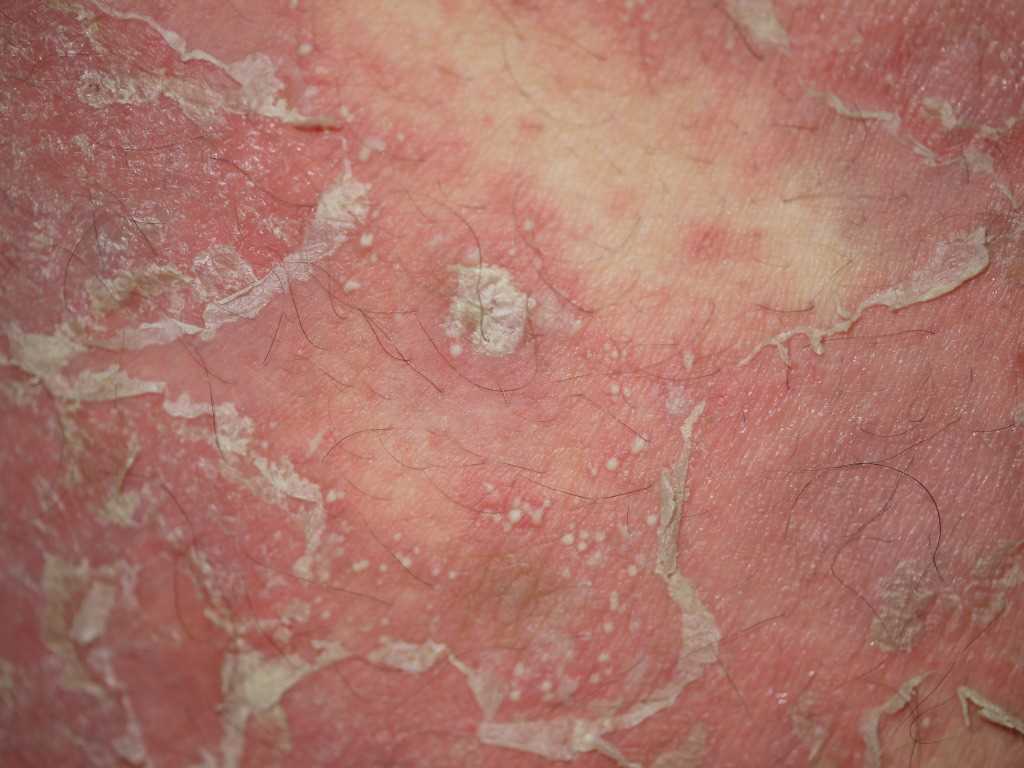
Pustular Psoriasis. Erythematous plaque with overlying sterile pustules and widespread desquamation, characteristic of generalized pustular psoriasis.
(Click Image to Enlarge)
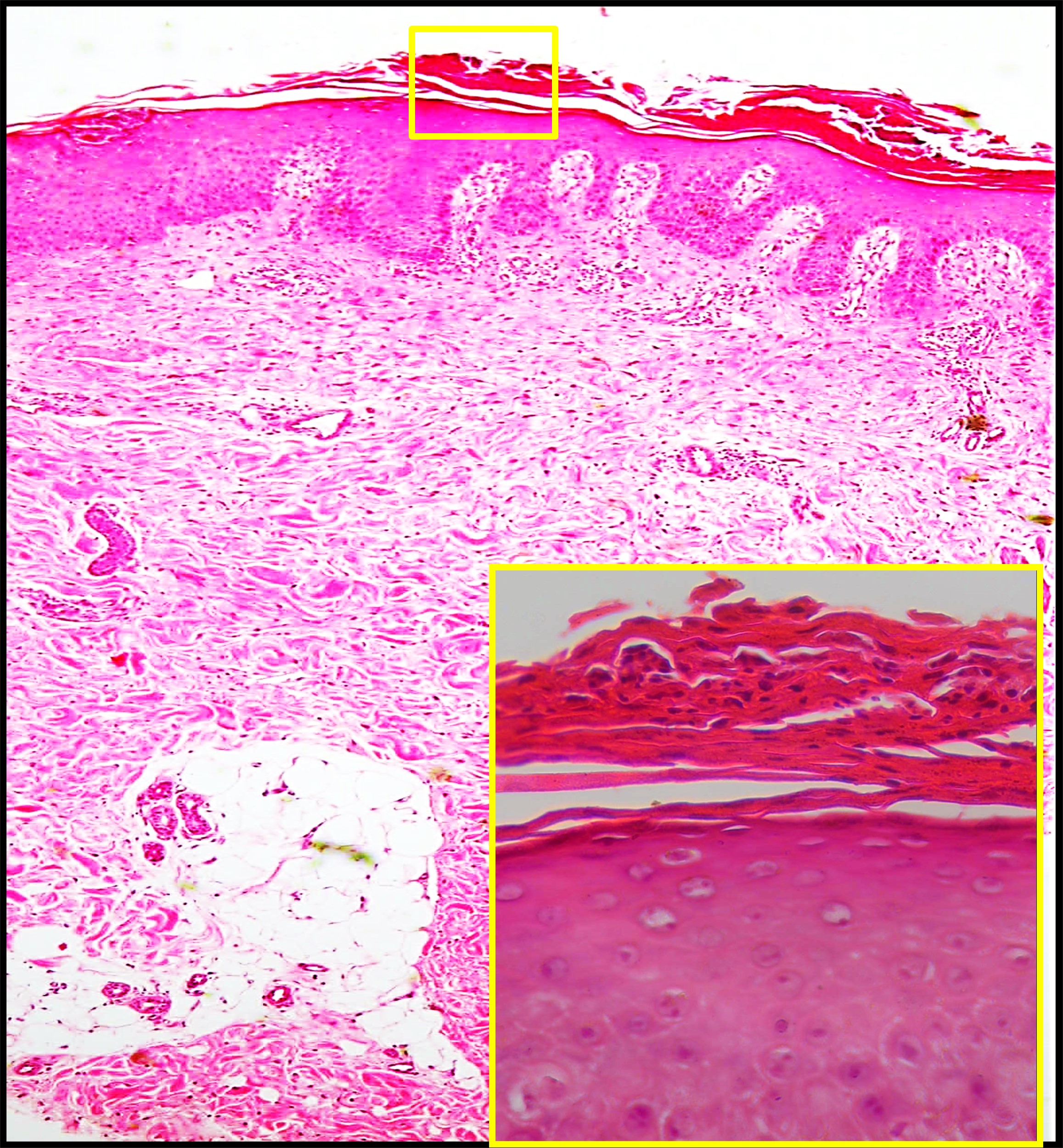
Histopathology of Pustular Psoriasis. Low-power view (H&E, ×100) shows regular psoriasiform acanthosis with overlying subcorneal neutrophilic collections. High-power inset (H&E, ×400) highlights neutrophilic microabscesses in the stratum corneum consistent with pustule formation.
Contributed by Mona Abdel-Halim Ibrahim, MD
(Click Image to Enlarge)
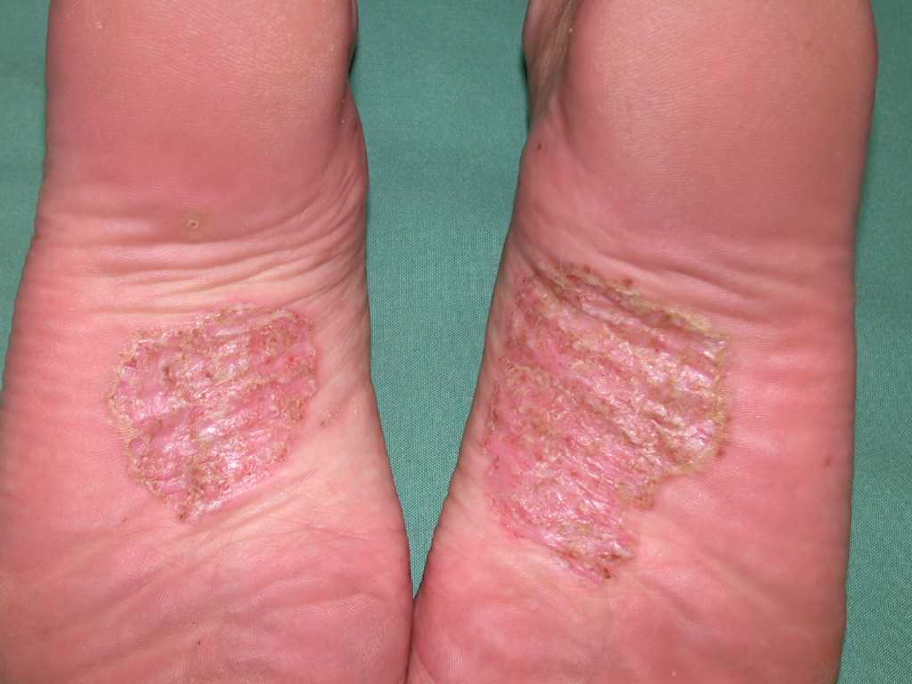
Pustulosis on the Soles. This photograph captures the localized inflammation and hyperkeratosis occurring on the arches of both feet. The clinical features include visible yellow-brown crusts and significant redness within the primary lesion sites.
References
Nagai M, Imai Y, Wada Y, Kusakabe M, Yamanishi K. Serum Procalcitonin and Presepsin Levels in Patients with Generalized Pustular Psoriasis. Disease markers. 2018:2018():9758473. doi: 10.1155/2018/9758473. Epub 2018 Dec 16 [PubMed PMID: 30647802]
Peccerillo F, Odorici G, Ciardo S, Conti A, Pellacani G. Evaluation of generalized pustular psoriasis by reflectance confocal microscopy. Skin research and technology : official journal of International Society for Bioengineering and the Skin (ISBS) [and] International Society for Digital Imaging of Skin (ISDIS) [and] International Society for Skin Imaging (ISSI). 2019 May:25(3):402-403. doi: 10.1111/srt.12655. Epub 2019 Jan 5 [PubMed PMID: 30614082]
Akiyama M. Early-onset generalized pustular psoriasis is representative of autoinflammatory keratinization diseases. The Journal of allergy and clinical immunology. 2019 Feb:143(2):809-810. doi: 10.1016/j.jaci.2018.11.009. Epub 2019 Jan 1 [PubMed PMID: 30606494]
Cassandra M, Conte E, Cortez B. Childhood pustular psoriasis elicited by the streptococcal antigen: a case report and review of the literature. Pediatric dermatology. 2003 Nov-Dec:20(6):506-10 [PubMed PMID: 14651571]
Level 3 (low-level) evidenceZhou X, An R, Qu P, Wang Q, Cheng W, Liu S, Zhao J, Jing H. RSV infection-associated pediatric generalized pustular psoriasis with innate immune activation: a case report. Frontiers in immunology. 2026:17():1808692. doi: 10.3389/fimmu.2026.1808692. Epub 2026 Apr 17 [PubMed PMID: 42079642]
Level 3 (low-level) evidenceKawamura A, Kinoshita MT, Suzuki H. Generalized pustular psoriasis with hypoparathyroidism. European journal of dermatology : EJD. 1999 Oct-Nov:9(7):574-6 [PubMed PMID: 10523741]
Brenner M, Molin S, Ruebsam K, Weisenseel P, Ruzicka T, Prinz JC. Generalized pustular psoriasis induced by systemic glucocorticosteroids: four cases and recommendations for treatment. The British journal of dermatology. 2009 Oct:161(4):964-6. doi: 10.1111/j.1365-2133.2009.09348.x. Epub 2009 Jun 11 [PubMed PMID: 19681858]
Level 3 (low-level) evidenceBorges-Costa J, Silva R, Gonçalves L, Filipe P, Soares de Almeida L, Marques Gomes M. Clinical and laboratory features in acute generalized pustular psoriasis: a retrospective study of 34 patients. American journal of clinical dermatology. 2011 Aug 1:12(4):271-6. doi: 10.2165/11586900-000000000-00000. Epub [PubMed PMID: 21495733]
Level 2 (mid-level) evidenceHong SB, Kim NI. Generalized pustular psoriasis following withdrawal of short-term cyclosporin therapy for psoriatic arthritis. Journal of the European Academy of Dermatology and Venereology : JEADV. 2005 Jul:19(4):522-3 [PubMed PMID: 15987319]
Duckworth L, Maheshwari MB, Thomson MA. A diagnostic challenge: acute generalized exanthematous pustulosis or pustular psoriasis due to terbinafine. Clinical and experimental dermatology. 2012 Jan:37(1):24-7. doi: 10.1111/j.1365-2230.2011.04129.x. Epub 2011 Jul 25 [PubMed PMID: 21790726]
Bachelez H. Pustular psoriasis and related pustular skin diseases. The British journal of dermatology. 2018 Mar:178(3):614-618. doi: 10.1111/bjd.16232. Epub 2018 Jan 15 [PubMed PMID: 29333670]
Mathieu RJ, Cobb CBC, Telang GH, Firoz EF. New-onset pustular psoriasis in the setting of severe acute respiratory syndrome coronavirus 2 infection causing coronavirus disease 2019. JAAD case reports. 2020 Dec:6(12):1360-1362. doi: 10.1016/j.jdcr.2020.10.013. Epub 2020 Oct 16 [PubMed PMID: 33102669]
Level 3 (low-level) evidenceOnsun N, Kaya G, Işık BG, Güneş B. A generalized pustular psoriasis flare after CoronaVac COVID-19 vaccination: Case report. Health promotion perspectives. 2021:11(2):261-262. doi: 10.34172/hpp.2021.32. Epub 2021 May 19 [PubMed PMID: 34195051]
Level 3 (low-level) evidenceKomatsuda S, Kamata M, Chijiwa C, Namiki K, Fukaya S, Hayashi K, Fukuyasu A, Tanaka T, Ishikawa T, Ohnishi T, Abe K, Yamamoto T, Aozasa N, Sugiura K, Tada Y. Gastrointestinal bleeding with severe mucosal involvement in a patient with generalized pustular psoriasis without IL36RN mutation. The Journal of dermatology. 2019 Jan:46(1):73-75. doi: 10.1111/1346-8138.14711. Epub 2018 Nov 26 [PubMed PMID: 30474867]
Boehner A, Navarini AA, Eyerich K. Generalized pustular psoriasis - a model disease for specific targeted immunotherapy, systematic review. Experimental dermatology. 2018 Oct:27(10):1067-1077. doi: 10.1111/exd.13699. Epub 2018 Jul 20 [PubMed PMID: 29852521]
Level 1 (high-level) evidenceViguier M, Bentayeb M, Azzi J, de Pouvourville G, Gloede T, Langellier B, Massol J, Medina P, Thoma C, Bachelez H. Generalized pustular psoriasis: A nationwide population-based study using the National Health Data System in France. Journal of the European Academy of Dermatology and Venereology : JEADV. 2024 Jun:38(6):1131-1139. doi: 10.1111/jdv.19901. Epub 2024 Feb 25 [PubMed PMID: 38404163]
Ohkawara A, Yasuda H, Kobayashi H, Inaba Y, Ogawa H, Hashimoto I, Imamura S. Generalized pustular psoriasis in Japan: two distinct groups formed by differences in symptoms and genetic background. Acta dermato-venereologica. 1996 Jan:76(1):68-71 [PubMed PMID: 8721499]
Chao PH, Cheng YW, Chung MY. Generalized pustular psoriasis in a 6-week-old infant. Pediatric dermatology. 2009 May-Jun:26(3):352-4. doi: 10.1111/j.1525-1470.2009.00918.x. Epub [PubMed PMID: 19706107]
Twelves S, Mostafa A, Dand N, Burri E, Farkas K, Wilson R, Cooper HL, Irvine AD, Oon HH, Kingo K, Köks S, Mrowietz U, Puig L, Reynolds N, Tan ES, Tanew A, Torz K, Trattner H, Valentine M, Wahie S, Warren RB, Wright A, Bata-Csörgő Z, Szell M, Griffiths CEM, Burden AD, Choon SE, Smith CH, Barker JN, Navarini AA, Capon F. Clinical and genetic differences between pustular psoriasis subtypes. The Journal of allergy and clinical immunology. 2019 Mar:143(3):1021-1026. doi: 10.1016/j.jaci.2018.06.038. Epub 2018 Jul 21 [PubMed PMID: 30036598]
Liao PB, Rubinson R, Howard R, Sanchez G, Frieden IJ. Annular pustular psoriasis--most common form of pustular psoriasis in children: report of three cases and review of the literature. Pediatric dermatology. 2002 Jan-Feb:19(1):19-25 [PubMed PMID: 11860564]
Level 3 (low-level) evidenceTosukhowong T, Kiratikanon S, Wonglamsam P, Netiviwat J, Innu T, Rujiwetpongstorn R, Tovanabutra N, Chiewchanvit S, Kwangsukstith C, Chuamanochan M. Epidemiology and clinical features of pustular psoriasis: A 15-year retrospective cohort. The Journal of dermatology. 2021 Dec:48(12):1931-1935. doi: 10.1111/1346-8138.16164. Epub 2021 Sep 16 [PubMed PMID: 34532894]
Level 2 (mid-level) evidenceBrunasso AM, Puntoni M, Aberer W, Delfino C, Fancelli L, Massone C. Clinical and epidemiological comparison of patients affected by palmoplantar plaque psoriasis and palmoplantar pustulosis: a case series study. The British journal of dermatology. 2013 Jun:168(6):1243-51. doi: 10.1111/bjd.12223. Epub [PubMed PMID: 23301847]
Level 2 (mid-level) evidenceMeier-Schiesser B, Feldmeyer L, Jankovic D, Mellett M, Satoh TK, Yerly D, Navarini A, Abe R, Yawalkar N, Chung WH, French LE, Contassot E. Culprit Drugs Induce Specific IL-36 Overexpression in Acute Generalized Exanthematous Pustulosis. The Journal of investigative dermatology. 2019 Apr:139(4):848-858. doi: 10.1016/j.jid.2018.10.023. Epub 2018 Nov 2 [PubMed PMID: 30395846]
Li L, You J, Fu X, Wang Z, Sun Y, Liu H, Zhang F. Variants of CARD14 are predisposing factors for generalized pustular psoriasis (GPP) with psoriasis vulgaris but not for GPP alone in a Chinese population. The British journal of dermatology. 2019 Feb:180(2):425-426. doi: 10.1111/bjd.17392. Epub 2018 Nov 29 [PubMed PMID: 30387497]
Bachelez H, Choon SE, Marrakchi S, Burden AD, Tsai TF, Morita A, Turki H, Hall DB, Shear M, Baum P, Padula SJ, Thoma C. Inhibition of the Interleukin-36 Pathway for the Treatment of Generalized Pustular Psoriasis. The New England journal of medicine. 2019 Mar 7:380(10):981-983. doi: 10.1056/NEJMc1811317. Epub [PubMed PMID: 30855749]
Saggini A, Chimenti S, Chiricozzi A. IL-6 as a druggable target in psoriasis: focus on pustular variants. Journal of immunology research. 2014:2014():964069. doi: 10.1155/2014/964069. Epub 2014 Jul 13 [PubMed PMID: 25126586]
Chen YL, Wang ZY, Ma L, Xu ZG. Three cases of IL36RN-associated pustulosis: An evolution of acrodermatitis continua of Hallopeau to generalized pustular psoriasis. Indian journal of dermatology, venereology and leprology. 2020 Sep-Oct:86(5):562-565. doi: 10.4103/ijdvl.IJDVL_581_19. Epub [PubMed PMID: 32769301]
Level 3 (low-level) evidenceArmstrong AW, Elston CA, Elewski BE, Ferris LK, Gottlieb AB, Lebwohl MG, Medical Board of the National Psoriasis Foundation. Generalized pustular psoriasis: A consensus statement from the National Psoriasis Foundation. Journal of the American Academy of Dermatology. 2024 Apr:90(4):727-730. doi: 10.1016/j.jaad.2023.09.080. Epub 2023 Oct 13 [PubMed PMID: 37838256]
Level 3 (low-level) evidenceHo PH, Tsai TF. Successful treatment of refractory juvenile generalized pustular psoriasis with secukinumab monotherapy: A case report and review of published work. The Journal of dermatology. 2018 Nov:45(11):1353-1356. doi: 10.1111/1346-8138.14636. Epub 2018 Sep 19 [PubMed PMID: 30230584]
Level 3 (low-level) evidenceOumeish OY, Parish JL. Impetigo herpetiformis. Clinics in dermatology. 2006 Mar-Apr:24(2):101-4 [PubMed PMID: 16487882]
Robinson A, Van Voorhees AS, Hsu S, Korman NJ, Lebwohl MG, Bebo BF Jr, Kalb RE. Treatment of pustular psoriasis: from the Medical Board of the National Psoriasis Foundation. Journal of the American Academy of Dermatology. 2012 Aug:67(2):279-88. doi: 10.1016/j.jaad.2011.01.032. Epub 2012 May 18 [PubMed PMID: 22609220]
Passeron T, Perrot JL, Jullien D, Goujon C, Ruer M, Boyé T, Villani AP, Quiles Tsimaratos N. Treatment of Severe Palmoplantar Pustular Psoriasis With Bimekizumab. JAMA dermatology. 2024 Feb 1:160(2):199-203. doi: 10.1001/jamadermatol.2023.5051. Epub [PubMed PMID: 38054800]
Ibis N, Hocaoglu S, Cebicci MA, Sutbeyaz ST, Calis HT. Palmoplantar pustular psoriasis induced by adalimumab: a case report and literature review. Immunotherapy. 2015:7(7):717-20. doi: 10.2217/imt.15.45. Epub 2015 Aug 7 [PubMed PMID: 26250408]
Level 3 (low-level) evidenceFukushima H, Iwata Y, Arima M, Tanaka Y, Sugiura K. Efficacy and safety of treatment with anti-tumor necrosis factor-α drugs for severe impetigo herpetiformis. The Journal of dermatology. 2021 Feb:48(2):207-210. doi: 10.1111/1346-8138.15635. Epub 2020 Oct 9 [PubMed PMID: 33035362]
Trivedi MK, Vaughn AR, Murase JE. Pustular psoriasis of pregnancy: current perspectives. International journal of women's health. 2018:10():109-115. doi: 10.2147/IJWH.S125784. Epub 2018 Feb 26 [PubMed PMID: 29520163]
Level 3 (low-level) evidenceKromer C, Loewe E, Schaarschmidt ML, Pinter A, Gerdes S, Celis D, Poortinga S, Wilsmann-Theis D, Mössner R. Treatment of acrodermatitis continua of Hallopeau: A case series of 39 patients. The Journal of dermatology. 2020 Sep:47(9):989-997. doi: 10.1111/1346-8138.15466. Epub 2020 Jul 6 [PubMed PMID: 32632983]
Level 2 (mid-level) evidenceHan GM, Yang WS, Yang B. Inhibition of Progression of Acrodermatitis Continua of Hallopeau With Baricitinib. JAMA dermatology. 2021 Apr 1:157(4):466-468. doi: 10.1001/jamadermatol.2021.0045. Epub [PubMed PMID: 33656520]
Kurihara Y, Nakano K, Eto A, Furue M. Successful treatment of acrodermatitis continua of Hallopeau with apremilast. The Journal of dermatology. 2019 Oct:46(10):e370-e371. doi: 10.1111/1346-8138.14927. Epub 2019 May 27 [PubMed PMID: 31131903]
Morita A, Strober B, Burden AD, Choon SE, Anadkat MJ, Marrakchi S, Tsai TF, Gordon KB, Thaçi D, Zheng M, Hu N, Haeufel T, Thoma C, Lebwohl MG. Efficacy and safety of subcutaneous spesolimab for the prevention of generalised pustular psoriasis flares (Effisayil 2): an international, multicentre, randomised, placebo-controlled trial. Lancet (London, England). 2023 Oct 28:402(10412):1541-1551. doi: 10.1016/S0140-6736(23)01378-8. Epub 2023 Sep 19 [PubMed PMID: 37738999]
Level 1 (high-level) evidenceMorita A, Choon SE, Bachelez H, Anadkat MJ, Marrakchi S, Zheng M, Tsai TF, Turki H, Hua H, Rajeswari S, Thoma C, Burden AD. Design of Effisayil™ 2: A Randomized, Double-Blind, Placebo-Controlled Study of Spesolimab in Preventing Flares in Patients with Generalized Pustular Psoriasis. Dermatology and therapy. 2023 Jan:13(1):347-359. doi: 10.1007/s13555-022-00835-6. Epub 2022 Nov 5 [PubMed PMID: 36333618]
Level 1 (high-level) evidenceJayasekera P, Parslew R, Al-Sharqi A. A case of tumour necrosis factor-α inhibitor- and rituximab-induced plantar pustular psoriasis that completely resolved with tocilizumab. The British journal of dermatology. 2014 Dec:171(6):1546-9. doi: 10.1111/bjd.13146. Epub 2014 Nov 3 [PubMed PMID: 24890762]
Level 2 (mid-level) evidencePalmou-Fontana N, Sánchez Gaviño JA, McGonagle D, García-Martinez E, Iñiguez de Onzoño Martín L. Tocilizumab-induced psoriasiform rash in rheumatoid arthritis. Dermatology (Basel, Switzerland). 2014:228(4):311-3. doi: 10.1159/000362266. Epub 2014 Jun 12 [PubMed PMID: 24942661]
Smieszek S, Przychodzen B, Tyner C, Johnson C, Bushman M, Brzezynski J, Reich A, Bachelez H, Yap EWY, Borlu M, Chaowattanapanit S, Denguezli M, Gudjonsson JE, Lizzul P, Dahl M, Parmley S, Randazzo B, Severtson A, Raina P, Saikali K, Xiao C, Polymeropoulos C, Birznieks G, Polymeropoulos M. Efficacy and Safety of Imsidolimab for Generalized Pustular Psoriasis. NEJM evidence. 2026 May:5(5):EVIDoa2500272. doi: 10.1056/EVIDoa2500272. Epub 2026 Apr 28 [PubMed PMID: 42047557]
Ansari MS, Heidari S, Pourgholi E, Bahramian S, Tootoonchi N, Vahabi SM. Janus Kinase Inhibitors for Treatment of Palmoplantar Pustulosis, Generalized Pustular Psoriasis, and Palmoplantar Pustular Psoriasis: A Systematic Review of the Literature. Health science reports. 2026 Apr:9(4):e72301. doi: 10.1002/hsr2.72301. Epub 2026 Apr 6 [PubMed PMID: 41953903]
Level 1 (high-level) evidenceChoon SE, van de Kerkhof P, Gudjonsson JE, de la Cruz C, Barker J, Morita A, Romiti R, Affandi AM, Asawanonda P, Burden AD, Gonzalez C, Marrakchi S, Mowla MR, Okubo Y, Oon HH, Terui T, Tsai TF, Callis-Duffin K, Fujita H, Jo SJ, Merola J, Mrowietz U, Puig L, Thaçi D, Velásquez M, Augustin M, El Sayed M, Navarini AA, Pink A, Prinz J, Turki H, Magalhães R, Capon F, Bachelez H, IPC Pustular Psoriasis Working Group. International Consensus Definition and Diagnostic Criteria for Generalized Pustular Psoriasis From the International Psoriasis Council. JAMA dermatology. 2024 Jul 1:160(7):758-768. doi: 10.1001/jamadermatol.2024.0915. Epub [PubMed PMID: 38691347]
Level 3 (low-level) evidenceSzatkowski J, Schwartz RA. Acute generalized exanthematous pustulosis (AGEP): A review and update. Journal of the American Academy of Dermatology. 2015 Nov:73(5):843-8. doi: 10.1016/j.jaad.2015.07.017. Epub 2015 Sep 6 [PubMed PMID: 26354880]
Cheng S, Edmonds E, Ben-Gashir M, Yu RC. Subcorneal pustular dermatosis: 50 years on. Clinical and experimental dermatology. 2008 May:33(3):229-33. doi: 10.1111/j.1365-2230.2008.02706.x. Epub 2008 Mar 18 [PubMed PMID: 18355359]
Zhou LL, Georgakopoulos JR, Ighani A, Yeung J. Systemic Monotherapy Treatments for Generalized Pustular Psoriasis: A Systematic Review. Journal of cutaneous medicine and surgery. 2018 Nov/Dec:22(6):591-601. doi: 10.1177/1203475418773358. Epub 2018 Apr 30 [PubMed PMID: 29707979]
Level 1 (high-level) evidenceHoegler KM, John AM, Handler MZ, Schwartz RA. Generalized pustular psoriasis: a review and update on treatment. Journal of the European Academy of Dermatology and Venereology : JEADV. 2018 Oct:32(10):1645-1651. doi: 10.1111/jdv.14949. Epub 2018 Apr 11 [PubMed PMID: 29573491]
Miyachi H, Konishi T, Kumazawa R, Matsui H, Shimizu S, Fushimi K, Matsue H, Yasunaga H. Treatments and outcomes of generalized pustular psoriasis: A cohort of 1516 patients in a nationwide inpatient database in Japan. Journal of the American Academy of Dermatology. 2022 Jun:86(6):1266-1274. doi: 10.1016/j.jaad.2021.06.008. Epub 2021 Jun 8 [PubMed PMID: 34116101]
Viguier M, Allez M, Zagdanski AM, Bertheau P, de Kerviler E, Rybojad M, Morel P, Dubertret L, Lémann M, Bachelez H. High frequency of cholestasis in generalized pustular psoriasis: Evidence for neutrophilic involvement of the biliary tract. Hepatology (Baltimore, Md.). 2004 Aug:40(2):452-8 [PubMed PMID: 15368450]
Jeon JJ, Lee S, Kim YS. Distinct Mortality Risks in Generalized Pustular Psoriasis and Palmoplantar Pustulosis: A Nationwide, Population-Based, Cohort Study. Dermatology (Basel, Switzerland). 2026:242(2):206-214. doi: 10.1159/000550019. Epub 2025 Dec 16 [PubMed PMID: 41401112]