 Tools
Tools
 Anatomy, Bony Pelvis and Lower Limb, Gemelli Muscles
Introduction
The superior and inferior gemelli are a pair of small, triangular muscles located within the posterior hip and gluteal musculature of the proximal posterior lower extremity and are collectively referred to as the "gemelli." These muscles constitute 2 of the 6 short external rotator muscles in the gluteal region of the proximal thigh (see Image. Landmarks and Muscle Attachments of the Femur). The remaining external rotators include the piriformis, obturator internus, quadratus femoris, and obturator externus muscles. The superior and inferior gemelli converge with the obturator internus to form a conjoined tendon, sometimes referred to as the "triceps coxae" (see Image. Short External Thigh Rotators).[1] The gemelli assist in the external rotation of the thigh and are implicated in sciatic nerve dysfunction, as in cases of deep gluteal space syndrome, also termed "gemelli-obturator syndrome."[2][3][4]
The gemelli serve as important surgical landmarks in posterior approaches to the hip, including the Kocher-Langenbeck technique. These muscles are essential for identifying and protecting the medial circumflex femoral artery, in conjunction with the obturator internus, during these procedures. These surgical approaches can facilitate surgical hip dislocation and are used in total hip arthroplasty, hip hemiarthroplasty, loose body removal, and management of posterior wall and posterior column acetabular fractures.[5]
Structure and Function
Register For Free And Read The Full Article
 Search engine and full access to all medical articles
- 10 free questions in your specialty
- Free CME/CE Activities
- Free daily question in your email
- Save favorite articles to your dashboard
- Emails offering discounts
Learn more about a Subscription to StatPearls Point-of-Care
Structure and Function
The superior gemellus originates from the ischial spine, whereas the inferior gemellus originates from the ischial tuberosity. Both gemelli insert on the medial surface of the greater trochanter of the femur in conjunction with the obturator internus (see Image. Right Proximal Femur Anatomy). The primary function of the gemelli is external (lateral) rotation of the thigh and stabilization of the hip joint. The muscles also contribute to hip abduction while in a flexed position. The gemelli work synergistically with the obturator internus and the remaining short external rotator to rotate the thigh externally and stabilize the femoral head in the acetabulum.[6]
Embryology
Limb buds appear at approximately 4 to 5 weeks of gestation as mesoderm migrates into the developing limb bud, forming posterior and anterior condensations that give rise to the muscular and skeletal components of the lower limb. Around the 4th to 5th week of gestation, myoblasts migrate from the somites into the limb bud. The posterior condensation, also referred to as the "dorsal muscle mass," develops into the extensor and abductor musculature of the lower limb, whereas the anterior condensation, also called the "ventral muscle mass," develops into the flexor and adductor musculature of the lower limb.[7]
Several factors influence the formation of limb bud musculature, including retinoic acid and sonic hedgehog (Shh), along with regulatory genes such as homeobox (HOX) genes. Development is further organized by embryologic regions, including the apical ectodermal ridge (AER) and the zone of polarizing activity (ZPA).
Retinoic acid functions as a global organizing gradient that initiates transcription factor production responsible for regional differentiation and limb polarization. The AER produces fibroblast growth factor (FGF), which promotes limb bud outgrowth through stimulation of mitosis.[8] Dependence on FGF and AER signaling is essential for continued limb development. Absence of appropriate signaling results in arrest of limb growth. FGF10 serves as the key FGF in hindlimb development and is upregulated by T-box transcription factor 4 (Tbx4).
The zone of polarizing activity produces Shh, which organizes the limb bud along the anterior-posterior axis. Shh activates specific HOX genes, including HOXD9, HOXD10, HOXD11, HOXD12, and HOXD13, which are essential for limb polarization and regional specification. These genes regulate patterning and thereby determine the morphology of the developing limb in the human embryo. Errors in expression, signaling, or maintenance of these pathways result in limb malformations.[9]
Blood Supply and Lymphatics
The inferior gluteal artery supplies the superior and inferior gemelli. This blood vessel arises from the anterior division of the internal iliac artery, passes between the 2nd and 3rd sacral nerve roots of the sacral plexus, and exits the greater sciatic foramen inferior to the piriformis to supply the gemelli (see Image. Internal Iliac Branches). Lymphatic vessels of the lower limb are divided into superficial and deep systems. Superficial lymphatic vessels are further divided into medial and lateral groups. The medial group follows the greater saphenous vein, whereas the lateral group follows the small saphenous vein. Deep lymphatic vessels, including the anterior tibial, posterior tibial, and peroneal vessels, accompany the corresponding arteries. Lymphatic drainage of the lower limb proceeds through the popliteal, superficial inguinal, deep inguinal, external iliac, and lumbar (aortic) lymph nodes.
Nerves
The nerve supply to the superior gemellus is provided by the nerve to the obturator internus (L4–S2), whereas the inferior gemellus receives innervation from the nerve to the quadratus femoris (L4–S1). Each nerve arises from the sacral plexus, formed by the ventral rami of L4 to S4.
The nerve to the obturator internus passes through the greater sciatic foramen inferior to the piriformis as it exits the pelvis. Innervation to the superior gemellus is provided during its course. The nerve then reenters the pelvis through the lesser sciatic foramen to supply the obturator internus.
The nerve to the quadratus femoris exits the pelvis via the greater sciatic foramen inferior to the piriformis. The nerve descends deep to the short external rotators and terminates on the deep surface of the quadratus femoris, providing innervation to the inferior gemellus during its course.
Physiologic Variants
The superior gemellus, inferior gemellus, and obturator internus were historically considered a single functional unit due to shared insertion and similar function. Current evidence supports classification as distinct entities based on differences in origin and innervation.[10] Reported physiologic variants include duplication of the superior gemellus, absence of both gemelli, and variations in origin and insertion patterns.[11] Awareness of potential anatomic variants may assist surgical exposure and dissection during procedures involving this region.
Surgical Considerations
The gemelli are most relevant surgically when a posterior approach to the hip is planned. A form of this technique, known as the Kocher-Langenbeck approach, is commonly used for total hip arthroplasty and hip hemiarthroplasty due to several advantages, including a clear anatomical plane, lower incidence of heterotopic ossification, and preservation of the hip abductors.[12] Following initial exposure, the triceps coxae are elevated from their insertion approximately 1 to 2 cm along the posterior aspect of the greater trochanter.
Direct elevation of the conjoined tendon from bone is also performed based on surgeon preference. Injury to these structures is generally unavoidable. However, controversy persists regarding methods to minimize injury, indications for repair, and the clinical impact of restoration. Recent literature highlights a significant reduction in posterior dislocation risk following meticulous posterior capsule repair, with particular attention to the deep external rotators, including the triceps coxae.[13] Therefore, effective reconstruction of the gemelli and obturator internus may be important in the early postoperative period to reduce dislocation risk and support postoperative recovery and rehabilitation.[14][15][16][17]
Clinical Significance
Posterior hip or gluteal pain and sciatica represent common diagnostic and therapeutic challenges for orthopedic surgeons and general medical providers. Historically, the term “piriformis syndrome” encompassed multiple pathologies in this region. Advances in imaging techniques and evaluation of muscle and nerve kinematics have led to the adoption of the preferred term “deep gluteal syndrome" (DGS). The condition is characterized by pain or dysesthesia in the buttock, hip, or posterior thigh, with or without radicular symptoms, due to nondiscogenic sciatic nerve entrapment.[18]
DGS includes several pathologic processes, including piriformis syndrome, gemelli-obturator syndrome, and tumoral compression. Gemelli-obturator syndrome arises from dynamic compression of the sciatic nerve when the gemelli-obturator internus complex is placed under stretch. The sciatic nerve courses deep to the piriformis and superficial to the triceps coxae. This anatomic relationship may result in sciatic nerve compression, producing sciatic pain and necessitating inclusion in the differential diagnosis of nondiscogenic sciatica.
Differentiation of causes of DGS relies on physical examination maneuvers and imaging modalities to support definitive diagnosis. Magnetic resonance imaging assists in the evaluation of anatomic variants and soft-tissue abnormalities. Treatment of DGS targets the underlying etiology and typically includes imaging-guided intra-articular or extra-articular injections, physical therapy, and administration of anti-inflammatory medications. Open or endoscopic surgical procedures may be considered in rare cases refractory to conservative management.
Media
(Click Image to Enlarge)
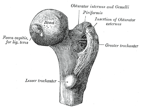
Right Proximal Femur Anatomy. This illustration shows the femoral head, fovea capitis, ligamentum teres, femoral neck, trochanteric fossa, trochanter, lesser trochanter, greater trochanter, intertrochanteric crest, and insertions for the obturator internus, gemelli, piriformis, obturator externus.
Henry Vandyke Carter, Public Domain, via Wikimedia Commons
(Click Image to Enlarge)
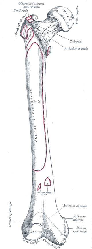
Landmarks and Muscle Attachments of the Femur. This anterior view of the right femur illustrates proximal landmarks, including the greater trochanter, fovea capitis, and tubercle, alongside insertion points for the piriformis, obturator internus, and gemelli. The shaft and distal regions display the extensive origins of the vastus lateralis and vastus intermedius, as well as the articularis genu, adductor tubercle, and medial epicondyle. The anatomy concludes at the knee joint with the medial condyle, lateral condyle, and patella.
Henry Vandyke Carter, Public Domain, via Wikimedia Commons
(Click Image to Enlarge)
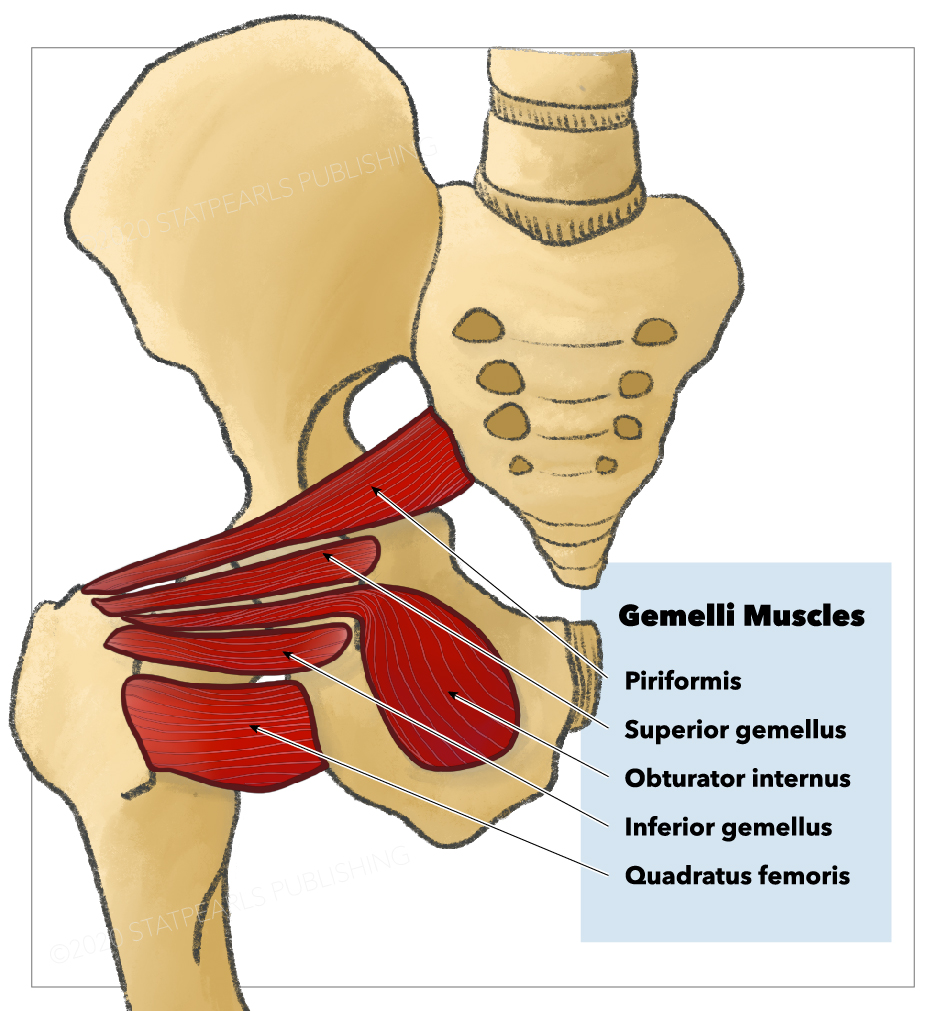
Short External Thigh Rotators. Shown in this illustration are the superior and inferior gemelli, quadratus femoris, obturator internus, inferior gemellus, superior gemellus, and piriformis.
StatPearls Publishing Illustration
(Click Image to Enlarge)
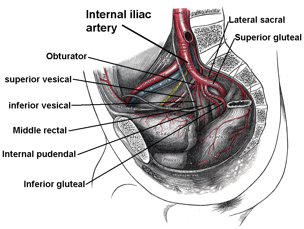
Internal Iliac Branches. This image shows that the internal iliac artery divides into 2 trunks, anterior and posterior. The anterior trunk gives off 8 further branches, while the posterior trunk has 3 branches.
Mikael Häggström, Public Domain, via Wikimedia Commons
References
Siebenrock KA, Keel MJB, Tannast M, Bastian JD. Surgical Hip Dislocation for Exposure of the Posterior Column. JBJS essential surgical techniques. 2019 Mar 26:9(1):e2. doi: 10.2106/JBJS.ST.18.00048. Epub 2019 Jan 23 [PubMed PMID: 31086720]
Carro LP, Hernando MF, Cerezal L, Navarro IS, Fernandez AA, Castillo AO. Deep gluteal space problems: piriformis syndrome, ischiofemoral impingement and sciatic nerve release. Muscles, ligaments and tendons journal. 2016 Jul-Sep:6(3):384-396. doi: 10.11138/mltj/2016.6.3.384. Epub 2016 Dec 21 [PubMed PMID: 28066745]
Martin HD, Reddy M, Gómez-Hoyos J. Deep gluteal syndrome. Journal of hip preservation surgery. 2015 Jul:2(2):99-107. doi: 10.1093/jhps/hnv029. Epub 2015 Jun 6 [PubMed PMID: 27011826]
Assi C, Bonnel F, Elkayem E, Mansour J, Mares O, Yammine K. The short external rotator muscles of the hip: a cadaveric study on 18 specimens with clinical implications. Surgical and radiologic anatomy : SRA. 2024 Jul:46(7):1109-1115. doi: 10.1007/s00276-024-03384-z. Epub 2024 May 26 [PubMed PMID: 38797739]
Tosounidis TH, Giannoudis VP, Kanakaris NK, Giannoudis PV. The Kocher-Langenbeck Approach: State of the Art. JBJS essential surgical techniques. 2018 Jun 27:8(2):e18. doi: 10.2106/JBJS.ST.16.00102. Epub 2018 Jun 13 [PubMed PMID: 30233990]
Balius R, Susín A, Morros C, Pujol M, Pérez-Cuenca D, Sala-Blanch X. Gemelli-obturator complex in the deep gluteal space: an anatomic and dynamic study. Skeletal radiology. 2018 Jun:47(6):763-770. doi: 10.1007/s00256-017-2831-2. Epub 2017 Dec 7 [PubMed PMID: 29218390]
Lezak B, Wehrle CJ, Summers S. Anatomy, Bony Pelvis and Lower Limb: Posterior Tibial Artery. StatPearls. 2026 Jan:(): [PubMed PMID: 30725666]
Mróz I, Kielczewski S, Pawlicki D, Kurzydło W, Bachul P, Konarska M, Bereza T, Walocha K, Kaythampillai LN, Depukat P, Pasternak A, Bonczar T, Chmielewski P, Mizia E, Skrzat J, Mazur M, Warchoł Ł, Tomaszewski K. Blood vessels of the shin - anterior tibial artery - anatomy and embryology - own studies and review of the literature. Folia medica Cracoviensia. 2016:56(1):33-47 [PubMed PMID: 27513837]
Gros J, Tabin CJ. Vertebrate limb bud formation is initiated by localized epithelial-to-mesenchymal transition. Science (New York, N.Y.). 2014 Mar 14:343(6176):1253-6. doi: 10.1126/science.1248228. Epub [PubMed PMID: 24626928]
Level 3 (low-level) evidenceShinohara H. Gemelli and obturator internus muscles: different heads of one muscle? The Anatomical record. 1995 Sep:243(1):145-50 [PubMed PMID: 8540628]
Arifoglu Y, Sürücü HS, Sargon MF, Tanyeli E, Yazar F. Double superior gemellus together with double piriformis and high division of the sciatic nerve. Surgical and radiologic anatomy : SRA. 1997:19(6):407-8 [PubMed PMID: 9479716]
Level 3 (low-level) evidenceHernández A, Nuñez JH, Mimendia I, Barro V, Azorin L. Early dislocation in primary total hip arthroplasty using a posterior approach with repair of capsule and external rotators. Revista espanola de cirugia ortopedica y traumatologia (English ed.). 2018 Nov-Dec:62(6):421-427. doi: 10.1016/j.recot.2018.03.006. Epub [PubMed PMID: 29776889]
Girdwood M, West L, Connell D, Brukner P. Contact-Related Strain of Quadratus Femoris, Obturator Externus, and Inferior Gemellus in an Australian Football Player: A Case Report. Journal of sport rehabilitation. 2019 Nov 1:28(8):887-890. doi: 10.1123/jsr.2018-0279. Epub [PubMed PMID: 30747560]
Level 3 (low-level) evidenceSodhi N, Chen Z, Bains SS, Garbarino LJ, Mont MA. An Updated Review on Layered Closure Techniques for Total Hip Arthroplasty. Surgical technology international. 2022 Nov 15:41():365-372. doi: 10.52198/22.STI.41.OS1611. Epub [PubMed PMID: 35801356]
Cherry A, Broderick JM, Thompson ZS, Pentland V, Dietz MJ, Khoshbin A, Atrey A. Trans-osseous repair of the posterior structures is superior to direct suturing in posterior approach total hip arthroplasty: a comparative cadaveric study. International orthopaedics. 2025 Sep:49(9):2055-2061. doi: 10.1007/s00264-025-06618-w. Epub 2025 Jul 26 [PubMed PMID: 40715845]
Level 2 (mid-level) evidenceArora S, Gupta P, Sharma M, Meena MK, Khan S, Kashyap A. Quadriceps Coxae-Sparing Modified Posterior Approach to the Hip Joint for Hemiarthroplasty. JBJS essential surgical techniques. 2026 Jan-Mar:16(1):. doi: 10.2106/JBJS.ST.25.00008. Epub 2026 Jan 14 [PubMed PMID: 41522696]
Won SH, Joh Y, Lim YW, Kwon SY, Kim YS, Kim SC. Can External Rotator Preservation Reduce Dislocation Risk Following Posterior Approach Total Hip Arthroplasty in High-Risk Patients? A Propensity-Matched Comparative Study. The Journal of arthroplasty. 2025 Dec 18:():. pii: S0883-5403(25)01573-6. doi: 10.1016/j.arth.2025.12.024. Epub 2025 Dec 18 [PubMed PMID: 41421705]
Level 2 (mid-level) evidenceWang T, Shao L, Xu W, Li F, Huang W. Surgical injury and repair of hip external rotators in THA via posterior approach: a three-dimensional MRI-evident quantitative prospective study. BMC musculoskeletal disorders. 2019 Jan 14:20(1):22. doi: 10.1186/s12891-018-2367-1. Epub 2019 Jan 14 [PubMed PMID: 30642331]