 Tools
Tools
 Eyelid Papilloma
Introduction
Despite the eyelid's small surface area, it plays an important role in protecting the eyeball. Skin on the eyelid has no subcutaneous fat and is the thinnest skin found in the body.[1] These characteristics, as well as the eyelids' location on the body, make them particularly sensitive to irritants and ultraviolet (UV) damage. As such, eyelids are prone to developing benign and malignant eyelid tumors. One study reported that 5% to 10% of all skin cancers occur on the eyelids.[2]
The term papilloma refers to a group of various benign epithelial proliferations that can affect the eyelid skin.[3] These lesions are not necessarily associated with the papillomavirus. Eyelid papillomas are not dangerous but can cause mild irritation or be cosmetically unfavorable for the patient (see Image. Eyelid Papilloma). Also, differentiating a benign lesion, eg, a papilloma, from a potentially malignant lesion on the eyelid is important. The lesions which fall into this classification include, but are not limited to, seborrheic keratosis, pseudoepitheliomatous hyperplasia, inverted follicular keratosis, verruca vulgaris, squamous papilloma (acrochordon/skin tag), basosquamous acanthoma, and squamous acanthoma.
Papilloma Subtypes
Types of papillomas include:
- Squamous papilloma: Also known as an acrochordon or skin tag. This is a soft, flesh-colored lesion that is smooth, round, and pedunculated.
- Seborrheic keratosis
- This is a benign proliferation of cells. Classically, seborrheic keratosis has a “stuck-on” appearance and can have varying degrees of pigmentation.
- Lesions tend to range from pink or flesh-colored to dark brown. These lesions are well-circumscribed and usually slightly elevated. Normally, these lesions are benign, but the sudden appearance of multiple seborrheic keratoses in a region of the body could indicate a paraneoplastic process. Seborrheic keratosis often has an inflamed base.
- The Leser-Trélat sign is a relatively rare paraneoplastic cutaneous marker of internal malignancy, characterized by the abrupt eruption of multiple seborrheic keratoses. Please see StatPearls' companion resource, "Leser-Trélat Sign," for further information.
- Pseudoepitheliomatous hyperplasia: These lesions are often reactive processes in response to things, eg, trauma, wound, burn, pharmaceutical agents, that can appear similar to basal cell or squamous cell carcinoma. These lesions are usually elevated on the skin with an irregular surface and occasionally ulceration or crusting.[1]
- Inverted follicular keratosis: This is normally a solitary lesion affecting the eyelid margin. Inverted follicular keratosis may or may not be pigmented and has been described as either nodular or papillary in appearance.[1]
- Verruca vulgaris: This is a flesh-colored skin growth caused by human papillomavirus and is rare on the eyelid.
Etiology
Register For Free And Read The Full Article
 Search engine and full access to all medical articles
- 10 free questions in your specialty
- Free CME/CE Activities
- Free daily question in your email
- Save favorite articles to your dashboard
- Emails offering discounts
Learn more about a Subscription to StatPearls Point-of-Care
Etiology
Papilloma etiology depends on the type of epithelial proliferation. Squamous papillomas and seborrheic keratosis are idiopathic benign cellular proliferations. There is no known definitive cause of these lesions. However, malignant skin lesions that can look like papillomas are often associated with chronic UV exposure and sun-damaged skin. Verruca vulgaris is caused by human papillomavirus type 6 or 11.
Epidemiology
One study found that, overall, epidermal tumors make up the majority of eyelid tumors. One study found seborrheic keratosis to be the most frequently encountered lesion, followed by squamous cell papilloma.[4] Another study, however, reported that squamous papillomas are the most common benign epithelial tumor of the eyelid.[1] Seborrheic keratosis is typically found in middle-aged or older adult patients. Squamous papillomas do not have a strong predilection for a particular race or sex. These papillomas tend to become more frequent with age, but they can occur at any age.
Histopathology
Seborrheic keratosis displays hyperkeratosis, acanthosis, and some degree of papillomatosis on histological preparation. By definition, the squamous cells comprising the lesion do not exhibit dysplasia. One characteristic finding in some seborrheic keratosis is the pseudo-horn cysts. These are circular collections of surface keratin in the acanthotic epithelium of the seborrheic keratosis lesion.
Squamous papillomas display benign squamous epithelium with variable acanthosis and hyperkeratosis, and focal parakeratosis, overlying a fibrovascular core. These have occasionally been shown to exhibit features of chronic inflammation.[1] Pseudoepitheliomatous hyperplasia demonstrates invasive, elongated processes of hyperplastic epithelium and often contains inflammatory cells; however, features of malignancy, eg, dysplasia and atypical mitoses, are absent.[1] Inverted follicular keratoses display endophytic proliferation of basal and squamous elements. Pigmentation, acantholysis, and chronic inflammation can occur in these lesions.[1]
History and Physical
Clinical History
When evaluating a skin lesion on the eyelid, the following questions are important to ask the patient:
- How long has the lesion been present?
- Have you noticed the lesion changing color, size, or shape?
- Does the lesion cause pain or irritation?
- Has the lesion bled or drained any fluid or purulent material?
- Do you have any other similar skin lesions on other parts of your body?
- Have you had a similar lesion in the past on the eyelid?
- Do you have a history of skin cancer?
When patients present with eyelid papillomas, they typically have been present for months to years. These lesions do not grow rapidly, and patients do not notice changes in their character over time. Occasionally, the patient can feel the papilloma from the weight of the eyelid, or the lesion may become mildly inflamed, but chronic or severe inflammation is uncharacteristic of a papilloma. Papillomas do not bleed or drain purulent material unless the patient picks at the lesion and it subsequently becomes infected. A history of skin cancer in a patient should prompt the clinician to monitor the lesion more closely for suspicious characteristics.
Physical Examination
When performing the physical exam, clinicians should perform the following evaluations:
- Examine the surrounding skin for additional skin lesions.
- Feel facial/neck lymph nodes to assess for lymphadenopathy.
- Using the slit lamp, examine the lesion for the destruction of nearby tissues (skin ulceration, destruction of Meibomian glands, or eyelash loss (madarosis). Note any telangiectasia of the lesion, whitening of nearby eyelashes (poliosis), and destruction of Meibomian glands. Evert eyelids to look for disruption of the palpebral conjunctiva.
The following physical exam findings for papillomas vary slightly by type:
- Squamous papilloma: This is a soft, flesh-colored lesion that is generally smooth, round, or pedunculated.
- Seborrheic keratosis: Classically has a “stuck on” appearance and can have varying degrees of pigmentation. Lesions tend to range from pink or flesh-colored to dark brown. These lesions are well-circumscribed and usually slightly elevated.
- Pseudoepitheliomatous hyperplasia: These lesions are often reactive processes in response to trauma, wounds, burns, and pharmaceutical agents that can appear similar to basal cell or squamous cell carcinoma. These lesions are usually elevated on the skin with an irregular surface and occasionally ulceration or crusting.[1]
- Inverted follicular keratosis: This is normally a solitary lesion affecting the eyelid margin. Inverted follicular keratosis may or may not be pigmented and has been described as either nodular or papillary in appearance.[1]
- Verruca vulgaris: Flesh-colored skin growth. The superior portion of the lesion can have very tiny, fingerlike projections that are sometimes visible without magnification.
Evaluation
Documenting the lesion with a photo allows for comparison of the size and appearance of the lesion over time. Measuring the size of the lesion at each visit can confirm that the lesion is growing over time.. If the lesion displays any characteristics that are concerning for malignancy, consider performing a biopsy. Typically, an incisional biopsy is performed in these cases.[5]
Treatment / Management
Lesions identified as eyelid papillomas can be observed. If the papilloma is causing irritation or is cosmetically unacceptable for the patient, it can be removed. Most papillomas can be removed with a bedside shave excision. Verruca vulgaris typically responds better to cryotherapy. Other modalities utilized to remove these types of lesions include radiofrequency and plasma exeresis.[6][7][5]
One study reported successful treatment of a large eyelid papilloma with intralesional interferon. A larger study of 64 patients with eyelid papilloma-like lesions found that radiofrequency lesion removal was safe and effective. In this study, 72% of the lesions treated were squamous papillomas.
Papilloma Excision Technique
The following steps are recommended when performing papilloma excision. First, topical tetracaine should be instilled in the ipsilateral eye to minimize irritation from the cleaning solution. Clean the papilloma and surrounding eyelid tissue with full-strength povidone-iodine solution, then place a small sterile drape with a fenestration to isolate the lesion.
Then inject 1 mL to 2 mL of lidocaine with epinephrine directly beneath the papilloma, using the minimal volume necessary to ensure patient comfort. Using 0.5 mm forceps, gently grasp and elevate the lesion while excising it with a 15-blade or iris scissors. Begin at the base to ensure complete removal without extending into deeper tissues. Achieve hemostasis with a handheld cautery device as needed. The residual defect generally remains small and does not require suturing to approximate the skin. Prescribe an antibiotic, eg, erythromycin ophthalmic ointment, applied 3 to 4 times daily for 1 to 2 weeks until healing occurs to reduce infection risk. Send the excised tissue for pathologic evaluation when any suspicious features are present.[6][8][9](B2)
Differential Diagnosis
Several benign and malignant eyelid lesions can resemble an eyelid papilloma and should be included in the differential diagnosis during evaluation, eg, chalazion/hordeolum, epidermal inclusion cyst, molluscum contagiosum, xanthelasma, squamous cell carcinoma, nevus, actinic keratosis, basal cell carcinoma, and sebaceous gland carcinoma.[10] Careful clinical examination and, when necessary, histopathologic analysis are important to ensure accurate diagnosis and appropriate management.
One common benign condition that may be mistaken for an eyelid papilloma is seborrheic keratosis, which presents as a well-demarcated, waxy, “stuck-on” lesion that can develop on the eyelids. Verruca vulgaris (common wart) may also appear similar, as this lesion presents with a rough, papillomatous surface due to human papillomavirus infection. Skin tags (acrochordons) are another benign lesion that can occur on the eyelids and present as soft, pedunculated growths. Although typically harmless, they may clinically resemble small papillomas.
Malignant lesions must also be considered, particularly because the eyelid is a common site for skin cancer. Basal cell carcinoma, the most common malignant eyelid tumor, may initially present as a small, nodular lesion with pearly borders and telangiectasia, sometimes mimicking benign growths. Squamous cell carcinoma can present as a scaly, ulcerated lesion and may occasionally resemble papillomatous growths in early stages. Sebaceous gland carcinoma, though less common, is an aggressive eyelid malignancy that may masquerade as a benign lesion or chronic inflammatory condition.
Because these conditions can have overlapping clinical features, lesions that demonstrate rapid growth, irregular borders, ulceration, bleeding, lash loss, or persistent recurrence should be evaluated carefully. When diagnostic uncertainty exists, biopsy and histopathologic examination are recommended to confirm the diagnosis and rule out malignancy. Accurate differentiation among these conditions is essential to ensure appropriate treatment and favorable patient outcomes.
Prognosis
The prognosis for eyelid papillomas is excellent, as these lesions are benign epithelial proliferations that rarely lead to significant complications. Most eyelid papillomas remain stable over time and do not undergo malignant transformation.[11] When treatment is desired due to cosmetic concerns, irritation, or diagnostic uncertainty, simple excision or minor procedural removal is typically curative. Recurrence after complete removal is uncommon but may occur if the lesion is incompletely excised.
Complications
Complications are rare but may include local irritation, bleeding, or infection following removal procedures. In most cases, patients experience good cosmetic outcomes when lesions are treated appropriately. The most important prognostic consideration is ensuring accurate differentiation from malignant eyelid tumors, eg, basal cell carcinoma or squamous cell carcinoma, which may initially resemble benign papillomas. Prompt evaluation of suspicious or changing lesions enables early diagnosis and treatment, contributing to favorable long-term outcomes and the preservation of eyelid function and appearance.
Deterrence and Patient Education
Eyelid papillomas are benign lesions that typically do not pose significant health risks, but patients should be educated to monitor any changes in eyelid growths. Individuals should be advised to seek medical evaluation if a lesion changes in size, color, or shape; becomes ulcerated; bleeds easily; causes lash loss; or produces persistent irritation. Because the eyelids are a common site of skin cancer, early evaluation of suspicious lesions is important for prompt diagnosis and treatment. Patients should avoid attempting to remove eyelid lesions themselves, as improper removal may lead to infection, scarring, or incomplete treatment.
Preventive education should also emphasize protecting the delicate eyelid skin from ultraviolet radiation by applying broad-spectrum sunscreen to the face, wearing sunglasses with UV protection, and wearing wide-brimmed hats when outdoors. Patients with a history of skin lesions or significant sun exposure should undergo regular skin and eyelid examinations by a healthcare professional. Educating patients to recognize concerning changes and maintain routine follow-up can support early detection of malignant lesions and promote optimal long-term outcomes.
Enhancing Healthcare Team Outcomes
Eyelid papillomas are benign epithelial proliferations occurring on the thin, UV-sensitive eyelid skin that lacks subcutaneous fat, making the region susceptible to both benign and malignant lesions. Common entities include squamous papilloma, seborrheic keratosis, pseudoepitheliomatous hyperplasia, inverted follicular keratosis, and verruca vulgaris. Clinically, these lesions are typically slow growing over months to years, often flesh-colored or variably pigmented, and may present as smooth, pedunculated, or “stuck-on” lesions without bleeding, ulceration, or rapid change. Evaluation requires careful history, slit-lamp examination, and assessment for malignant features such as madarosis, ulceration, telangiectasia, or tissue destruction, with biopsy reserved for suspicious lesions. Management ranges from observation to excision or cryotherapy, with emphasis on distinguishing benign from malignant pathology to prevent delayed cancer diagnosis and unnecessary procedures.
Interprofessional collaboration improves diagnostic accuracy, procedural safety, and continuity of care. Physicians and advanced practitioners perform lesion assessment, risk stratification, biopsy decisions, and excision when indicated. Primary care clinicians support early detection, monitoring, and referral. Nurses assist with patient education, periprocedural preparation, wound care, and complication surveillance. Pharmacists ensure safe and appropriate use of topical and ophthalmic medications, including post-procedural antibiotic therapy. Pathologists provide definitive histologic diagnosis to guide further management. Coordinated communication and shared decision-making across the care team enhance timely referral, reduce diagnostic error, prevent complications, and improve both functional and cosmetic outcomes.
Media
(Click Image to Enlarge)
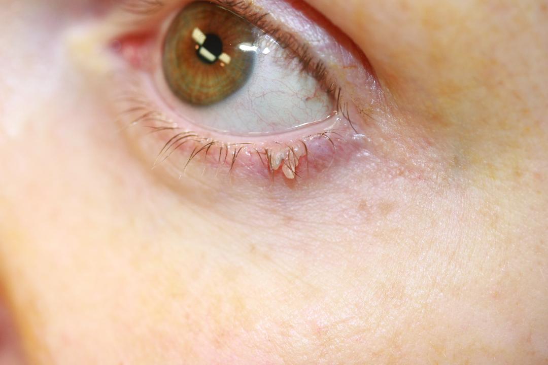
Eyelid Papilloma. Image of a patient with an eyelid papilloma.
Contributed by JA Foster, MD, and CN Czyz, DO
References
Pe'er J. Pathology of eyelid tumors. Indian journal of ophthalmology. 2016 Mar:64(3):177-90. doi: 10.4103/0301-4738.181752. Epub [PubMed PMID: 27146927]
Huang YY, Liang WY, Tsai CC, Kao SC, Yu WK, Kau HC, Liu CJ. Comparison of the Clinical Characteristics and Outcome of Benign and Malignant Eyelid Tumors: An Analysis of 4521 Eyelid Tumors in a Tertiary Medical Center. BioMed research international. 2015:2015():453091. doi: 10.1155/2015/453091. Epub 2015 Nov 8 [PubMed PMID: 26634208]
Lai CC, Lee WA. Eyelid Papilloma. Ophthalmology. 2024 Feb:131(2):226. doi: 10.1016/j.ophtha.2023.05.005. Epub 2023 Jun 1 [PubMed PMID: 37269263]
Yu SS, Zhao Y, Zhao H, Lin JY, Tang X. A retrospective study of 2228 cases with eyelid tumors. International journal of ophthalmology. 2018:11(11):1835-1841. doi: 10.18240/ijo.2018.11.16. Epub 2018 Nov 18 [PubMed PMID: 30450316]
Level 2 (mid-level) evidenceBrown R, Fard S, Feng P, Kerr PE. Evaluation and management of benign tumors of the eye and eyelid. Clinics in dermatology. 2024 Jul-Aug:42(4):343-350. doi: 10.1016/j.clindermatol.2024.01.005. Epub 2024 Jan 26 [PubMed PMID: 38281689]
Eshraghi B, Torabi HR, Kasaie A, Rajabi MT. The use of a radiofrequency unit for excisional biopsy of eyelid papillomas. Ophthalmic plastic and reconstructive surgery. 2010 Nov-Dec:26(6):448-9. doi: 10.1097/IOP.0b013e3181d8e126. Epub [PubMed PMID: 20736876]
Ucar F, Unluzeybek M. Plasma Exeresis for the Treatment of Benign Eyelid Lesions: A New Surgical Approach. Ophthalmic plastic and reconstructive surgery. 2024 Sep-Oct 01:40(5):533-537. doi: 10.1097/IOP.0000000000002635. Epub 2024 Feb 27 [PubMed PMID: 38427826]
Lee BJ, Nelson CC. Intralesional interferon for extensive squamous papilloma of the eyelid margin. Ophthalmic plastic and reconstructive surgery. 2012 Mar-Apr:28(2):e47-8. doi: 10.1097/IOP.0b013e31821f1df3. Epub [PubMed PMID: 21659914]
Level 3 (low-level) evidenceDíez-Montero C, González González D, Pérez Martínez E, Schellini S, Galindo-Ferreiro A. Periocular inverted follicular keratosis: a retrospective series over 17 years. Japanese journal of ophthalmology. 2019 Mar:63(2):210-214. doi: 10.1007/s10384-018-00650-7. Epub 2019 Jan 2 [PubMed PMID: 30604112]
Level 2 (mid-level) evidenceGundogan FC, Yolcu U, Tas A, Sahin OF, Uzun S, Cermik H, Ozaydin S, Ilhan A, Altun S, Ozturk M, Sahin F, Erdem U. Eyelid tumors: clinical data from an eye center in Ankara, Turkey. Asian Pacific journal of cancer prevention : APJCP. 2015:16(10):4265-9 [PubMed PMID: 26028084]
Jara K, Youhnovska E, Marcotte E, Marroquín L, Villalobos J, Alvarez Y, Kawaguchi A, Urbano E, Velasco-Stoll J, Muro P, El-Hadad C, Arthurs B, Burnier MN Jr. Clinicopathological comparison of benign eyelid tumours in tertiary care centers in Lima and Montreal from 2010 to 2019. Canadian journal of ophthalmology. Journal canadien d'ophtalmologie. 2025 Aug:60(4):e589-e597. doi: 10.1016/j.jcjo.2025.01.008. Epub 2025 Feb 25 [PubMed PMID: 39900113]