 Tools
Tools
 Blepharoconjunctivitis
Introduction
Blepharoconjunctivitis is an ophthalmic disorder characterized by concurrent inflammation of the eyelid margins and conjunctiva (see Image. Blepharoconjunctivitis). The condition encompasses overlapping entities such as anterior and posterior blepharitis, meibomian gland dysfunction (MGD), and various infectious or immune-mediated forms of conjunctivitis.[1][2] Inflammation involves the eyelid margin (blepharitis; see Images. Anterior Blepharitis; Illustration of Blepharitis) and the adjacent conjunctiva (conjunctivitis; see Image. Bacterial Conjunctivitis). The eyelids and conjunctiva are continuous at the mucocutaneous junction, facilitating the extension of inflammation from the lid margin to the conjunctival epithelium and, in severe or chronic cases, the corneal surface.
Patients typically report ocular burning, foreign body sensation, tearing, pruritus, photophobia, and fluctuating vision. Clinical findings include lid margin erythema, collarettes, conjunctival injection, papillary or follicular reactions, and superficial punctate keratopathy. Blepharoconjunctivitis is closely related to blepharitis and often regarded as a progressive or advanced stage of the latter. When blepharitis remains untreated, inflammation may extend to the adjacent conjunctiva, resulting in blepharoconjunctivitis. Several classification systems have been proposed based on etiology, anatomic involvement, clinical features, and standardized photographic grading scales.[3][4]
A mechanistic link in blepharoconjunctivitis involves inflammation induced by microbial colonization (eg, Staphylococcus spp.), viral agents (eg, adenovirus, herpes simplex virus or HSV, varicella-zoster virus or VZV, molluscum contagiosum), parasitic infestation (Demodex), rosacea, allergic sensitization, or epithelial toxicity, all of which can compromise the tear film and alter meibum composition.[5][6][7][8][9] Culture-independent sequencing has substantially advanced understanding of ocular surface dysbiosis in this condition, revealing changes in α- and β-diversity and enrichment of specific genera relative to healthy controls in both adult and pediatric populations.[10] Studies utilizing 16S rRNA amplicon sequencing in patients with blepharoconjunctivitis demonstrate significant differences in microbial diversity compared with healthy individuals, with multiple taxa enriched, supporting a dysbiosis model with potential diagnostic and therapeutic relevance.[11][12]
Blepharoconjunctivitis is commonly classified according to the American Academy of Ophthalmology (AAO) Preferred Practice Pattern (PPP) recommendations, which distinguish anterior from posterior blepharoconjunctivitis based on the anatomical structures involved.[13] Anterior blepharoconjunctivitis is typically associated with colonization of the eyelashes and eyelid margins (eg, by Staphylococcus aureus), leading to toxin-mediated keratinization and the formation of collarettes. Posterior blepharoconjunctivitis is primarily linked to MGD, characterized by altered meibum quality and expressibility, orifice protrusion, and telangiectatic changes of the eyelid margins.
Chronic conjunctival inflammation induces papillary or follicular reactions. Persistent disease may result in corneal complications, including punctate epithelial erosions, marginal infiltrates, and neovascularization, potentially progressing to scarring and decreased visual acuity.
Both etiology and host factors influence the natural history of blepharoconjunctivitis. Many acute cases resolve within days to weeks with supportive care and appropriately tailored medication. In contrast, chronic or recurrent blepharoconjunctivitis may persist for months to years, often with intermittent exacerbations.
Pediatric cases require particular attention, as repeated inflammation can induce corneal astigmatism, increase the risk of amblyopia, and potentially result in vision loss if treated inadequately or misdiagnosed. In adults, persistent MGD frequently coexists with ocular rosacea and dry eye disease, intensifying symptoms and negatively impacting quality of life. Recurrence is common across all age groups when lid hygiene and trigger management are inconsistent, Demodex infestation persists, or systemic factors, such as atopy, dermatologic rosacea, or dupilumab-associated ocular surface disease (OSD), remain unaddressed.[14]
Current evidence-based guidelines recommend systematic assessment, maintenance of lid hygiene, reduction of environmental triggers, and the selective use of topical steroids, antibiotics, or antiseptics. A stepwise therapeutic strategy, individualized according to etiology and disease severity, is essential for optimal management of blepharoconjunctivitis.[15][16]
Etiology
Register For Free And Read The Full Article
 Search engine and full access to all medical articles
- 10 free questions in your specialty
- Free CME/CE Activities
- Free daily question in your email
- Save favorite articles to your dashboard
- Emails offering discounts
Learn more about a Subscription to StatPearls Point-of-Care
Etiology
Distinguishing the causes of blepharoconjunctivitis from those of blepharitis is challenging due to the anatomical proximity of ocular structures and the frequent, rapid involvement of the conjunctiva in blepharitis.[17] The condition is often multifactorial, arising from multiple, intersecting etiologies.
Infectious causes include bacteria, most commonly Staphylococcus aureus and epidermidis; viruses such as adenoviruses, HSV, VZV, Epstein-Barr virus, enteroviruses, and molluscum contagiosum; and, less frequently, Chlamydiae and zoonotic parasites. Bacterial colonization of the lid margin produces lipases that hydrolyze meibomian lipids, generating proinflammatory free fatty acids and disrupting the tear film.[18] Adenoviral infections typically cause acute follicular conjunctivitis with preauricular lymphadenopathy and may present with pronounced lid edema.[19] Herpetic infections manifest as lid vesicles and dendritic epitheliopathy.[20] Chronic follicular blepharoconjunctivitis can result from molluscum contagiosum shedding until the lesion is removed.[21]
Inflammatory or immune-mediated etiologies include allergic blepharoconjunctivitis and ocular rosacea, each associated with specific triggers and comorbidities.[22][23] Parasitic infestation by Demodex species—D. folliculorum at the lash base and D. brevis within meibomian glands—is recognized as a cause or exacerbating factor in chronic blepharoconjunctivitis.[24] Clinical signs include cylindrical dandruff (collarettes) and eyelash misdirection, with infestation density correlating with disease severity and persistence. Additional causes include pubic lice and medications such as dupilumab.[25][26][27][28]
Acute onset with ulcerative features may indicate an infectious etiology, with Staphylococcus species being the most frequently implicated organism, particularly when the anterior eyelid is affected.[29] Acute onset without ulceration generally suggests an allergic process. However, the absence of ulceration does not definitively exclude infection, as this finding is not entirely specific. Chronic blepharoconjunctivitis may be categorized based on the anatomical region involved—posterior, angular, or anterior. MGD is a common underlying cause, primarily affecting the posterior eyelid.[30] Involvement of the canthal angle defines angular blepharoconjunctivitis, which may result from infectious agents, such as Moraxella, or, in rare instances, from vitamin B6 deficiency.[31]
Epidemiology
Epidemiological data for blepharoconjunctivitis are difficult to distinguish from those of blepharitis due to the clinical overlap between the 2 conditions. Furthermore, reliable population-based prevalence data for blepharitis are limited, as most studies have focused on patients attending eye clinics.
A U.S. survey of ophthalmologists and optometrists reported that 37% to 47% of patients in their clinics exhibited signs of blepharitis.[32] In a single-center study of 90 patients, the mean patient age was 50 years.[33] Another study found that infectious (staphylococcal) blepharitis was more prevalent in female patients, with a mean age of onset of 42 years, whereas seborrheic blepharitis had a mean age of approximately 50 years, with no significant sex differences.[34] A 2021 meta-analysis estimated the global prevalence of MGD, which is commonly associated with blepharoconjunctivitis, at approximately 36% (95% CI, 24%-50%).[35]
Pathophysiology
The pathophysiology of blepharoconjunctivitis is incompletely understood and is likely multifactorial. Contributing factors include inflammatory skin conditions, mild chronic bacterial infections, and parasitic infestation. The condition reflects inflammatory pathways triggered by microbial toxins or enzymes, mechanical and antigenic stimulation from Demodex, allergic sensitization, or epithelial toxicity, all of which may compromise the tear film and damage the ocular surface.
In anterior disease, bacterial lipases generate free fatty acids that irritate the epithelium and induce keratinization. In posterior forms, obstructive or qualitative changes in the meibomian glands increase tear evaporation and hyperosmolar stress, exacerbating cytokine-mediated inflammatory cascades. Experimental immune-mediated models demonstrate conjunctival leukocyte infiltration, characterized by a dynamic equilibrium of T cells and macrophages, alongside increased levels of interferon γ and interleukin 2, indicating an immune environment dominated by T-helper 1 and T-helper 17 cells, characteristic of delayed-type hypersensitivity responses.[36]
Histopathology
Chronic anterior blepharoconjunctivitis can exhibit, at the microscopic level, epidermal hyperkeratinization, acanthosis, perifollicular inflammation, and, occasionally, ulceration at sites of crust detachment. Lash follicles may show distention and reactive changes resulting from Demodex egg deposition and mechanical trauma.
Posterior lid involvement may manifest as hyperkeratinization of the meibomian gland orifices, ductal dilatation or obstruction, acinar atrophy, and periductal inflammatory infiltrates. Levels of matrix metalloproteinase 8 may be elevated in this form of inflammation.[37] Sebaceous metaplasia and gland dropout also increase with disease duration, correlating with findings from in vivo meibography.
In allergic blepharoconjunctivitis, histological examination typically demonstrates subepithelial edema with mixed inflammatory infiltrates. Eosinophils predominate during the acute phase, whereas chronic cases show predominantly lymphocytic patterns. Severe atopic conditions may also involve goblet cell alterations.
History and Physical
Patients with blepharoconjunctivitis typically report a blepharitic pattern of ocular irritation, including foreign body sensation, eyelid crusting, and conjunctival hyperemia. Symptoms are often more pronounced in the morning, with some patients experiencing eyelid adhesion upon awakening. Both eyes are usually affected, and symptom severity may fluctuate over time.
A comprehensive history should include the onset of symptoms (acute versus chronic), potential exposures (eg, sick contacts, swimming pools, cosmetics), laterality, diurnal variation, and identifiable triggers, such as allergens, contact lens use, digital screen time, and exposure to wind or smoke. Medication history is essential, as topical glaucoma agents, preservatives (eg, benzalkonium chloride), retinoids, isotretinoin, and biologics such as dupilumab can induce or exacerbate ocular surface inflammation.[38][39]
Dermatological and systemic comorbidities must also be documented, particularly atopy, seborrheic dermatitis, rosacea, acne, and autoimmune mucocutaneous disorders.[40] In pediatric patients, the history should explore styes or chalazia, photosensitivity, recurrent conjunctivitis, and visual behaviors indicative of amblyogenic blur, such as squinting or head tilting.[41] Travel and pet exposure histories are rarely conclusive but may be relevant in atypical or treatment-resistant cases, including infections such as dirofilariasis.[42]
The physical examination begins with an external assessment for periorbital erythema, telangiectasia, signs of rosacea, and molluscum lesions. Slit-lamp biomicroscopy typically reveals lid margin erythema, scaling or collarettes, lash misdirection or madarosis, “pouting” meibomian orifices, and telangiectasia. Palpation and expression are used to assess meibum quality and expressibility.
Conjunctival examination includes evaluation for papillae, which suggest allergic or staphylococcal etiology, and follicles, which are associated with viral, chlamydial, or molluscum infections. The character of discharge, whether mucous or mucopurulent, should also be noted.[43] Corneal assessment may reveal punctate keratopathy, marginal infiltrates, phlyctenules, or dendritic lesions.
Demodex infestation is indicated by cylindrical dandruff at the lash base and may be confirmed by epilating several lashes for light microscopy or examining them through in vivo confocal microscopy. Higher mite density correlates with treatment resistance and recurrence.[44] Fluorescein staining under blue light often demonstrates a reduced tear film break-up time (<10 seconds) and may reveal corneal erosions or ulceration. Scleral and conjunctival inspection typically shows varying degrees of conjunctival injection.
When underlying blepharitis is anterior in origin, the eyelid margin typically appears edematous and erythematous, with telangiectasia often present. Crusting along the lid margin may be observed, with collarette formation at the base of the eyelashes. Chronic cases can involve alterations of the eyelashes, including poliosis (depigmentation), trichiasis (misdirection of lashes), or madarosis (reduction in lash number). Eyelid contour may be distorted, with ectropion or entropion visible.
In posterior blepharitis, the meibomian glands are often dilated, with orifices clogged by a visible “head” or “cap” of thickened meibum. On slit-lamp examination, these caps resemble a string of pearls along the eyelid margin. Surrounding eyelid tissue may appear scarred or inflamed.
Clinical signs can aid in determining etiology. Hyperacute purulent discharge accompanied by chemosis and eyelid edema is typical of gonococcal infection. Vesicular rashes characterize herpes zoster ophthalmicus, which may present with corneal epithelial defects, infiltration, and severe ocular pain. Preauricular lymphadenopathy and eyelid vesicles suggest viral infection, whereas focal umbilicated eyelid papules indicate molluscum contagiosum. In children, significant photophobia and reduced vision with persistent redness necessitate evaluation for juvenile blepharokeratoconjunctivitis with corneal involvement, as untreated cases may result in amblyopia.[45]
Evaluation
Blepharoconjunctivitis is a clinical diagnosis based on the patient’s history, characteristic signs, and symptom timeline. Slit-lamp examination and assessment of the meibomian glands may include evaluation of tear film break-up time, tear meniscus height, and corneal and conjunctival staining. Meibography can provide additional structural information but is not required for diagnosis. Evelid biopsy should be considered to exclude malignancy in refractory cases. Fluorescein staining under blue light is useful for detecting corneal erosions or ulcers, which are frequently associated with chronic, poorly controlled blepharoconjunctivitis.
Lash sampling for Demodex and standardized meibomian gland expression can facilitate disease phenotyping.[46] Point-of-care testing for tear osmolarity or matrix metalloproteinase-9 may indicate ocular surface inflammation in chronic cases.[47][48] Microbiologic evaluation is warranted in severe, atypical, or treatment-refractory presentations. Bacterial culture is recommended for hyperacute purulence, ulceration, or immunocompromised patients. In suspected adenoviral outbreaks, rapid antigen testing may be employed if available, although management remains primarily supportive.
The AAO PPP guidelines on blepharitis and conjunctivitis provide evidence-based recommendations for patient history, clinical examination, selective use of routine cultures, and infection prevention.[49] The guidelines advise against the routine use of antibiotics for uncomplicated viral infections. The AAO Blepharitis PPP emphasizes a systematic eyelid assessment, highlights the identification of Demodex infestation and MGD characteristics, and recommends targeted microbiologic testing for severe or atypical cases.
Pediatric consensus initiatives from 2023 to 2024 have advocated for standardized diagnostic criteria for blepharokeratoconjunctivitis in this group, incorporating corneal findings and refractive consequences to facilitate earlier and more consistent diagnosis, as well as improved comparability in research studies. Teleophthalmology and smartphone-based slit-lamp attachments can document baseline and follow-up findings, enhancing triage and continuity of care, particularly when in-person slit-lamp evaluation is limited.
Treatment / Management
Management of blepharoconjunctivitis is guided by the underlying etiology, disease severity, and duration, and is implemented incrementally, with patient education on eyelid hygiene and trigger avoidance as foundational components. Fundamental interventions include warm compresses, eyelid massage, daily lid cleansing with diluted cleansers or hypochlorous acid solutions, reduction of preservative exposure, and tear supplementation. These measures reduce microbial bioburden, soften meibomian gland secretions (meibum), and alleviate symptoms.
Meticulous lid hygiene is the mainstay of treatment. Application of a warm compress for 5 to 10 minutes softens meibum within the meibomian glands, facilitating expression via lid massage. Eyelash scrubs with mild shampoos can remove debris. Maintenance of lid hygiene is recommended even after acute exacerbations, due to the chronic and recurrent nature of the condition. Artificial tears may be used to relieve associated dry eye symptoms.
Topical antibiotics should be considered when lid hygiene alone is insufficient, particularly in bacterial etiologies such as staphylococcal infection.[50] Bacterial blepharoconjunctivitis generally responds to short-term topical antibiotics, including erythromycin or bacitracin ointment. Topical corticosteroids may be employed during acute inflammatory exacerbations.[51] Seborrheic blepharitis frequently coexists with seborrheic dermatitis, and concurrent treatment of the underlying dermatologic condition is recommended. In posterior blepharitis, systemic therapy with oral azithromycin or tetracyclines, such as doxycycline, can be beneficial.[52][53](A1)
Allergic blepharoconjunctivitis is effectively managed with dual-action antihistamine-mast cell stabilizers and short courses of low- to moderate-potency topical corticosteroids during exacerbations. Topical calcineurin inhibitors may be used off-label as steroid-sparing agents in selected chronic atopic cases. Viral blepharoconjunctivitis is typically managed supportively. For HSV or VZV, immediate systemic antiviral therapy is indicated in the presence of high-risk features, including corneal involvement. Adenoviral infections are addressed with eyelid hygiene, cold compresses, and patient counseling.
Demodex blepharoconjunctivitis now has an on-label therapy approved by the U.S. Food and Drug Administration with lotilaner 0.25% ophthalmic solution (XDEMVY), administered twice daily for approximately 6 weeks. Phase 3 randomized, double-masked trials demonstrated significant collarette resolution and mite eradication compared with vehicle controls.[54][55] Management may also include in-office microblepharo-exfoliation and lid scrapes with tea tree oil, supplemented, in select cases, with topical ivermectin or metronidazole gels.[56] The role of ω-3 and ω-6 fatty acid supplementation for alleviating dry eye symptoms remains controversial. A recent Cochrane systematic review suggested potential benefit, though the quality of evidence was rated as low.[57](A1)
Pediatric management depends on the severity and extent of corneal involvement.[58] Despite limited high-quality randomized data in children, treatment strategies may include lid hygiene, topical antibiotics or anti-inflammatory agents, and carefully dosed systemic macrolides or tetracyclines (age-appropriate) for severe MGD-driven disease, aiming to protect the cornea and prevent amblyopia.
Differential Diagnosis
Principal alternatives to blepharoconjunctivitis, defined as inflammation of both the conjunctiva and eyelids, include isolated blepharitis without significant conjunctival involvement, pure conjunctivitis of viral, allergic, or bacterial origin, ocular rosacea, phlyctenular keratoconjunctivitis, marginal keratitis secondary to staphylococcal hypersensitivity, chlamydial inclusion conjunctivitis, and toxic reactions to topical agents.
Herpetic disease should be suspected in the presence of lid vesicles and cutaneous involvement along the trigeminal dermatomes, specifically herpes zoster ophthalmicus.[59] HSV infection may initially present as nonspecific blepharoconjunctivitis. Corneal staining can subsequently reveal dendritiform lesions following the initial signs and symptoms.[60]
Molluscum contagiosum can produce persistent unilateral follicular conjunctivitis accompanied by umbilicated eyelid papules. Remission typically follows lesion curettage or cryotherapy. Parasitic or zoonotic causes are uncommon but clinically important in recalcitrant or migratory cases, such as subcutaneous dirofilariasis, which generally requires surgical excision for resolution.[61]
Systemic or dermatologic mimickers of blepharoconjunctivitis include atopic keratoconjunctivitis, contact dermatitis, seborrheic dermatitis, and cicatrizing disorders such as ocular cicatricial pemphigoid and Stevens-Johnson syndrome. These conditions can result in lid margin and conjunctival scarring.[62][63][64][65][66]
Malignancy must be excluded in patients who are refractory to standard therapy, particularly when eyelash changes are observed. Neoplastic lesions typically affect the lower eyelid, and a high index of suspicion is warranted for unilateral presentations. Basal cell carcinoma and sebaceous carcinoma may ulcerate the eyelid margin, mimicking chronic unilateral blepharitis. Biopsy is indicated for nonhealing or suspicious lesions.[67]
Dupilumab-induced OSD presents with conjunctivitis, blepharitis, and dry eye symptoms in patients receiving biologic therapy for atopic dermatitis. Management requires collaboration between dermatology and ophthalmology to adjust systemic treatment while protecting the ocular surface.[68]
Staging
No globally standardized staging system exists for blepharoconjunctivitis. Clinicians commonly categorize disease severity using composite clinical scores that assess lid margin hyperemia and scaling, meibomian gland orifice appearance, meibum quality and expressibility, conjunctival injection, papillae or follicles, and corneal staining.
Instruments for evaluating MGD and OSD include the Ocular Surface Disease Index (OSDI) questionnaire, noninvasive tear break-up time (NIBUT), meibography grading, and the Standard Patient Evaluation of Eye Dryness (SPEED) questionnaire. These tools facilitate objective measurement of disease severity and monitoring of treatment response. Composite scoring systems, such as the Composite Ocular Symptom Score (COTE) and Oxford staining scores, are frequently used in conjunction with imaging-based meibography to evaluate structural and functional changes following therapy.
Prognosis
The prognosis is generally favorable with accurate etiological diagnosis, adherence to eyelid hygiene, and timely, targeted therapy. Recurrences and resistance to treatment are common in patients with chronic MGD, concomitant ocular rosacea, OSD, and Demodex-related conditions.
In pediatric cases, prognosis depends on prompt management of corneal inflammation to prevent scarring and amblyopia. Recurrences are frequent, and monitoring for progression of astigmatism is critical to safeguard visual development.[69] Targeted treatment for Demodex increases the likelihood of collarette resolution and symptom control.
Viral blepharoconjunctivitis, such as adenoviral infection, generally resolves spontaneously within 2 to 3 weeks. Subepithelial corneal infiltrates may persist in some cases, resulting in glare and photophobia for several months.
Complications
Uncontrolled or recurrent blepharoconjunctivitis may result in corneal complications, including persistent punctate erosions, marginal infiltrates or ulcers, phlyctenules, and pannus formation. In pediatric patients, severe scarring increases the risk of secondary astigmatism and amblyopia.
The eyelid margin may become distorted, producing trichiasis, ectropion or entropion, poliosis, notching, or madarosis. Demodex-associated disease can lead to recurrent chalazia or hordeola due to glandular obstruction and granulomatous inflammation.
Chronic MGD may cause evaporative dry eye, characterized by tear hyperosmolarity and ocular surface inflammation, which further exacerbates surface instability and symptoms. Ulceration or corneal perforation necessitates urgent, aggressive intervention to prevent permanent vision loss.
Deterrence and Patient Education
Patients should be informed that blepharoconjunctivitis is a chronic condition requiring ongoing maintenance therapy, primarily through consistent lid hygiene, to prevent acute exacerbations. Education should include recognition of complication-related symptoms. Patients must be advised to seek prompt medical attention in the event of vision deterioration or new-onset ocular pain.
Prevention of blepharoconjunctivitis relies on proactive measures, patient education, and reinforcement of lifestyle modifications that reduce the risk of recurrence. Patients should be instructed on proper eyelid care, including the daily use of warm compresses, lid washes, or commercially available cleansing pads to limit bacterial proliferation and debris accumulation along the lash line.
Education must emphasize that blepharoconjunctivitis is a chronic condition requiring long-term management rather than short-term therapy to control symptoms and prevent exacerbations. Patients should be advised to avoid eye makeup during active disease, replace cosmetic products regularly, and refrain from sharing personal items such as towels or applicators.
Strict adherence to treatment protocols, including topical antibiotics, lubricants, or anti-inflammatory drops, is essential for reducing relapses and improving quality of life. Environmental modifications, such as minimizing exposure to smoke, avoiding allergens, and discontinuing contact lens use during flares, should also be included in patient instructions. The use of artificial tears and humidifiers can help prevent ocular surface dryness, enhancing tear film stability, particularly in individuals with concomitant MGD. Clear, structured guidance from clinicians can significantly reduce recurrence and promote favorable long-term outcomes.
In addition to eyelid hygiene, targeted patient education regarding comorbid conditions is essential, as blepharoconjunctivitis often coexists with systemic or dermatologic disorders, including rosacea, seborrheic dermatitis, and atopy. Patients with rosacea should receive guidance on modifiable dietary and environmental factors. Individuals with seborrheic dermatitis may benefit from scalp and facial treatment using medicated shampoos.
The potential role of Demodex infestation should be considered, and patients informed of the efficacy of tea tree oil or ivermectin-based therapies for mite eradication. Pediatric patients with recurrent blepharoconjunctivitis require parental involvement to ensure adherence, particularly for consistent eyelid hygiene practices.
Patient education must also correct common misconceptions, including the belief that antibiotics alone provide a long-term cure, emphasizing the necessity of ongoing eyelid maintenance. Instruction regarding warning signs of complications, such as increased pain, decreased vision, or corneal involvement, enables prompt medical evaluation and helps prevent long-term sequelae.
Pearls and Other Issues
Effective management of blepharoconjunctivitis requires recognition of its multifactorial etiology, necessitating a tailored, multimodal therapeutic approach rather than reliance on a single treatment modality. Patient adherence to eyelid hygiene is the most important predictor of long-term treatment success. Clinicians should prioritize patient education and reinforcement of compliance.
Topical antibiotics and anti-inflammatory agents are frequently indicated. However, prolonged or excessive antibiotic use should be avoided due to the risk of microbial resistance and ocular surface toxicity. A comprehensive strategy that combines pharmacologic therapy with mechanical lid care and management of comorbid conditions yields superior outcomes.
Special attention is required for contact lens users, as poor lens hygiene or extended wear may exacerbate blepharoconjunctivitis, necessitating temporary discontinuation. Pediatric presentations often differ, with increased prevalence of chalazia and phlyctenular keratoconjunctivitis, requiring closer clinical monitoring.
Clinicians frequently underestimate the impact of blepharoconjunctivitis on patients’ quality of life, often dismissing it as a minor or purely cosmetic condition. Inadequate treatment or management can result in persistent discomfort, recurrent infections, and corneal complications, including keratitis or ulceration. Another limitation is the underrecognition of associated systemic disorders, particularly rosacea, which may necessitate systemic therapy for comprehensive management.
Preventive strategies should emphasize continuous maintenance rather than solely addressing acute episodes. Regular follow-up appointments are essential to monitor disease progression. Complications may be reduced through management of modifiable risk factors, including avoidance of unnecessary steroid use, adherence to proper contact lens hygiene, and refraining from harmful behaviors such as excessive eye rubbing.
Enhancing Healthcare Team Outcomes
The management of blepharoconjunctivitis benefits significantly from an interprofessional team approach due to the chronic, recurrent nature of the condition and its relevance to ophthalmology, dermatology, and general medical care. Ophthalmologists and optometrists are central to diagnosing the condition, assessing severity, and coordinating treatment. Nurses and advanced practice providers play key roles in patient education, reinforcing eyelid hygiene practices, monitoring adherence, and evaluating therapeutic response. Pharmacists contribute by ensuring appropriate use of topical and systemic medications, counseling on potential adverse effects, and preventing drug interactions, particularly in patients requiring systemic tetracyclines or ivermectin.
Dermatologists often collaborate in managing concurrent rosacea or seborrheic dermatitis, while allergists may be involved in cases of atopic blepharoconjunctivitis. Efficient communication among specialists ensures coordinated care, reduces fragmented management, and lowers the risk of chronic complications.
Pediatric care requires collaboration between family physicians, pediatricians, and eye specialists to facilitate early detection and timely intervention. A patient-centered approach, in which all clinicians deliver consistent guidance regarding disease management and self-care, is essential for optimizing outcomes.
Strategies to optimize healthcare team outcomes include structured communication systems, integrated electronic health records, and the use of standardized patient education resources among clinicians. Encouraging active patient engagement is both an ethical responsibility and a practical measure that improves outcomes.
By leveraging the combined expertise of physicians, advanced practice providers, nurses, pharmacists, dermatologists, and allied health professionals, the interprofessional team can deliver patient-centered care, enhance quality of life, and reduce the risk of long-term complications in the management of blepharoconjunctivitis.
Media
(Click Image to Enlarge)
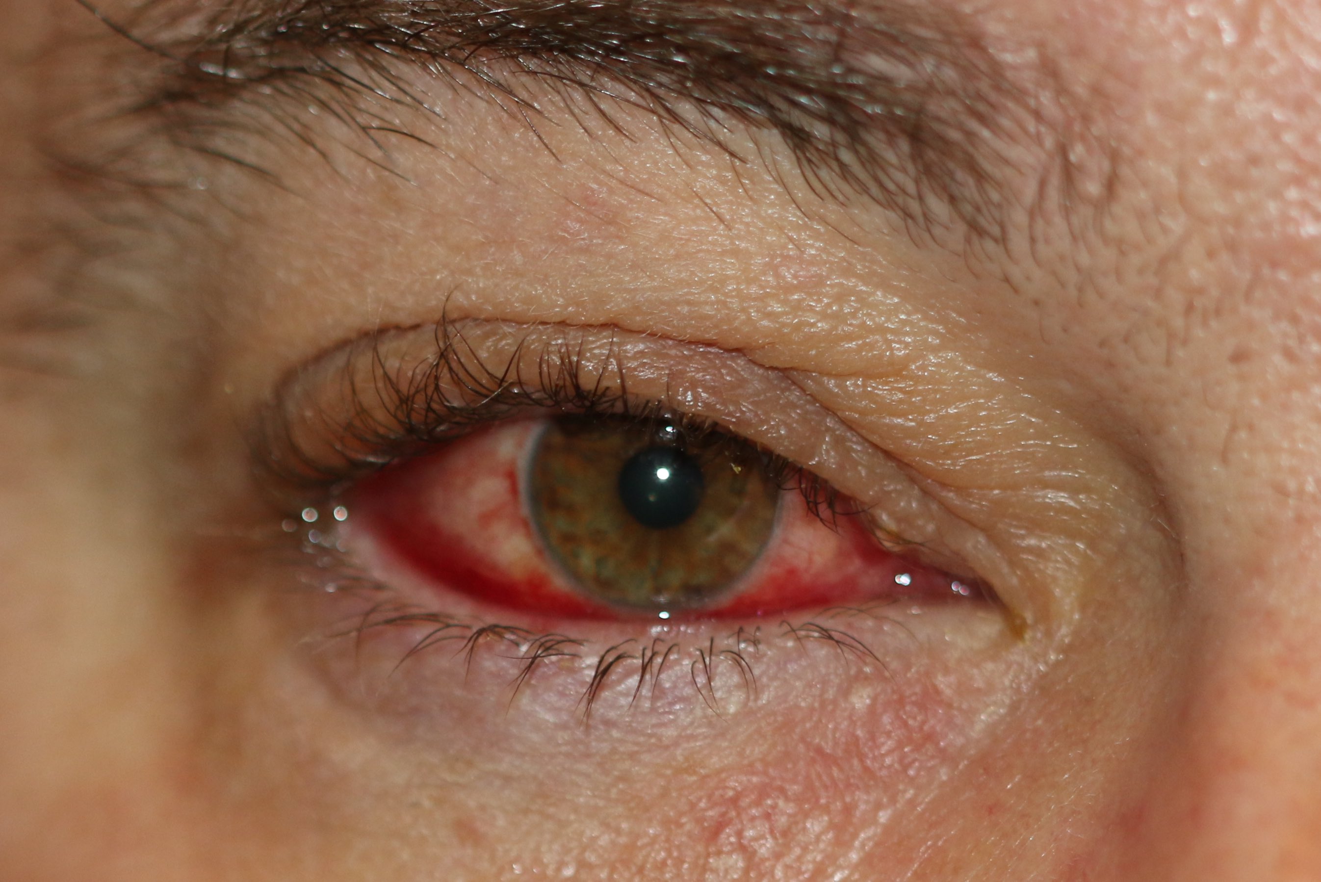
Blepharoconjunctivitis. Conjunctival injection and eyelid margin erythema are present, indicating active blepharoconjunctivitis with inflammation of ocular surface structures.
Contributed by Prof. Bhupendra C. K. Patel MD, FRCS
(Click Image to Enlarge)
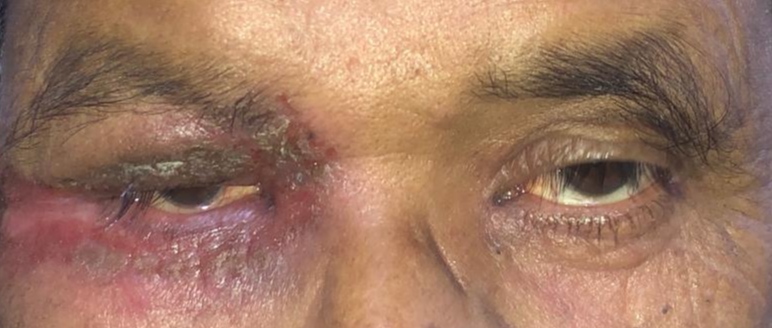
Anterior Blepharitis. The eyelid margin exhibits inflammatory changes with crusting and hyperemia characteristic of anterior blepharitis.
Contributed by S Shah, MS, FICO, MRCS
(Click Image to Enlarge)
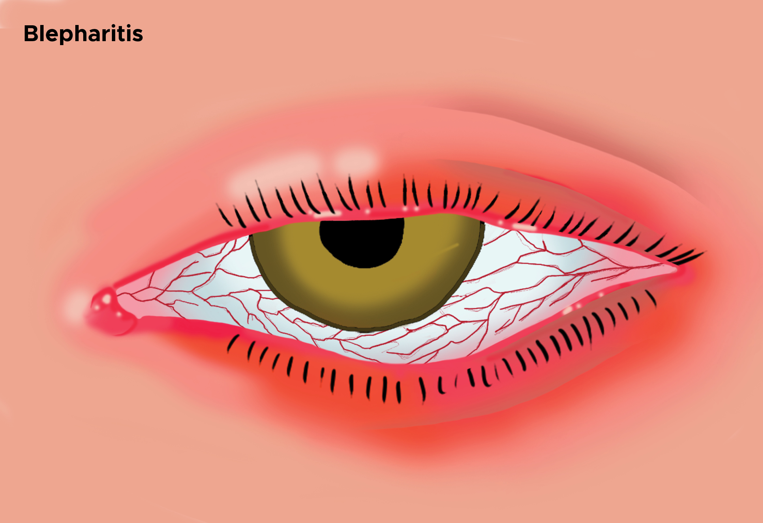
Illustration of Blepharitis. This image depicts blepharitis, showing a red, inflamed eyelid with crusting and irritation around the eyelashes.
Illustration by C Rowe
(Click Image to Enlarge)
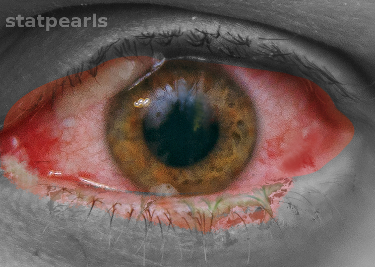
Bacterial Conjunctivitis. The eye exhibits conjunctival hyperemia and mucopurulent discharge consistent with bacterial conjunctivitis.
Contributed by O Chaigasame, MD
References
THYGESON P. The etiology and treatment of blepharitis; a study in military personnel. Military surgeon. 1946 Mar:98():191-203 [PubMed PMID: 21017790]
McCulley JP, Dougherty JM, Deneau DG. Classification of chronic blepharitis. Ophthalmology. 1982 Oct:89(10):1173-80 [PubMed PMID: 6218459]
Huber-Spitzy V, Baumgartner I, Böhler-Sommeregger K, Grabner G. Blepharitis--a diagnostic and therapeutic challenge. A report on 407 consecutive cases. Graefe's archive for clinical and experimental ophthalmology = Albrecht von Graefes Archiv fur klinische und experimentelle Ophthalmologie. 1991:229(3):224-7 [PubMed PMID: 1831177]
Level 3 (low-level) evidenceMathers WD, Choi D. Cluster analysis of patients with ocular surface disease, blepharitis, and dry eye. Archives of ophthalmology (Chicago, Ill. : 1960). 2004 Nov:122(11):1700-4 [PubMed PMID: 15534133]
Musa M, Bale BI, Suleman A, Aluyi-Osa G, Chukwuyem E, D'Esposito F, Gagliano C, Longo A, Russo A, Zeppieri M. Possible viral agents to consider in the differential diagnosis of blepharoconjunctivitis. World journal of virology. 2024 Dec 25:13(4):97867. doi: 10.5501/wjv.v13.i4.97867. Epub [PubMed PMID: 39722756]
Stepinska P, Zawadzinska-Halat K, Pastuszczak M. Pretreatment Demodex infestation predicts dupilumab-associated blepharoconjunctivitis in atopic dermatitis. The Journal of dermatological treatment. 2025 Dec:36(1):2537754. doi: 10.1080/09546634.2025.2537754. Epub 2025 Jul 29 [PubMed PMID: 40728232]
Kumar D, Agstam S. Acute allergic blepharoconjunctivitis during percutaneous coronary intervention. The Journal of invasive cardiology. 2025 Oct:37(10):. doi: 10.25270/jic/25.00097. Epub [PubMed PMID: 40359580]
Leonardi A, Bozkurt B, Silva D, Mortz CG, Baudouin C, Atanaskovic-Markovic M, Sharma V, Doan S, Agarwal S, Pérez-Formigo D, Vasconcelos MJ, Baudouin FB, Chorzepa G, Fauquert JL, Calder V, Demoly P, Delgado L. Drug-Induced Periocular and Ocular Surface Disorders: An EAACI Position Paper. Allergy. 2025 Nov:80(11):2953-2972. doi: 10.1111/all.70074. Epub 2025 Sep 30 [PubMed PMID: 41025570]
Tavassoli S, Wong N, Chan E. Ocular manifestations of rosacea: A clinical review. Clinical & experimental ophthalmology. 2021 Mar:49(2):104-117. doi: 10.1111/ceo.13900. Epub 2021 Feb 3 [PubMed PMID: 33403718]
Trojacka E, Izdebska J, Szaflik J, Przybek-Skrzypecka J. The Ocular Microbiome: Micro-Steps Towards Macro-Shift in Targeted Treatment? A Comprehensive Review. Microorganisms. 2024 Nov 4:12(11):. doi: 10.3390/microorganisms12112232. Epub 2024 Nov 4 [PubMed PMID: 39597621]
Çakır B, Sönmezoğlu BG, Şahin EÖ, Köroğlu M, Aksoy NÖ. Evaluation of ocular surface microbiota in children with blepharoconjunctivitis. Graefe's archive for clinical and experimental ophthalmology = Albrecht von Graefes Archiv fur klinische und experimentelle Ophthalmologie. 2025 Aug:263(8):2359-2367. doi: 10.1007/s00417-025-06836-3. Epub 2025 Apr 23 [PubMed PMID: 40266380]
Wang C, Dou X, Li J, Wu J, Cheng Y, An N. Composition and Diversity of the Ocular Surface Microbiota in Patients With Blepharitis in Northwestern China. Frontiers in medicine. 2021:8():768849. doi: 10.3389/fmed.2021.768849. Epub 2021 Dec 7 [PubMed PMID: 34950683]
Amescua G, Akpek EK, Farid M, Garcia-Ferrer FJ, Lin A, Rhee MK, Varu DM, Musch DC, Dunn SP, Mah FS, American Academy of Ophthalmology Preferred Practice Pattern Cornea and External Disease Panel. Blepharitis Preferred Practice Pattern®. Ophthalmology. 2019 Jan:126(1):P56-P93. doi: 10.1016/j.ophtha.2018.10.019. Epub 2018 Oct 23 [PubMed PMID: 30366800]
Thormann K, Lüthi AS, Deniau F, Heider A, Cazzaniga S, Radonjic-Hoesli S, Lehmann M, Schlapbach C, Herzog EL, Kreuzer M, Zinkernagel MS, Akdis CA, Zysset-Burri DC, Simon HU, Simon D. Dupilumab-associated ocular surface disease is characterized by a shift from Th2/Th17 toward Th1/Th17 inflammation. Allergy. 2024 Apr:79(4):937-948. doi: 10.1111/all.16045. Epub 2024 Feb 5 [PubMed PMID: 38317432]
Pelletier JS, Stewart KP, Capriotti K, Capriotti JA. Rosacea Blepharoconjunctivitis Treated with a Novel Preparation of Dilute Povidone Iodine and Dimethylsulfoxide: a Case Report and Review of the Literature. Ophthalmology and therapy. 2015 Dec:4(2):143-50. doi: 10.1007/s40123-015-0040-4. Epub 2015 Nov 2 [PubMed PMID: 26525679]
Level 3 (low-level) evidenceLibrando A, Carlesimo SC, Albanese G, Albanese GM, Migliorini R, Pacella E. Effectiveness of 0.1% topical salicylic acid on blepharoconjunctivitis affecting glaucoma patients treated with topical prostaglandin analogues: a prospective randomized trial. International journal of ophthalmology. 2018:11(12):1936-1940. doi: 10.18240/ijo.2018.12.10. Epub 2018 Dec 18 [PubMed PMID: 30588426]
Level 1 (high-level) evidencePutnam CM. Diagnosis and management of blepharitis: an optometrist's perspective. Clinical optometry. 2016:8():71-78. doi: 10.2147/OPTO.S84795. Epub 2016 Aug 8 [PubMed PMID: 30214351]
Level 3 (low-level) evidenceWoldemariam M, Aklilu A, Manilal A, Mengistu M, Tadesse D, Siraj M, Hatamleh AA, Alnafisi BK, Idhayadhulla A. Microbial profile and associated factors of external ocular bacterial and fungal infections in Arba Minch General Hospital: a cross-sectional study. Scientific reports. 2024 Nov 20:14(1):28744. doi: 10.1038/s41598-024-77723-w. Epub 2024 Nov 20 [PubMed PMID: 39567575]
Level 2 (mid-level) evidenceFardous J, Goutam A, Rahman T, Khan ZH, Nahin S, Hussain S, Khan MSB, Hawlader MDH, Amin MA. Adenovirus Disease and Ocular Symptoms in Children: Diagnosis and Prognostic Considerations. Case reports in infectious diseases. 2025:2025():2621782. doi: 10.1155/crdi/2621782. Epub 2025 Aug 6 [PubMed PMID: 40810057]
Level 3 (low-level) evidencePereira SM, Lima RV, Muniz MCR, Araújo MBF, de Moraes Ferreira Júnior L, de Queiroz Sales Martins JT, Luz CFC, Cid DAC, da Rocha Lucena D. Congenital herpes simplex with ophthalmic and multisystem features: a case report. BMC pediatrics. 2023 Dec 4:23(1):611. doi: 10.1186/s12887-023-04423-1. Epub 2023 Dec 4 [PubMed PMID: 38044450]
Level 3 (low-level) evidenceNaseer S, Mian SI, Hakim FE. Ocular and Periorbital Manifestations of Molluscum Contagiosum: A 20-year Systematic Review. International ophthalmology clinics. 2025 Apr 1:65(2):12-19. doi: 10.1097/IIO.0000000000000559. Epub 2025 Mar 21 [PubMed PMID: 40116404]
Level 1 (high-level) evidenceVillegas BV, Benitez-Del-Castillo JM. Current Knowledge in Allergic Conjunctivitis. Turkish journal of ophthalmology. 2021 Feb 25:51(1):45-54. doi: 10.4274/tjo.galenos.2020.11456. Epub [PubMed PMID: 33631915]
Machalińska A, Zakrzewska A, Markowska A, Safranow K, Wiszniewska B, Parafiniuk M, Machaliński B. Morphological and Functional Evaluation of Meibomian Gland Dysfunction in Rosacea Patients. Current eye research. 2016 Aug:41(8):1029-1034 [PubMed PMID: 26644191]
Shah PP, Stein RL, Perry HD. Update on the Management of Demodex Blepharitis. Cornea. 2022 Aug 1:41(8):934-939. doi: 10.1097/ICO.0000000000002911. Epub 2021 Nov 3 [PubMed PMID: 34743107]
Yao B, Hu C, Yue X, Liu G, Wang B. Clinical Characteristics and Prognosis of Pediatric Phthirus pubis Coinfestation of the Eyelashes and Scalp Hairs. The American journal of tropical medicine and hygiene. 2023 Sep 6:109(3):686-689. doi: 10.4269/ajtmh.23-0071. Epub 2023 Jul 31 [PubMed PMID: 37524328]
Paulose SA, Sherman SW, Dagi Glass LR, Suh LH. Dupilumab-associated blepharoconjunctivitis. American journal of ophthalmology case reports. 2019 Dec:16():100550. doi: 10.1016/j.ajoc.2019.100550. Epub 2019 Sep 5 [PubMed PMID: 31535057]
Level 3 (low-level) evidenceLiu J, Sheha H, Tseng SC. Pathogenic role of Demodex mites in blepharitis. Current opinion in allergy and clinical immunology. 2010 Oct:10(5):505-10. doi: 10.1097/ACI.0b013e32833df9f4. Epub [PubMed PMID: 20689407]
Level 3 (low-level) evidenceBaskan C, Duman R, Balci M, Ozdogan S. A rare cause of blepharoconjunctivitis: Phthiriasis palpebrarum. Nigerian journal of clinical practice. 2014 Nov-Dec:17(6):817-8. doi: 10.4103/1119-3077.144419. Epub [PubMed PMID: 25385926]
Level 3 (low-level) evidenceMcCulley JP. Blepharoconjunctivitis. International ophthalmology clinics. 1984 Summer:24(2):65-77 [PubMed PMID: 6233233]
Suzuki T. Inflamed Obstructive Meibomian Gland Dysfunction Causes Ocular Surface Inflammation. Investigative ophthalmology & visual science. 2018 Nov 1:59(14):DES94-DES101. doi: 10.1167/iovs.17-23345. Epub [PubMed PMID: 30481812]
IRINODA K, MIKAMI H. Angular blepharoconjunctivitis and pyridoxine (vitamin B6) deficiency. A.M.A. archives of ophthalmology. 1958 Aug:60(2):303-11 [PubMed PMID: 13558802]
Lemp MA, Nichols KK. Blepharitis in the United States 2009: a survey-based perspective on prevalence and treatment. The ocular surface. 2009 Apr:7(2 Suppl):S1-S14 [PubMed PMID: 19383269]
Level 3 (low-level) evidenceSchaumberg DA, Nichols JJ, Papas EB, Tong L, Uchino M, Nichols KK. The international workshop on meibomian gland dysfunction: report of the subcommittee on the epidemiology of, and associated risk factors for, MGD. Investigative ophthalmology & visual science. 2011 Mar:52(4):1994-2005. doi: 10.1167/iovs.10-6997e. Epub 2011 Mar 30 [PubMed PMID: 21450917]
McCulley JP, Dougherty JM. Blepharitis associated with acne rosacea and seborrheic dermatitis. International ophthalmology clinics. 1985 Spring:25(1):159-72 [PubMed PMID: 3156100]
Hassanzadeh S, Varmaghani M, Zarei-Ghanavati S, Heravian Shandiz J, Azimi Khorasani A. Global Prevalence of Meibomian Gland Dysfunction: A Systematic Review and Meta-Analysis. Ocular immunology and inflammation. 2021 Jan 2:29(1):66-75. doi: 10.1080/09273948.2020.1755441. Epub 2020 Jun 26 [PubMed PMID: 32589483]
Level 1 (high-level) evidenceHou A, Tin MQ, Fenner B, Liu YC, Tong L. Conjunctival infiltrates and cytokines in an experimental immune-mediated blepharoconjunctivitis rat model. Frontiers in medicine. 2023:10():1200589. doi: 10.3389/fmed.2023.1200589. Epub 2023 Jun 28 [PubMed PMID: 37448795]
Määttä M, Kari O, Tervahartiala T, Wahlgren J, Peltonen S, Kari M, Rytilä P, Saari M, Sorsa T. Elevated expression and activation of matrix metalloproteinase 8 in tear fluid in atopic blepharoconjunctivitis. Cornea. 2008 Apr:27(3):297-301. doi: 10.1097/ICO.0b013e31815c18d6. Epub [PubMed PMID: 18362656]
Popiela MZ, Barbara R, Turnbull AMJ, Corden E, Martinez-Falero BS, O'Driscoll D, Ardern-Jones MR, Hossain PN. Dupilumab-associated ocular surface disease: presentation, management and long-term sequelae. Eye (London, England). 2021 Dec:35(12):3277-3284. doi: 10.1038/s41433-020-01379-9. Epub 2021 Jan 28 [PubMed PMID: 33504973]
Costedoat I, Wallaert M, Gaultier A, Vasseur R, Vanhaecke C, Viguier M, Cordelette C, Denoyer A, Ferrier le Bouëdec MC, Coutu A, Lamiaux M, Tran THC, Lacour JP, Elmaleh V, Tetart F, Gueudry J, Tauber M, Giordano-Labadie F, Cassagne M, Nosbaum A, Ouilhon C, Jachiet M, Tadayoni R, Dezoteux F, Staumont-Salle D, Bouleau J, Labalette P, Doan S, Soria A, Mortemousque B, Seneschal J, Barbarot S, FRench Atopic DErmatitis Network from the Groupe de Recherche sur l'Eczema Atopique (GREAT), France. Multicenter prospective observational study of dupilumab-induced ocular events in atopic dermatitis patients. Journal of the European Academy of Dermatology and Venereology : JEADV. 2023 May:37(5):1056-1063. doi: 10.1111/jdv.18932. Epub 2023 Feb 16 [PubMed PMID: 36732052]
Level 2 (mid-level) evidenceFelfeli T, Georgakopoulos JR, Jo CE, Mimouni M, Piguet V, Drucker AM, Yeung J, Chan CC. Prevalence and Characteristics of Dupilumab-Induced Ocular Surface Disease in Adults With Atopic Dermatitis. Cornea. 2022 Oct 1:41(10):1242-1247. doi: 10.1097/ICO.0000000000002866. Epub 2021 Oct 19 [PubMed PMID: 34843182]
Chong-Neto HJ, Rosario C, Leonardi A, Filho NAR. Ocular allergy in children and adolescents. Allergologia et immunopathologia. 2022:50(S Pt 1):30-36. doi: 10.15586/aei.v50iSP1.536. Epub 2022 May 20 [PubMed PMID: 35726488]
Morosanu S, Don R, Morosanu V. Human Subcutaneous Dirofilariasis Behind a Blepharoconjunctivitis: A Case Report and Review of the Literature. Cureus. 2024 May:16(5):e60208. doi: 10.7759/cureus.60208. Epub 2024 May 13 [PubMed PMID: 38868288]
Level 3 (low-level) evidenceRitterband DC, Friedberg DN. Virus infections of the eye. Reviews in medical virology. 1998 Oct:8(4):187-201 [PubMed PMID: 10398508]
Liang L, Liu Y, Ding X, Ke H, Chen C, Tseng SCG. Significant correlation between meibomian gland dysfunction and keratitis in young patients with Demodex brevis infestation. The British journal of ophthalmology. 2018 Aug:102(8):1098-1102. doi: 10.1136/bjophthalmol-2017-310302. Epub 2017 Oct 21 [PubMed PMID: 29055903]
Morales-Mancillas NR, Velazquez-Valenzuela F, Kinoshita S, Suzuki T, Dahlmann-Noor AH, Dart JKG, Hingorani M, Ali A, Fung S, Akova YA, Doan S, Gupta N, Hammersmith KM, Tan DTH, Paez-Garza JH, Rodriguez-Garcia A. Definition and Diagnostic Criteria for Pediatric Blepharokeratoconjunctivitis. JAMA ophthalmology. 2024 Jan 1:142(1):39-47. doi: 10.1001/jamaophthalmol.2023.5750. Epub [PubMed PMID: 38127333]
Fromstein SR, Harthan JS, Patel J, Opitz DL. Demodex blepharitis: clinical perspectives. Clinical optometry. 2018:10():57-63. doi: 10.2147/OPTO.S142708. Epub 2018 Jul 4 [PubMed PMID: 30214343]
Level 3 (low-level) evidenceRokohl AC, Wall K, Trester M, Wawer Matos PA, Guo Y, Adler W, Pine KR, Heindl LM. Novel point-of-care biomarkers of the dry anophthalmic socket syndrome: tear film osmolarity and matrix metalloproteinase 9 immunoassay. Graefe's archive for clinical and experimental ophthalmology = Albrecht von Graefes Archiv fur klinische und experimentelle Ophthalmologie. 2023 Mar:261(3):821-831. doi: 10.1007/s00417-022-05895-0. Epub 2022 Nov 11 [PubMed PMID: 36357674]
Kang MJ, Kim HS, Kim MS, Kim EC. The Correlation between Matrix Metalloproteinase-9 Point-of-Care Immunoassay, Tear Film Osmolarity, and Ocular Surface Parameters. Journal of ophthalmology. 2022:2022():6132016. doi: 10.1155/2022/6132016. Epub 2022 Apr 11 [PubMed PMID: 35450324]
Cheung AY, Choi DS, Ahmad S, Amescua G, Jhanji V, Lin A, Mian SI, Rhee MK, Viriya ET, Mah FS, Varu DM, American Academy of Ophthalmology Preferred Practice Pattern Cornea/External Disease Panel. Conjunctivitis Preferred Practice Pattern. Ophthalmology. 2024 Apr:131(4):P134-P204. doi: 10.1016/j.ophtha.2023.12.037. Epub 2024 Feb 12 [PubMed PMID: 38349304]
Raskin EM, Speaker MG, Laibson PR. Blepharitis. Infectious disease clinics of North America. 1992 Dec:6(4):777-87 [PubMed PMID: 1460262]
O'Gallagher M, Bunce C, Hingorani M, Larkin F, Tuft S, Dahlmann-Noor A. Topical treatments for blepharokeratoconjunctivitis in children. The Cochrane database of systematic reviews. 2017 Feb 7:2(2):CD011965. doi: 10.1002/14651858.CD011965.pub2. Epub 2017 Feb 7 [PubMed PMID: 28170093]
Level 1 (high-level) evidenceGreene JB, Jeng BH, Fintelmann RE, Margolis TP. Oral azithromycin for the treatment of meibomitis. JAMA ophthalmology. 2014 Jan:132(1):121-2. doi: 10.1001/jamaophthalmol.2013.5295. Epub [PubMed PMID: 24201556]
Level 2 (mid-level) evidenceStarosta DA, Lorenz B. [Pediatric ocular rosacea effectively treated with topical 1.5% azithromycin eye drops]. Der Ophthalmologe : Zeitschrift der Deutschen Ophthalmologischen Gesellschaft. 2021 Jan:118(1):68-73. doi: 10.1007/s00347-020-01279-z. Epub [PubMed PMID: 33296015]
Yeu E, Wirta DL, Karpecki P, Baba SN, Holdbrook M, Saturn I Study Group. Lotilaner Ophthalmic Solution, 0.25%, for the Treatment of Demodex Blepharitis: Results of a Prospective, Randomized, Vehicle-Controlled, Double-Masked, Pivotal Trial (Saturn-1). Cornea. 2023 Apr 1:42(4):435-443. doi: 10.1097/ICO.0000000000003097. Epub 2022 Aug 10 [PubMed PMID: 35965392]
Level 1 (high-level) evidenceSharma A, Sharma PK, Kompella UB. Lotilaner for Demodex Blepharitis: The Journey from Veterinary Use to Human Medicine. Journal of ocular pharmacology and therapeutics : the official journal of the Association for Ocular Pharmacology and Therapeutics. 2025 May:41(4):173-186. doi: 10.1089/jop.2024.0145. Epub 2025 Mar 13 [PubMed PMID: 40080410]
Rhee MK, Yeu E, Barnett M, Rapuano CJ, Dhaliwal DK, Nichols KK, Karpecki P, Mah FS, Chan A, Mun J, Gaddie IB. Demodex Blepharitis: A Comprehensive Review of the Disease, Current Management, and Emerging Therapies. Eye & contact lens. 2023 Aug 1:49(8):311-318. doi: 10.1097/ICL.0000000000001003. Epub 2023 Jun 2 [PubMed PMID: 37272680]
Downie LE, Ng SM, Lindsley KB, Akpek EK. Omega-3 and omega-6 polyunsaturated fatty acids for dry eye disease. The Cochrane database of systematic reviews. 2019 Dec 18:12(12):CD011016. doi: 10.1002/14651858.CD011016.pub2. Epub 2019 Dec 18 [PubMed PMID: 31847055]
Level 1 (high-level) evidenceProzornaya LP, Brzheskiy VV. [Ophthalmoferon efficacy in the treatment of children with chronic blepharoconjunctivitis]. Vestnik oftalmologii. 2017:133(5):49-55. doi: 10.17116/oftalma2017133549-55. Epub [PubMed PMID: 29165413]
Litt J, Cunningham AL, Arnalich-Montiel F, Parikh R. Herpes Zoster Ophthalmicus: Presentation, Complications, Treatment, and Prevention. Infectious diseases and therapy. 2024 Jul:13(7):1439-1459. doi: 10.1007/s40121-024-00990-7. Epub 2024 Jun 4 [PubMed PMID: 38834857]
Valerio GS, Lin CC. Ocular manifestations of herpes simplex virus. Current opinion in ophthalmology. 2019 Nov:30(6):525-531. doi: 10.1097/ICU.0000000000000618. Epub [PubMed PMID: 31567695]
Level 3 (low-level) evidenceCamacho M, Antonietti M, Sayegh Y, Colson JD, Kunkler AL, Clauss KD, Muniz-Castro H, Lee WW, Yoo SH, Johnson TE, Dubovy SR. Ocular Dirofilariasis: A Clinicopathologic Case Series and Literature Review. Ocular oncology and pathology. 2024 Apr:10(1):43-52. doi: 10.1159/000533340. Epub 2023 Dec 9 [PubMed PMID: 38751499]
Level 2 (mid-level) evidenceJabbehdari S, Starnes TW, Kurji KH, Eslani M, Cortina MS, Holland EJ, Djalilian AR. Management of advanced ocular surface disease in patients with severe atopic keratoconjunctivitis. The ocular surface. 2019 Apr:17(2):303-309. doi: 10.1016/j.jtos.2018.12.002. Epub 2018 Dec 4 [PubMed PMID: 30528292]
Erdinest N, Nche E, London N, Solomon A. Ocular allergic contact dermatitis from topical drugs. Current opinion in allergy and clinical immunology. 2020 Oct:20(5):528-538. doi: 10.1097/ACI.0000000000000677. Epub [PubMed PMID: 32739980]
Level 3 (low-level) evidenceAlofi RM, Alrohaily LS, Alharthi NN, Almouteri MM. Ocular Manifestations in Seborrheic Dermatitis Epidemiology, Clinical Features, and Management: A Comprehensive Review. Cureus. 2024 Sep:16(9):e70335. doi: 10.7759/cureus.70335. Epub 2024 Sep 27 [PubMed PMID: 39463532]
Rimoni O, Ghanem W, Marcovich A, Einan-Lifshitz A. [OCULAR CICATRICIAL PEMPHIGOID]. Harefuah. 2025 Mar:164(3):163-168 [PubMed PMID: 40134155]
Kittipibul T, Puangsricharern V. The Ocular Microbiome in Stevens-Johnson Syndrome. Frontiers in medicine. 2021:8():645053. doi: 10.3389/fmed.2021.645053. Epub 2021 May 7 [PubMed PMID: 34026783]
Watson AH, Akbani S, Homer N, Somogyi M, Durairaj V. Adnexal squamous cell carcinoma: incidence of eyelid margin involvement. Orbit (Amsterdam, Netherlands). 2023 Jun:42(3):269-272. doi: 10.1080/01676830.2022.2092156. Epub 2022 Jun 29 [PubMed PMID: 35766103]
Neagu N, Dianzani C, Avallone G, Dell'Aquila C, Morariu SH, Zalaudek I, Conforti C. Dupilumab ocular side effects in patients with atopic dermatitis: a systematic review. Journal of the European Academy of Dermatology and Venereology : JEADV. 2022 Jun:36(6):820-835. doi: 10.1111/jdv.17981. Epub 2022 Feb 17 [PubMed PMID: 35122335]
Level 1 (high-level) evidenceAwan R, Khan S, Khan WA. Pediatric blepharokeratoconjunctivitis: review of epidemiology, pathophysiology, and current treatments. Current opinion in ophthalmology. 2025 Jul 1:36(4):314-321. doi: 10.1097/ICU.0000000000001141. Epub 2025 Apr 21 [PubMed PMID: 40279250]
Level 3 (low-level) evidence