 Tools
Tools
 Arterial Gas Embolism
Introduction
An arterial gas embolism (AGE) occurs when air enters the systemic arterial circulation. This remains a rare but potentially severe condition requiring prompt recognition and early intervention. Recent advances have improved understanding of its pathophysiology, supported treatment aligned with updated guidelines, helped resolve controversies regarding patient positioning, reduced iatrogenic risks, and facilitated investigation of novel treatments. This article focuses on the etiology, epidemiology, clinical manifestations, and management of AGE. Other types of embolism, such as venous air, thrombotic, carbon dioxide, or fat embolism, are beyond the scope of this discussion. Please see StatPearls' companion reference, "Carbon Dioxide Embolism," for further information.[1]
Etiology
Register For Free And Read The Full Article
 Search engine and full access to all medical articles
- 10 free questions in your specialty
- Free CME/CE Activities
- Free daily question in your email
- Save favorite articles to your dashboard
- Emails offering discounts
Learn more about a Subscription to StatPearls Point-of-Care
Etiology
Introducing air into the venous system results in a pulmonary embolism, whereas air entering the systemic arterial circulation causes an AGE. Air can enter the bloodstream through 2 main mechanisms. First, air can enter a blood vessel directly from the atmosphere, often during a surgical procedure involving an incision. Second, pressure gradients can cause air to enter the circulation indirectly, for example during diving. Air can cause arterial embolism through direct entry into the arterial circulation or indirect transfer from the venous circulation. Table 1 outlines these mechanisms.
Table 1. Mechanisms and Examples of Arterial Gas Embolism
| Etiology | Example |
| Direct entry of gas into an artery |
|
| Indirect entry of air through the pulmonary circulation or right-to-left shunt [14] |
|
AGE can result from several additional causes. One common etiology is severe decompression sickness during diver ascent.[23] Other mechanisms include ruptured alveoli caused by lung barotrauma or by migration of gas from the venous circulation through a preexisting right-to-left shunt, such as a ventricular or atrial septal defect.[24] In addition to environmental factors, arterial gas embolism may result from iatrogenic causes related to medical and surgical procedures. Examples include percutaneous lung biopsy, tumor ablation, and arterial catheterization. These procedures can inadvertently introduce air into the arterial circulation, leading to arterial gas embolism.
Potential causes during arterial catheterization include improper catheter flushing and balloon rupture.[25] Moreover, blunt and penetrating thoracic trauma may also cause AGE, likely through bronchial injury with associated pulmonary vein injury.[26] Catheter ablation procedures for atrial fibrillation have also been associated with arterial embolism.[27] Awareness of these iatrogenic causes is crucial for healthcare professionals seeking to minimize risk and promptly recognize and treat AGE during procedures.
Epidemiology
The entry of air into the arterial circulation is typically rare because of the protective high pressure within the arterial vascular system. Because clinical presentations and disease etiologies vary and cases may remain unrecognized or undocumented, the true incidence of AGE remains unknown. In scuba diving, the incidence of arterial gas embolism ranges from 0.4 to 1 per 100,000 dives.[28] The incidence may increase with deeper and longer dives.
Although diving-related arterial gas embolism remains widely acknowledged, iatrogenic factors have emerged as the leading cause in medical settings. Procedures such as endoscopy, hemodialysis, thoracentesis, tissue biopsy, angiography, and central or peripheral venous access have surpassed surgical procedures and trauma as major contributors to vascular air embolism. In the United States, approximately 20,000 cases of cerebral air embolism are reported each year.[29]
In patients undergoing cardiac bypass surgical procedures, the prevalence of AGE ranges from 0.003% to 0.007%; however, approximately 50% of these events may result in serious adverse outcomes.[30] The overall incidence of significant air embolism is approximately 0.2% and varies by procedure, such as percutaneous transluminal coronary angioplasty or diagnostic coronary angiography. The incidence may also vary according to the experience of the healthcare professionals involved.[31]
Percutaneous transthoracic lung biopsy is an established iatrogenic cause, with symptomatic air embolism occurring at a rate of 0.08%. However, systematic whole-thorax CT findings demonstrated a significantly higher radiological incidence of approximately 4.8%, with most emboli remaining asymptomatic.[32] Approximately one-third of symptomatic cases result in complications or death.[33] Independent risk factors include a needle path through ventilated lung tissue, use of a needle 19 gauge or larger, prone positioning, an increased number of biopsy samples collected, anesthesia with intubation, and lesions situated above the left atrium.[34] Positioning the patient with the lesion below the left atrium during the biopsy (ipsilateral-dependent positioning) reduced the incidence of systemic air embolism from 3.77% to 0.16% (odds ratio, 0.04; P < .001) in study findings.
Pathophysiology
The pathophysiological effects of AGE depend on the specific location of the embolism within the arterial system. The degree of impairment is influenced by several factors, including the type of gas involved (room air, carbon dioxide, helium, or nitrous oxide), gas volume, the rate of embolism, the presence of collateral circulation, and intracardiac or intrapulmonary shunts. The detrimental effects of AGE extend beyond reduced blood flow to the affected areas.
Understanding of the pathophysiology of AGE has progressed beyond a model based solely on mechanical obstruction of blood vessels. Air bubbles initiate immediate thromboinflammatory responses at the endothelial level. These responses involve neutrophil activation, complement cascade engagement, platelet aggregation, and endothelial cell detachment from the basement membrane.[29]
Therefore, large arterial bubbles can cause arterial blockage, ischemia, and infarction and produce secondary cerebral effects resembling stroke pathophysiology, such as excitatory neurotransmitter release, oxidative stress, and immune-mediated damage. Unlike solid clots, obstructing bubbles may dissolve or redistribute spontaneously or with recompression; however, these processes may cause additional endothelial damage, inflammation, and plasma leakage.[23] The reported lethal volume of an air embolism ranges from 3 to 5 mL/kg.[30] Furthermore, estimates suggest that the introduction of 300 to 500 mL of gas at a rate of 100 mL/s constitutes a fatal dose.[30]
Iatrogenic AGE occurs when air enters the arterial circulation during a medical or surgical procedure, potentially leading to significant complications and adverse outcomes. During an iatrogenic event, gas may migrate from the venous to the arterial circulation, resulting in tissue ischemia, including cardiac or cerebral infarction.[35] The clinical presentation may resemble that of pulmonary embolism.
Air in the pulmonary artery increases pulmonary arterial and right ventricular pressures. Increased right ventricular pressure may lead to right-sided heart failure, reduced cardiac output, and arrhythmias. Additionally, air in the pulmonary artery causes ventilation-perfusion mismatch, leading to intrapulmonary shunting and increased alveolar dead space. Air in the left ventricle may impair diastolic filling, leading to complete cardiovascular compromise. Air may also pass from the left ventricle into the coronary vasculature, leading to myocardial ischemia.[36]
Numerous processes contribute to the formation of arterial gas embolism during diving. Lung barotrauma during rapid ascent can cause alveolar rupture. Please see StatPearls' companion reference, "Barotrauma," for further information. During rapid ascent from deep water or from sea level to high altitude, dissolved gas can expand and form bubbles that occlude small vessels.[37] Bubbles that form in the arterial circulation can cause end-organ damage.
In addition, venous gas emboli can develop during rapid ascent from sea level to high altitude. The resulting venous gas emboli are usually very small, measuring less than 700 μm,[38] and can therefore be filtered by pulmonary capillaries.[39] However, some larger venous gas emboli can reach the arterial circulation via right-to-left shunts or cardiac septal defects, such as a patent foramen ovale or an atrial septal defect.[40]
The presence of an atrial septal defect or patent foramen ovale increases the possibility of decompression sickness presentations, circulation disruption, and impaired cerebral blood flow resulting in brain hemorrhage and ischemia.[40][41] Cerebral AGE may cause ischemic stroke, whereas coronary AGE may cause myocardial infarction, cardiogenic shock, or cardiac arrest. Unlike thrombotic stroke, arterial gas embolism–related stroke may resolve spontaneously or after recompression therapy when the gas is reabsorbed, although inflammation and endothelial injury may persist.[42]
Histopathology
An AGE occurs when gas bubbles directly injure the endothelium of postcapillary vessels, thereby activating cellular and humoral responses. The resulting endothelial injury initiates a cascade of events that contributes to the pathogenesis of arterial gas embolism.
History and Physical
The signs and symptoms of arterial gas embolism vary depending on the underlying cause and the affected organ system. The Image. Rapid Recognition of Arterial Gas Embolism shows that arterial gas embolism commonly occurs during diver ascent or within 10 minutes after reaching the surface.[43] The classic presentation is often catastrophic because gas bubbles obstruct blood vessels, leading to infarction and sudden collapse.
Symptoms may develop rapidly and vary in intensity according to the affected organ system. In diving-related cases, 92% of symptoms appear within 5 minutes after surfacing, and symptom onset more than 10 minutes after surfacing is uncommon.[23] The most frequent initial symptoms include loss of consciousness (39%), confusion (37%), dizziness or presyncope (30%), hemiplegia (27%), visual disturbances (21%), headache (20%), and seizures (11% to 47%).[44]
Diagnosing iatrogenic arterial gas embolism can be challenging, and the condition may remain unrecognized unless air entrainment is directly witnessed. Clinicians should suspect arterial gas embolism in patients who experience a stroke-like event, do not regain consciousness, or develop seizures or delirium during or immediately after a high-risk procedure.[23] In severe cases, arterial gas embolism can lead to cardiovascular collapse, characterized by a sudden decrease in blood pressure, an irregular heartbeat, and signs of shock. Gas bubbles may also affect the pulmonary circulation, causing respiratory symptoms such as shortness of breath, rapid breathing, chest pain, and hemoptysis.
When evaluating a patient with suspected arterial gas embolism, the clinical history should include the onset, location, and duration of symptoms; potential precipitating events; and factors that may worsen or alleviate symptoms. Obtaining a detailed history may be challenging in patients with cerebral involvement because of neurologic symptoms and potential loss of consciousness. Many cases of arterial gas embolism have an iatrogenic etiology. Therefore, clinicians should consider arterial gas embolism when symptoms develop during invasive medical procedures, such as central venous catheter placement, invasive monitoring, endoscopic procedures, and surgical interventions.[45]
In patients with delayed symptom onset, the history should primarily focus on identifying recent procedures or events associated with arterial gas embolism. Relevant events include lung biopsy, tumor ablation, arterial catheterization, and recent pulmonary or thoracic trauma that could lead to fistula formation between the airways and pulmonary vasculature. Clinicians should also ask whether symptoms began after rapid ascent during diving because rapid ascent may cause gas bubbles to form and enter the bloodstream.
The physical examination should be thorough but must not delay emergency intervention for compromised airway, breathing, or circulation. Clinicians should monitor the patient's vital signs, including blood pressure, heart rate, respiratory rate, and oxygen saturation, as these measurements indicate hemodynamic stability and respiratory function. A complete cardiovascular evaluation is warranted to assess for murmurs, signs of heart failure, bradycardia, hypotension, or pulse deficits.
A comprehensive pulmonary examination should focus on detecting abnormal or diminished breath sounds and signs of respiratory distress. A neurological examination should focus on cranial nerve deficits, motor weakness, and sensory loss. Assessment using the Glasgow Coma Scale is important for determining the level of consciousness in patients with altered mental status. The gastrointestinal examination should assess for signs of mesenteric ischemia, including abdominal tenderness, guarding, rebound tenderness, and abnormal bowel sounds. Other examination components can be performed as necessary based on the patient's chief concern.
Evaluation
The diagnosis of arterial gas embolism is based primarily on the clinical presentation and patient history. No specific laboratory test confirms the presence of arterial gas embolism. However, laboratory testing can help assess the affected organ systems and provide supportive information.
Relevant Laboratory Investigations
- Blood gas analysis: Arterial blood gas analysis can help evaluate the patient's oxygen and carbon dioxide levels and acid-base status. Arterial blood gas results may show hypoxemia and respiratory alkalosis caused by embolism-related impairment of gas exchange.
- Cardiac biomarkers: Measurement of cardiac biomarkers, such as troponin, may be considered to assess for cardiac injury or myocardial infarction, particularly when coronary artery involvement is suspected.
- Complete blood count: A complete blood count may be ordered to assess for evidence of infection or anemia, which could be associated with specific causes or complications of arterial gas embolism.
- Coagulation profile: Coagulation studies, including prothrombin time, activated partial thromboplastin time, and platelet count, may be considered to evaluate for coagulopathy that may contribute to or result from the embolic event.
Imaging Studies
Magnetic resonance imaging (MRI) and CT, can provide valuable information during the evaluation of arterial gas embolism. However, these modalities have limited sensitivity for directly detecting an air embolism.[46] Imaging techniques include the following:
- Magnetic resonance imaging: MRI can help assess organ damage and detect secondary effects of arterial gas embolism, such as ischemic changes or infarction; however, this modality has limited sensitivity for directly visualizing air bubbles. MRI is particularly useful for evaluating central nervous system involvement and identifying areas of cerebral infarction or edema.
- Computed tomography: CT may reveal indirect signs of arterial gas embolism, such as gas in blood vessels or air in unusual locations. However, CT has limited sensitivity for directly detecting small air bubbles. CT angiography may be used to evaluate the vasculature and identify potential causes of gas embolism, such as vascular injury or iatrogenic sources. Results from a 2026 case series suggested that general anesthesia during CT-guided lung procedures might mask early signs of air embolism, supporting the use of routine single-lung isolation, minimal continuous positive airway pressure during puncture, and immediate postprocedure CT monitoring. In addition, obtaining CT may be challenging because affected patients often require extensive resuscitation and stabilization before diagnostic imaging can be performed.[47]
- Other imaging techniques, such as bedside transthoracic echocardiography, may be used to detect air bubbles, assess cardiac function, and identify abnormal blood flow patterns. Chest radiography may be helpful after invasive thoracic procedures to determine whether symptoms are related to lung collapse. Likewise, supine chest radiography or chest ultrasonography may be used to rule out pneumothorax.[48] Rare chest radiography findings include air in the cardiac chambers and hepatic circulation. Intraoperatively, transesophageal echocardiography is the most sensitive imaging modality for detecting air embolism in the cardiovascular system.[49]
- Electrocardiography may reveal signs of coronary infarction or ischemia, bradycardia, and ventricular arrhythmias.[50]
2025 American Heart Association Guidelines for Treating Gas Embolism During Resuscitation
- Standard resuscitation: Standard cardiopulmonary resuscitation is not contraindicated in patients with gas embolism, and some patients recover with this intervention alone.[42]
- Hyperbaric oxygen therapy: It is recommended for gas embolism based on findings from observational studies, case series, and animal research. This treatment compresses gas bubbles, enhances nitrogen diffusion from the bubbles, oxygenates nearby tissues, and mitigates inflammatory responses.[42]
- Positioning: The Undersea and Hyperbaric Medical Society and the United States Navy Diving Manual recommend supine positioning for patients with normal mental status and the recovery position for patients with altered mental status. Neither organization currently endorses Trendelenburg positioning. Research findings regarding head-down positioning are inconsistent, and limited animal studies suggested that this position may worsen cerebral edema.
- Gas aspiration: Aspiration of gas from the right ventricle using a needle or catheter has been described in case reports and animal studies; however, the success rate and safety of this intervention without imaging guidance remain uncertain.[42]
- Oxygen administration: Immediate administration of 100% oxygen is crucial to create a diffusion gradient that promotes nitrogen removal from gas bubbles.[23]
Treatment / Management
The treatment approach for arterial gas embolism involves 3 main goals: maintaining hemodynamic stability, eliminating the source of air, and reducing gas bubble size. In an unstable patient with arterial gas embolism, the immediate priority is to stabilize the airway, ensure adequate breathing, and maintain circulation. Recommended patient positioning for arterial gas embolism differs from positioning for venous air embolism.
Patients with arterial gas embolism should remain supine instead of being placed in the Trendelenburg or left lateral decubitus position commonly used for venous air embolism.[51][52] Supine positioning is based primarily on the understanding that arterial blood flow propels air forward, unlike venous blood flow. Placing patients in the Trendelenburg position, with the head down and feet elevated, can potentially worsen cerebral edema and impede the movement of air through the circulatory system.(B3)
Oxygen therapy is a crucial component of treatment for patients with suspected arterial gas embolism. Administering oxygen with a high fraction of inspired oxygen (FiO2) serves multiple purposes. High FiO2 administration increases the partial pressure of oxygen in the blood, thereby improving tissue oxygenation and counteracting hypoxemia resulting from impaired gas exchange caused by gas bubbles.
A high FiO2 also reduces the partial pressure of nitrogen in the blood, promoting nitrogen reabsorption from the bubbles and reducing their size. Oxygen therapy can facilitate gas bubble resolution by promoting gas elimination through the lungs. The increased oxygen concentration in the blood enhances gas diffusion across the alveolar-capillary membrane, facilitating the elimination of nitrogen and other gases trapped within the bubbles.[53] Finally, patients requiring anesthesia with suspected air embolism or an increased risk of this complication should not receive nitrous oxide, as it can increase gas bubble size.[54]
Hemodynamic support is crucial for treating arterial gas embolism, particularly when hypotension or cardiovascular collapse occurs. Intravenous fluids may be administered to patients with hypotension to restore intravascular volume and improve cardiac output.[49] The choice of intravenous fluids may depend on the patient's clinical condition and underlying comorbidities.
Crystalloid solutions, such as normal saline or balanced electrolyte solutions, are commonly used to maintain euvolemia and optimize hemodynamic status. Vasopressors are reserved for patients who remain hypotensive despite adequate intravenous fluid administration. These medications increase systemic vascular resistance and improve blood pressure. Vasopressor selection and dosing should be individualized and guided by the patient's response to fluid therapy, ongoing hemodynamic monitoring, and the expertise of the healthcare team.
Hyperbaric oxygen therapy (HBOT) is an important treatment modality for arterial gas embolism. HBOT involves the administration of 100% oxygen at a pressure greater than atmospheric pressure, typically in a hyperbaric chamber. This treatment reduces the size of air emboli and promotes their reabsorption.[49] HBOT is the preferred treatment when available and feasible.[27][55] HBOT works through the following mechanisms:(B3)
- Increased oxygenation: HBOT provides a higher FiO2 than standard atmospheric conditions. The increased oxygen concentration improves tissue oxygenation, especially in areas affected by ischemia or compromised blood flow due to air emboli.
- Increased oxygen dissolution: The increased pressure during HBOT facilitates oxygen dissolution in the bloodstream. Increased oxygen dissolution may reduce gas bubble size because oxygen replaces nitrogen or other gases trapped within the bubbles.
- Promotion of gas bubble elimination: Elevated oxygen partial pressure and increased blood oxygen content during HBOT promote gas bubble elimination by enhancing gas diffusion across the alveolar-capillary membrane. Enhanced diffusion supports the reabsorption and clearance of air emboli from the circulation.
HBOT should be administered to patients with the following findings:
- Evidence of hemodynamic or cardiopulmonary compromise
- Presence of neurologic deficits
- Evidence of end-organ damage [56]
HBOT should be initiated early when indicated. Results from a 2023 meta-analysis of individual patient data from 10 studies involving 263 patients with iatrogenic cerebral arterial gas embolism demonstrated the importance of early HBOT intervention: Patients with positive outcomes received HBOT 2.4 hours sooner than patients with less favorable outcomes. The likelihood of a positive outcome decreased from approximately 65% when HBOT was administered immediately to approximately 30% when treatment was delayed by 15 hours. After adjustment for symptom severity, the association between HBOT timing and patient outcomes remained significant (P = .041).[57] (A1)
Additional interventions may be considered for patients with arterial gas embolism and concurrent coronary artery involvement. For example, nitrates, such as nitroglycerin, can dilate coronary arteries and improve blood flow to the myocardium. Coronary vasodilation may relieve coronary artery spasm or ischemia associated with the embolic event.
Gas aspiration using thrombectomy catheters may be considered when arterial gas embolism occurs during coronary angiography. Thrombectomy catheters are designed to remove blood clots or emboli from blood vessels. Catheter aspiration aims to remove or reduce gas emboli and restore blood flow through the affected coronary artery.[50] (A1)
In unstable individuals with a pulse and arterial gas embolism, closed-chest cardiac massage may be considered as part of cardiopulmonary resuscitation. Clinicians may perform this intervention as a last resort when the patient's condition deteriorates rapidly, and other measures have not restored stability. Importantly, diagnostic testing should not delay definitive treatment; however, chest radiography or ultrasonography is recommended to exclude pneumothorax before recompression.[23]
Differential Diagnosis
The differential diagnosis for AGE is extensive because the signs and symptoms may resemble those of arterial occlusion. In addition, the clinical presentation varies according to the specific location and affected organ system.
- Central nervous system: Vestibular migraine, alternobaric vertigo, seizure, cerebrovascular accident, transient ischemic attack, syncope, altered mental status, spinal cord infarction, decompression sickness, and carotid or vertebral dissection [23]
- Inner ear barotrauma: Inner ear barotrauma, including ear pain during descent
- Cardiovascular: Myocardial infarction, cardiac arrest, ventricular arrhythmia, acute extremity ischemia, and cardiac tamponade
- Gastrointestinal tract: Mesenteric ischemia
- Renal: Renal infarction and acute kidney injury
- Metabolic: Hypoglycemia and respiratory or metabolic acidosis [58]
Prognosis
The prognosis for patients with arterial gas embolism is based on limited evidence from studies that primarily included case series of patients selected for hyperbaric oxygen therapy. Certain factors have been identified as potential indicators of a poor prognosis in patients with arterial gas embolism. These factors include:
- Positive Babinski sign at presentation
- Focal neurological deficits
- Acute kidney injury
- Cardiac arrest at presentation
- Advanced age
- Mechanical ventilation for more than 5 days
The outcomes of cerebral arterial gas embolism vary considerably, primarily depending on the timeliness of diagnosis and treatment. Results from a Mayo Clinic retrospective study of 15 patients showed that 26% achieved full recovery, defined as a modified Rankin Scale score of 0, within 1 year, with functional improvements often noted even in severe cases.[44] Results from a 2024 European study of 11 patients showed a mortality rate of 46%, with 18% of patients experiencing severe disability and 27% experiencing mild disability.[59] Early prognostication should be approached with caution because improvements may occur later. Results from a specific cohort showed that age, Glasgow Coma Scale nadir, and use of hyperbaric oxygen therapy were not significantly associated with 1-year functional outcomes, possibly due to the small sample size.[44]
Complications
Complications associated with arterial gas embolism vary according to the specific organ systems involved. Results from a case series of 119 patients with venous or arterial gas embolism who received hyperbaric oxygen therapy were assessed at 6 months and 1 year. Among survivors, 43% had neurological sequelae at discharge.[60]
The most commonly reported complications included visual field deficits, motor deficits, cognitive impairment, and seizures. However, most patients experienced functional improvement over time. At 6 months, three-fourths of the patients had mild or no disability.[60] These findings underscore the potential for recovery and rehabilitation after arterial gas embolism, especially with appropriate treatment.
Deterrence and Patient Education
Prevention of arterial gas embolism involves identifying procedures or interventions that increase the risk of air embolism and implementing measures to minimize it. One example is the use of lung-protective ventilation strategies at the initiation of mechanical ventilation to reduce the risk of pulmonary barotrauma. In addition, specific precautions should be taken in patients with subclavian or jugular central venous catheters to minimize the risk of air embolism during catheter removal. These precautions include placing the patient in the Trendelenburg position and asking the patient to perform a Valsalva maneuver. However, patients with femoral central venous catheters do not require Trendelenburg positioning; supine positioning is sufficient.
Particular attention should be given to patient positioning during neurosurgical procedures. The sitting position, in which the patient is seated upright, can increase the risk of air embolism, particularly during procedures involving the brain and cerebral vessels. Alternatively, clinicians may use a modified position known as the park bench position. In the park bench position, the patient is positioned semisitting, with the head elevated at an angle to facilitate surgical access while minimizing the risk of air embolism.
Enhancing Healthcare Team Outcomes
Prompt recognition of the signs and symptoms of arterial gas embolism is crucial for effective treatment and favorable patient outcomes. Clinicians must maintain a high index of suspicion and remain aware of the risk factors and causes of arterial gas embolism. To prevent iatrogenic arterial gas embolism, all clinicians involved in procedures at risk of air embolism should understand the potential risks and preventive measures.
The interdisciplinary team may include clinicians, surgeons, anesthesiologists, advanced practice clinicians, and nurses. Furthermore, healthcare personnel responsible for equipment used during interventional procedures require appropriate training and education. Nurses, technicians, and other staff members may assist with setting up, maintaining, and handling devices such as central venous catheters, arterial lines, and other invasive instruments. Proper training ensures that all personnel understand the appropriate handling, care, and maintenance of these devices, thereby reducing the risk of arterial gas embolism.
Institutional protocols and guidelines should be developed and regularly updated to address the prevention of arterial gas embolism. Preventive measures include proper priming, flushing, and secure catheter attachment, as well as adherence to recommended techniques for device insertion, removal, and care. Regular audits, quality improvement initiatives, and ongoing education can help reinforce best practices and ensure adherence to established guidelines. An interprofessional healthcare team approach may optimize patient outcomes.
Individuals engaging in recreational diving should thoroughly understand the risks associated with vascular air embolism. Divers should use dive computers, charts, or tables that provide guidelines for safe diving practices to minimize the risk of arterial gas embolism. These tools help divers limit dive depth and duration by accounting for time spent at different depths and required decompression stops to avoid decompression sickness. In addition, healthcare organizations should establish arrangements that provide timely access to a hyperbaric oxygen facility, enabling prompt intervention for arterial gas embolism.
Media
(Click Image to Enlarge)
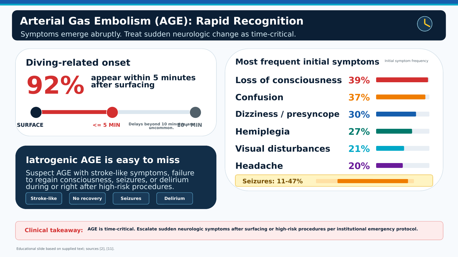
Rapid Recognition of Arterial Gas Embolism. The abrupt onset and most common initial manifestations of arterial gas embolism, particularly after diving or high-risk medical procedures. Most diving-related symptoms occur within 5 minutes after surfacing and may include loss of consciousness, confusion, dizziness, hemiplegia, visual disturbances, headache, and seizures.
Contributed by A Sankari, MD
References
Dudney TM, Elliott CG. Pulmonary embolism from amniotic fluid, fat, and air. Progress in cardiovascular diseases. 1994 May-Jun:36(6):447-74 [PubMed PMID: 8184098]
Dube L, Soltner C, Daenen S, Lemarieé J, Asfar P, Alquier P. Gas embolism: an exceptional complication of radial arterial catheterization. Acta anaesthesiologica Scandinavica. 2004 Oct:48(9):1208-10 [PubMed PMID: 15352970]
Level 3 (low-level) evidenceSCHMIDT PJ, KEVY SV. Air embolism; a hazard during phlebotomy. The New England journal of medicine. 1958 Feb 27:258(9):424-7 [PubMed PMID: 13517496]
Leclercq F, Kassnasrallah S, Cesari JB, Blard JM, Macia JC, Messner-Pellenc P, Mariottini CJ, Grolleau-Raoux R. Transcranial Doppler detection of cerebral microemboli during left heart catheterization. Cerebrovascular diseases (Basel, Switzerland). 2001:12(1):59-65 [PubMed PMID: 11435681]
Phillips YY. Primary blast injuries. Annals of emergency medicine. 1986 Dec:15(12):1446-50 [PubMed PMID: 3535591]
Wherrett CG, Mehran RJ, Beaulieu MA. Cerebral arterial gas embolism following diagnostic bronchoscopy: delayed treatment with hyperbaric oxygen. Canadian journal of anaesthesia = Journal canadien d'anesthesie. 2002 Jan:49(1):96-9 [PubMed PMID: 11782337]
Level 3 (low-level) evidencevan der Zee MP, Koene BM, Mariani MA. Fatal air embolism during cardiopulmonary bypass: analysis of an incident and prevention measures. Interactive cardiovascular and thoracic surgery. 2014 Nov:19(5):875-7. doi: 10.1093/icvts/ivu242. Epub 2014 Jul 31 [PubMed PMID: 25082835]
Level 3 (low-level) evidenceRehwald R, Loizides A, Wiedermann FJ, Grams AE, Djurdjevic T, Glodny B. Systemic air embolism causing acute stroke and myocardial infarction after percutaneous transthoracic lung biopsy - a case report. Journal of cardiothoracic surgery. 2016 May 6:11(1):80. doi: 10.1186/s13019-016-0478-z. Epub 2016 May 6 [PubMed PMID: 27154545]
Level 3 (low-level) evidenceHemmerling TM, Schmidt J, Bosert C, Klein P. Systemic air embolism during wedge resection of the lung. Anesthesia and analgesia. 2001 Nov:93(5):1135-6, table of contents [PubMed PMID: 11682382]
Level 3 (low-level) evidenceMorris WP, Butler BD, Tonnesen AS, Allen SJ. Continuous venous air embolism in patients receiving positive end-expiratory pressure. The American review of respiratory disease. 1993 Apr:147(4):1034-7 [PubMed PMID: 8466103]
Level 3 (low-level) evidenceSharma P, Pilling JE, Awad WI. Cerebral air embolism after noninvasive ventilation postpulmonary wedge resection. The Journal of thoracic and cardiovascular surgery. 2007 Jul:134(1):262-3 [PubMed PMID: 17599531]
Level 3 (low-level) evidenceHalpern P, Greenstein A, Melamed Y, Taitelman U, Sznajder I, Zveibil F. Arterial air embolism after penetrating lung injury. Critical care medicine. 1983 May:11(5):392-3 [PubMed PMID: 6839793]
Level 3 (low-level) evidenceHwang SL, Lieu AS, Lin CL, Liu GC, Howng SL, Kuo TH. Massive cerebral air embolism after cardiopulmonary resuscitation. Journal of clinical neuroscience : official journal of the Neurosurgical Society of Australasia. 2005 May:12(4):468-9 [PubMed PMID: 15925785]
Level 3 (low-level) evidenceButler BD, Hills BA. Transpulmonary passage of venous air emboli. Journal of applied physiology (Bethesda, Md. : 1985). 1985 Aug:59(2):543-7 [PubMed PMID: 4030608]
Level 3 (low-level) evidencePinho J, Amorim JM, Araújo JM, Vilaça H, Ribeiro M, Pereira J, Ferreira C. Cerebral gas embolism associated with central venous catheter: Systematic review. Journal of the neurological sciences. 2016 Mar 15:362():160-4. doi: 10.1016/j.jns.2016.01.043. Epub 2016 Jan 22 [PubMed PMID: 26944140]
Level 1 (high-level) evidenceRoberts S, Johnson M, Davies S. Near-fatal air embolism: fibrin sheath as the portal of air entry. Southern medical journal. 2003 Oct:96(10):1036-8 [PubMed PMID: 14570352]
Level 3 (low-level) evidenceLjubkovic M, Zanchi J, Breskovic T, Marinovic J, Lojpur M, Dujic Z. Determinants of arterial gas embolism after scuba diving. Journal of applied physiology (Bethesda, Md. : 1985). 2012 Jan:112(1):91-5. doi: 10.1152/japplphysiol.00943.2011. Epub 2011 Oct 13 [PubMed PMID: 21998270]
Ghannam M, Beran A, Ghazaleh D, Ferderer T, Berry B, Banna MA, Mohl L, Streib C, Thacker T, Matos I. Cerebral Air Embolism after Esophagogastroduodenoscopy: Insight on Pathophysiology, Epidemiology, Prevention and Treatment. Journal of stroke and cerebrovascular diseases : the official journal of National Stroke Association. 2019 Dec:28(12):104403. doi: 10.1016/j.jstrokecerebrovasdis.2019.104403. Epub 2019 Sep 26 [PubMed PMID: 31563566]
Groenman FA, Peters LW, Rademaker BM, Bakkum EA. Embolism of air and gas in hysteroscopic procedures: pathophysiology and implication for daily practice. Journal of minimally invasive gynecology. 2008 Mar-Apr:15(2):241-7. doi: 10.1016/j.jmig.2007.10.010. Epub [PubMed PMID: 18313001]
Bernhardt TL, Goldmann RW, Thombs PA, Kindwall EP. Hyperbaric oxygen treatment of cerebral air embolism from orogenital sex during pregnancy. Critical care medicine. 1988 Jul:16(7):729-30 [PubMed PMID: 3371050]
Level 3 (low-level) evidenceJadik S, Wissing H, Friedrich K, Beck J, Seifert V, Raabe A. A standardized protocol for the prevention of clinically relevant venous air embolism during neurosurgical interventions in the semisitting position. Neurosurgery. 2009 Mar:64(3):533-8; discussion 538-9. doi: 10.1227/01.NEU.0000338432.55235.D3. Epub [PubMed PMID: 19240616]
Moon RE. Hyperbaric treatment of air or gas embolism: current recommendations. Undersea & hyperbaric medicine : journal of the Undersea and Hyperbaric Medical Society, Inc. 2019 Sep - Dec - Fourth Quarter:46(5):673-683 [PubMed PMID: 31683367]
Mitchell SJ, Bennett MH, Moon RE. Decompression Sickness and Arterial Gas Embolism. The New England journal of medicine. 2022 Mar 31:386(13):1254-1264. doi: 10.1056/NEJMra2116554. Epub [PubMed PMID: 35353963]
Wilmshurst P. Risk mitigation in divers with persistent (patent) foramen ovale. Diving and hyperbaric medicine. 2019 Jun 30:49(2):77-78. doi: 10.28920/dhm49.2.77-78. Epub [PubMed PMID: 31177512]
Malik N, Claus PL, Illman JE, Kligerman SJ, Moynagh MR, Levin DL, Woodrum DA, Arani A, Arunachalam SP, Araoz PA. Air embolism: diagnosis and management. Future cardiology. 2017 Jul:13(4):365-378. doi: 10.2217/fca-2017-0015. Epub 2017 Jun 23 [PubMed PMID: 28644058]
Kandori K, Ishii W, Iiduka R. Massive systemic arterial air embolism caused by an air shunt after blunt chest trauma: A case report. International journal of surgery case reports. 2018:51():368-371. doi: 10.1016/j.ijscr.2018.09.014. Epub 2018 Sep 21 [PubMed PMID: 30268062]
Level 3 (low-level) evidenceBrull SJ, Prielipp RC. Vascular air embolism: A silent hazard to patient safety. Journal of critical care. 2017 Dec:42():255-263. doi: 10.1016/j.jcrc.2017.08.010. Epub 2017 Aug 7 [PubMed PMID: 28802790]
Hubbard M, Davis FM, Malcolm K, Mitchell SJ. Decompression illness and other injuries in a recreational dive charter operation. Diving and hyperbaric medicine. 2018 Dec 24:48(4):218-223. doi: 10.28920/dhm48.4.218-223. Epub [PubMed PMID: 30517953]
Marsh PL, Moore EE, Moore HB, Bunch CM, Aboukhaled M, Condon SM 2nd, Al-Fadhl MD, Thomas SJ, Larson JR, Bower CW, Miller CB, Pearson ML, Twilling CL, Reser DW, Kim GS, Troyer BM, Yeager D, Thomas SG, Srikureja DP, Patel SS, Añón SL, Thomas AV, Miller JB, Van Ryn DE, Pamulapati SV, Zimmerman D, Wells B, Martin PL, Seder CW, Aversa JG, Greene RB, March RJ, Kwaan HC, Fulkerson DH, Vande Lune SA, Mollnes TE, Nielsen EW, Storm BS, Walsh MM. Iatrogenic air embolism: pathoanatomy, thromboinflammation, endotheliopathy, and therapies. Frontiers in immunology. 2023:14():1230049. doi: 10.3389/fimmu.2023.1230049. Epub 2023 Sep 19 [PubMed PMID: 37795086]
Gordy S, Rowell S. Vascular air embolism. International journal of critical illness and injury science. 2013 Jan:3(1):73-6. doi: 10.4103/2229-5151.109428. Epub [PubMed PMID: 23724390]
Khan M, Schmidt DH, Bajwa T, Shalev Y. Coronary air embolism: incidence, severity, and suggested approaches to treatment. Catheterization and cardiovascular diagnosis. 1995 Dec:36(4):313-8 [PubMed PMID: 8719380]
Level 3 (low-level) evidenceLee JH, Yoon SH, Hong H, Rho JY, Goo JM. Incidence, risk factors, and prognostic indicators of symptomatic air embolism after percutaneous transthoracic lung biopsy: a systematic review and pooled analysis. European radiology. 2021 Apr:31(4):2022-2033. doi: 10.1007/s00330-020-07372-w. Epub 2020 Oct 13 [PubMed PMID: 33051730]
Level 1 (high-level) evidenceMonnin-Bares V, Chassagnon G, Vernhet-Kovacsik H, Zarqane H, Vanoverschelde J, Picot MC, Bommart S. Systemic air embolism depicted on systematic whole thoracic CT acquisition after percutaneous lung biopsy: Incidence and risk factors. European journal of radiology. 2019 Aug:117():26-32. doi: 10.1016/j.ejrad.2019.05.016. Epub 2019 May 22 [PubMed PMID: 31307649]
Level 1 (high-level) evidenceWu T, Li S, Gao M, Yang B, Wang Y, Xie T. Risk factors associated with air embolism following computed tomography-guided percutaneous lung biopsy: a retrospective case-control study. PeerJ. 2024:12():e18232. doi: 10.7717/peerj.18232. Epub 2024 Oct 15 [PubMed PMID: 39430567]
Level 2 (mid-level) evidenceLi Z, Li G, Li Y, Chen Y, Li J, Chen H. Flow field around bubbles on formation of air embolism in small vessels. Proceedings of the National Academy of Sciences of the United States of America. 2021 Jun 29:118(26):. doi: 10.1073/pnas.2025406118. Epub [PubMed PMID: 34155104]
He R, Huang Q, Yan X, Liu Y, Yang J, Chen X. A Case of Paradoxical Embolism Causing Anterior Spinal Cord Syndrome and Acute Myocardial Infarction Following the Intradiscal Oxygen-Ozone Therapy. Frontiers in neurology. 2019:10():137. doi: 10.3389/fneur.2019.00137. Epub 2019 Feb 22 [PubMed PMID: 30853936]
Level 3 (low-level) evidenceLee JI, Yim BS, Kim JM. Effect of dissolved-gas concentration on bulk nanobubbles generation using ultrasonication. Scientific reports. 2020 Nov 2:10(1):18816. doi: 10.1038/s41598-020-75818-8. Epub 2020 Nov 2 [PubMed PMID: 33139819]
Sánchez-Villalobos JM, Fortuna-Alcaraz ML, Serrano-Velasco L, Pujante-Escudero Á, Garnés-Sánchez CM, Pérez-Garcilazo JE, Olea-González A, Pérez-Vicente JA. Breath-Hold Diving-Related Decompression Sickness with Brain Involvement: From Neuroimaging to Pathophysiology. Tomography (Ann Arbor, Mich.). 2022 Apr 19:8(3):1172-1183. doi: 10.3390/tomography8030096. Epub 2022 Apr 19 [PubMed PMID: 35645382]
Hills BA, Butler BD. Size distribution of intravascular air emboli produced by decompression. Undersea biomedical research. 1981 Sep:8(3):163-70 [PubMed PMID: 7292785]
Level 3 (low-level) evidenceBanham ND, Saw J, Hankey GJ, Ghia D. Cerebral arterial gas embolism proven by computed tomography following transthoracic echocardiography using bubble contrast. Diving and hyperbaric medicine. 2020 Sep 30:50(3):300-302. doi: 10.28920/dhm50.3.300-302. Epub [PubMed PMID: 32957135]
Helps SC, Gorman DF. Air embolism of the brain in rabbits pretreated with mechlorethamine. Stroke. 1991 Mar:22(3):351-4 [PubMed PMID: 2003304]
Level 3 (low-level) evidenceCao D, Arens AM, Chow SL, Easter SR, Hoffman RS, Lagina AT 3rd, Lavonas EJ, Patil KD, Sutherland LD, Tijssen JA, Wang GS, Zelop CM, Rodriguez AJ, Drennan IR, McBride ME. Part 10: Adult and Pediatric Special Circumstances of Resuscitation: 2025 American Heart Association Guidelines for Cardiopulmonary Resuscitation and Emergency Cardiovascular Care. Circulation. 2025 Oct 21:152(16_suppl_2):S578-S672. doi: 10.1161/CIR.0000000000001380. Epub 2025 Oct 22 [PubMed PMID: 41122889]
Leitch DR, Green RD. Pulmonary barotrauma in divers and the treatment of cerebral arterial gas embolism. Aviation, space, and environmental medicine. 1986 Oct:57(10 Pt 1):931-8 [PubMed PMID: 3778391]
Brown AE, Rabinstein AA, Braksick SA. Clinical Characteristics, Imaging Findings, and Outcomes of Cerebral Air Embolism. Neurocritical care. 2023 Feb:38(1):158-164. doi: 10.1007/s12028-022-01664-3. Epub 2023 Jan 10 [PubMed PMID: 36627433]
Shah J, Jiwa N, Mamdani N, Hill D. Venous and arterial air embolism: a rare phenomenon with fatal consequences. BMJ case reports. 2016 Dec 5:2016():. doi: 10.1136/bcr-2016-217550. Epub 2016 Dec 5 [PubMed PMID: 27920021]
Level 3 (low-level) evidenceHatling D, Høgset A, Guttormsen AB, Müller B. Iatrogenic cerebral gas embolism-A systematic review of case reports. Acta anaesthesiologica Scandinavica. 2019 Feb:63(2):154-160. doi: 10.1111/aas.13260. Epub 2018 Sep 10 [PubMed PMID: 30203491]
Level 1 (high-level) evidenceAdamu M, Skillicorn C, Stone T, Moudgil H, Abayaratne C. Systemic arterial air embolism following computed tomography (CT)-guided percutaneous lung biopsy: Case series and review of underlying risk factors, treatment and preventive strategies. Clinical medicine (London, England). 2026 Mar:26(2):100530. doi: 10.1016/j.clinme.2025.100530. Epub 2025 Nov 17 [PubMed PMID: 41260265]
Level 2 (mid-level) evidenceRuiz Avila HA, García-Araque HF, Acosta-Gutiérrez E. Paradoxical venous air embolism detected with point-of-care ultrasound: a case report. The ultrasound journal. 2022 May 18:14(1):19. doi: 10.1186/s13089-022-00265-7. Epub 2022 May 18 [PubMed PMID: 35583704]
Level 3 (low-level) evidenceShaikh N, Ummunisa F. Acute management of vascular air embolism. Journal of emergencies, trauma, and shock. 2009 Sep:2(3):180-5. doi: 10.4103/0974-2700.55330. Epub [PubMed PMID: 20009308]
Jorens PG, Van Marck E, Snoeckx A, Parizel PM. Nonthrombotic pulmonary embolism. The European respiratory journal. 2009 Aug:34(2):452-74. doi: 10.1183/09031936.00141708. Epub [PubMed PMID: 19648522]
Alvaran SB, Toung JK, Graff TE, Benson DW. Venous air embolism: comparative merits of external cardiac massage, intracardiac aspiration, and left lateral decubitus position. Anesthesia and analgesia. 1978 Mar-Apr:57(2):166-70 [PubMed PMID: 565152]
Level 3 (low-level) evidenceDexter F, Hindman BJ. Computer simulation of microscopic cerebral air emboli absorption during cardiac surgery. Undersea & hyperbaric medicine : journal of the Undersea and Hyperbaric Medical Society, Inc. 1998 Spring:25(1):43-50 [PubMed PMID: 9566086]
Mirski MA, Lele AV, Fitzsimmons L, Toung TJ. Diagnosis and treatment of vascular air embolism. Anesthesiology. 2007 Jan:106(1):164-77 [PubMed PMID: 17197859]
Berlot G, Rinaldi A, Moscheni M, Ferluga M, Rossini P. Uncommon Occurrences of Air Embolism: Description of Cases and Review of the Literature. Case reports in critical care. 2018:2018():5808390. doi: 10.1155/2018/5808390. Epub 2018 Jul 8 [PubMed PMID: 30073096]
Level 3 (low-level) evidenceLeach RM, Rees PJ, Wilmshurst P. Hyperbaric oxygen therapy. BMJ (Clinical research ed.). 1998 Oct 24:317(7166):1140-3 [PubMed PMID: 9784458]
Fakkert RA, Karlas N, Schober P, Weber NC, Preckel B, van Hulst RA, Weenink RP. Early hyperbaric oxygen therapy is associated with favorable outcome in patients with iatrogenic cerebral arterial gas embolism: systematic review and individual patient data meta-analysis of observational studies. Critical care (London, England). 2023 Jul 12:27(1):282. doi: 10.1186/s13054-023-04563-x. Epub 2023 Jul 12 [PubMed PMID: 37434172]
Level 1 (high-level) evidenceKhalid F, Rehman S, AbdulRahman R, Gupta S. Fatal air embolism following local anaesthetisation: does needle size matter? BMJ case reports. 2018 Feb 5:2018():. pii: bcr-2017-222254. doi: 10.1136/bcr-2017-222254. Epub 2018 Feb 5 [PubMed PMID: 29437732]
Level 3 (low-level) evidenceČerveňák V, Všianský V, Cviková M, Brichta J, Vinklárek J, Štefela J, Haršány M, Hájek M, Herzig R, Kouřil D, Bárková V, Filip P, Aulický P, Weiss V. Cerebral air embolism: neurologic manifestations, prognosis, and outcome. Frontiers in neurology. 2024:15():1417006. doi: 10.3389/fneur.2024.1417006. Epub 2024 Jun 19 [PubMed PMID: 38962484]
Sen S, Sen S. Therapeutic effects of hyperbaric oxygen: integrated review. Medical gas research. 2021 Jan-Mar:11(1):30-33. doi: 10.4103/2045-9912.310057. Epub [PubMed PMID: 33642335]