 Tools
Tools
 Anatomy, Bony Pelvis and Lower Limb: Metatarsal Bones
Introduction
Metatarsal bones constitute the osseous structures of the forefoot, connecting the distal aspects of the medial, intermediate, and lateral cuneiform bones, as well as the cuboid bone, to the bases of the 5 phalanges of the foot (see Image. Foot Bones). Five metatarsal bones are present, numbered 1 to 5 from the hallux (great toe) to the 5th digit (small toe). These bones serve as key sites for the origin and insertion of multiple lower limb and foot muscles and contribute to the proximal component of the metatarsophalangeal (MTP) joints. Vascular supply to the metatarsal region is derived primarily from branches of the dorsalis pedis and plantar arterial arches, providing an extensive anastomotic network. Innervation is supplied predominantly through branches of the deep fibular (peroneal) nerve on the dorsum and the medial (MPN) and lateral (LPN) plantar nerves on the plantar aspect.
Metatarsal pathology is clinically significant due to the high incidence of fractures, stress injuries, Lisfranc injuries, and degenerative or ischemic conditions such as Freiberg disease, all of which commonly present with forefoot pain and weight-bearing dysfunction. Management of metatarsal pathology may involve surgical procedures, such as intramedullary screw fixation for 5th-metatarsal fractures, open reduction and internal fixation (ORIF) or primary arthrodesis for Lisfranc injuries, and corrective osteotomies or arthroplasty in advanced Freiberg disease. Detailed knowledge of metatarsal anatomy and biomechanics supports accurate injury localization, improves interpretation of imaging and physical examination findings, and guides appropriate selection between conservative and operative management strategies.
Structure and Function
Register For Free And Read The Full Article
 Search engine and full access to all medical articles
- 10 free questions in your specialty
- Free CME/CE Activities
- Free daily question in your email
- Save favorite articles to your dashboard
- Emails offering discounts
Learn more about a Subscription to StatPearls Point-of-Care
Structure and Function
The 5 metatarsal bones are numbered 1 through 5 from medial to lateral, beginning at the hallux (great toe). Each metatarsal bone articulates proximally with a tarsal bone and distally with 1 of the 5 phalanges of the foot, forming the MTP joint. The proximal articulations between the metatarsal bones and tarsal bones form the tarsometatarsal (TMT) joint complex, commonly referred to as the "Lisfranc joint." Disruption of the articulation between the medial cuneiform and the base of the 2nd metatarsal is commonly referred to as a "Lisfranc injury."
The TMT joint complex is divided into medial, middle, and lateral columns. The medial column includes the 1st metatarsal and the medial cuneiform. The 1st metatarsal is the shortest and widest metatarsal, articulating proximally with the medial cuneiform and distally with the proximal phalanx of the hallux. The middle column of the TMT joint complex consists of the 2nd and 3rd metatarsals, associated with the intermediate and lateral cuneiforms, respectively. The 2nd metatarsal is the longest metatarsal bone, articulating with the intermediate cuneiform, while the 3rd metatarsal articulates with the lateral cuneiform. The lateral column of the TMT joint complex comprises the 4th and 5th metatarsals and the cuboid. Two sesamoid bones are associated with the medial column, located plantar to the 1st metatarsal head within the flexor hallucis brevis tendon. Hallux sesamoid bones are among the most clinically significant sesamoid bones in the human body, alongside the patella and sesamoids of the hand.[1]
The metatarsals serve as important sites for the insertion and origin of multiple muscles of the lower limb and foot. The specific insertions and origins on each metatarsal are as follows:
- Insertions
- 1st metatarsal: fibularis (peroneus) longus, tibialis anterior
- 2nd metatarsal: tibialis posterior
- 3rd metatarsal: tibialis posterior
- 4th metatarsal: tibialis posterior
- 5th metatarsal: fibularis (peroneus) brevis, fibularis (peroneus) tertius
- Origins
- 1st metatarsal: none
- 2nd metatarsal: adductor hallucis (oblique head), dorsal interossei
- 3rd metatarsal: adductor hallucis (oblique head), plantar interossei, dorsal interossei
- 4th metatarsal: adductor hallucis (oblique head), plantar interossei, dorsal interossei
- 5th metatarsal: plantar interossei, dorsal interossei
In addition to serving as insertion sites for multiple muscles of the lower limb and foot, the metatarsals constitute an important component of the arches of the foot. The arches function by absorbing force, supporting structure, and acting as a rigid lever during gait propulsion. Three principal arches are recognized: the medial longitudinal, lateral longitudinal, and transverse arches. The medial longitudinal arch is formed by the first 3 metatarsals, their respective tarsal articulations, and the calcaneus. The lateral longitudinal arch is formed by the 4th and 5th metatarsal heads, the cuboid, and the calcaneus. The transverse arch is formed by the bases of the metatarsals, the cuboid, and the 3 cuneiform bones. Support of the arches is provided by musculotendinous units of the anterior and posterior leg compartments, as well as the intrinsic muscles and ligaments of the midfoot and hindfoot (see Image. Ligaments of the Lateral Aspect of the Right Foot and Ankle).
The metatarsals are essential for normal biomechanical gait, forming a major distal weight-bearing region at their heads. The 1st metatarsal bears approximately 30% to 50% of the load during the gait cycle. The 1st and 5th metatarsal column complexes are mobile, permitting superior-inferior motion to accommodate uneven surfaces. The remaining metatarsals generally do not exhibit this motion, as fixation at their bases limits mobility.[2]
Embryology
Limb bud formation begins approximately 4 to 5 weeks after fertilization as mesoderm migrates into the limb bud, forming anterior, posterior, and lateral condensations that ultimately give rise to the muscular and skeletal components of the lower limb.[3] The primary contribution to the skeletal elements of the lower limb derives from the lateral plate mesoderm, which forms the ilium, ischium, pubis, femur, tibia, fibula, tarsals, metatarsals, and phalanges. Lateral condensations undergo chondrification to form hyaline cartilage during weeks 6 to 7. Primary ossification commences during the fetal period and continues until birth.[4]
Blood Supply and Lymphatics
The foot's blood supply is primarily provided by the anterior tibial and posterior tibial arteries, which are terminal branches of the popliteal artery. The anterior tibial artery courses distally and terminates at the anterior ankle, midway between the medial and lateral malleoli, as the dorsalis pedis artery. The dorsalis pedis artery continues distally between the extensor hallucis longus and extensor digitorum longus tendons, where the pulse is commonly palpated during clinical physical examination. The dorsalis pedis artery gives off the lateral tarsal artery, forming an anastomosis that contributes to the dorsal metatarsal arteries. The dorsalis pedis artery terminates as the deep plantar artery.
The posterior tibial artery gives rise to the medial and lateral plantar arteries, which anastomose to form the deep plantar arch. The dorsalis pedis artery gives rise to the deep plantar artery, which communicates with the deep plantar arch, establishing an anastomosis between the anterior tibial and posterior tibial arterial systems.
The lymphatic vessels of the lower limb are divided into 2 major groups: superficial and deep. The superficial lymphatic vessels of the lower limb are further subdivided into a medial group, which follows the greater saphenous vein, and a lateral group, which follows the small saphenous vein. Deep lymphatic vessels include the anterior tibial, posterior tibial, and fibular (peroneal) vessels, which follow the course of the corresponding blood vessels. Lymphatic drainage of the lower limb proceeds to the popliteal, superficial inguinal, deep inguinal, external iliac, and lumbar or aortic lymph nodes.[5]
Nerves
The tibial nerve divides into the 2 major nerves of the plantar foot—the MPN and LPN. The MPN supplies the 1st lumbrical, abductor hallucis, flexor digitorum brevis, and flexor hallucis brevis. The LPN innervates all intrinsic foot muscles except the 4 muscles listed above.
The dorsum of the foot contains 2 muscles: the extensor digitorum brevis and the extensor hallucis brevis. Both muscles receive innervation from the deep fibular nerve, a branch of the common fibular nerve, which also supplies all muscles of the anterior compartment of the lower limb.
Surgical Considerations
The 5th metatarsal is the most commonly fractured metatarsal in adults. Conservative management, including protected weight-bearing in a stiff-soled shoe, boot, or cast, or non–weight-bearing for 6 to 8 weeks, is generally the preferred treatment for zone 1 injuries (see Clinical Significance). Fractures in zones 2 and 3 in elite or competitive athletes are typically managed surgically with intramedullary screw fixation to reduce the risk of nonunion and prolonged restriction from activity.[6][7][8][9]
Lisfranc injuries may be managed operatively through ORIF, primary arthrodesis of the involved TMT joints, or midfoot arthrodesis. ORIF is indicated in the presence of instability. ORIF in Lisfranc injuries tends to yield better outcomes in bony fracture-dislocations compared with strictly ligamentous injuries. Primary arthrodesis may be considered in cases of instability as an alternative to ORIF and has demonstrated equivalent functional outcomes, with a reduced rate of return to the operating room for hardware removal. Midfoot arthrodesis is considered in chronic Lisfranc injuries that have failed conservative therapy or in the setting of midfoot collapse.[10][11]
Surgical intervention may be required in severe cases of Freiberg disease. Surgical options include MTP arthrotomy with removal of loose bodies, dorsal closing-wedge osteotomy, and DuVries arthroplasty (partial metatarsal head resection). The first option is rarely required, whereas the remaining procedures may be indicated when the disease involves a greater proportion of the dorsal bone and cartilage or in later-stage disease.[12]
Clinical Significance
Metatarsal injuries are common, with metatarsal fractures, stress fractures, and Lisfranc injuries among the most frequently documented. A brief overview of clinical conditions involving the metatarsal bones is provided below.
Metatarsal fractures are some of the most common injuries of the foot. The 1st metatarsal is most frequently fractured in children younger than 5 years, whereas the 5th metatarsal is the most commonly fractured metatarsal in older children, adolescents, and adults.[13] Two principal mechanisms of metatarsal fractures are recognized: direct crush injuries and indirect rotational mechanisms. Indirect mechanisms occur more frequently than direct crush injuries and typically involve hindfoot inversion, forefoot adduction, or repetitive microtrauma. Clinical manifestations include pain over the affected border of the forefoot, limitation of weight-bearing, and possible resistance to foot eversion on physical examination. Manual palpation of the affected region typically elicits significant pain.
Most metatarsal fractures heal with nonoperative management. Open fractures, displaced fractures, and multiple fractures of the central metatarsals constitute indications for surgical fixation. Malunion of metatarsal fractures may result in transfer metatarsalgia, defined as forefoot pain secondary to dysfunction in an adjacent region of the forefoot.[14]
Fifth-metatarsal fractures are the most common metatarsal fractures in adults. From proximal to distal, the 5th metatarsal is divided into the tuberosity, base, shaft, neck, and head. Fractures of the 5th metatarsal are classified according to the location, or zone, of the fracture.
Fractures proximal to the region between the 4th and 5th metatarsals (4th–5th metatarsal articulation) are classified as zone 1 fractures, also known as “pseudo-Jones” fractures. Fractures at the metadiaphyseal junction involving the 4th–5th metatarsal articulation are classified as zone 2 fractures, or “Jones” fractures. Owing to the vascular watershed region at this junction, Jones fractures are associated with an increased risk of nonunion, reported in approximately 15% to 30% of cases (see Image. Radiograph of a Classic Jones Fracture). Zone 3 fractures are located distal to the 4th–5th metatarsal articulation and may result from direct trauma or stress injury in the setting of antecedent pain.
Clinical examination typically demonstrates pain and swelling along the lateral border of the foot. Radiographs are often sufficient for diagnosis. Zone 1 injuries are managed nonoperatively with a hard-sole shoe or controlled ankle motion walking boot. Injuries in zones 2 and 3 may be initially managed nonoperatively with non–weight-bearing in a short leg splint, followed by transition to a short leg cast for 6 to 8 weeks. In elite or competitive athletes, zone 2 fractures may be managed with intramedullary screw fixation to reduce the risk of nonunion and facilitate earlier return to sport.[15]
Stress fractures of the metatarsals are not uncommon. Also known as fatigue fractures, repeated strain cycles lead to microfractures within the bone. Continued repetitive loading may cause microfracture formation to exceed the rate of bone healing, resulting in progression to a complete fracture. The metatarsals are some of the most common sites of stress fractures in the body, accounting for nearly half of all stress fractures. Treatment is usually conservative, consisting of activity modification for 6 to 12 weeks and use of a hard-soled shoe or cast.[16]
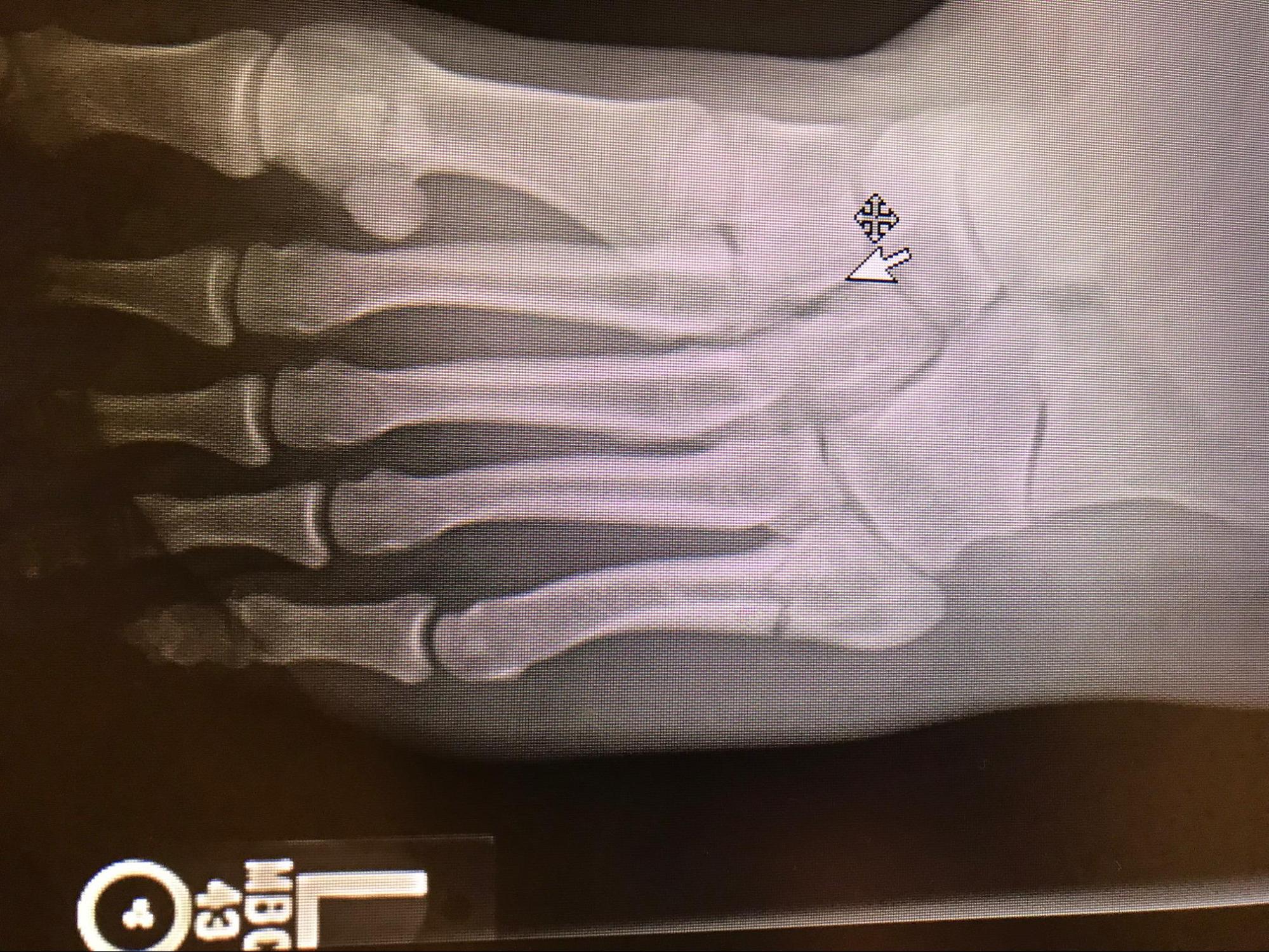
Lisfranc injuries are characterized by disruption of the articulation between the medial cuneiform and the base of the 2nd metatarsal, resulting in disruption of the TMT joint complex (see Image. Lisfranc Fracture-Dislocation of the Left Foot). This injury most commonly results from high-energy trauma, including motor vehicle accidents, falls from height, or athletic activities involving axial loading of a hyperplantarflexed forefoot. Lisfranc injuries may be managed nonoperatively with cast immobilization when weight-bearing or stress radiographs demonstrate no displacement, or no bony injury is identified on CT. Operative management commonly involves ORIF or, more frequently, primary arthrodesis of the involved TMT joints. Recent studies demonstrate that primary arthrodesis is associated with lower rates of revision surgery and hardware removal, while maintaining equivalent functional outcomes.[17][18][19]
Lastly, Freiberg disease is a rare condition characterized by infarction and fragmentation of the metatarsal head (see Image. Radiograph of Freiberg Disease in the Right Foot). Freiberg disease is believed to result from disruption of the blood supply in the setting of microtrauma or osteonecrosis, leading to subchondral collapse. The condition most commonly affects adolescent female athletes and typically involves the dorsal aspect of the 2nd metatarsal head. Treatment may be nonoperative in early stages, including activity modification, intake of nonsteroidal anti-inflammatory medications, and immobilization with a short cast or stiff-soled shoe for 4 to 6 weeks. Severe cases may require surgical intervention.
Media
(Click Image to Enlarge)
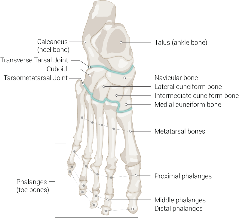
Foot Bones. Anatomy of the foot including talus (ankle bone), navicular bone, lateral cuneiform bone, intermediate cuneiform bone, medial cuneiform bone, metatarsal bones, proximal phalanges, middle phalanges, distal phalanges, phalanges (toe bones), tarsometatarsal joint, cuboid, transverse tarsal joint, and calcaneus (heel bone).
Illustrated by B Palmer
(Click Image to Enlarge)
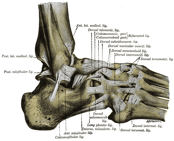
Ligaments of the Lateral Aspect of the Right Foot and Ankle. This anatomical illustration details the complex connective tissues of the foot, including the anterior and posterior lateral malleolar ligaments and the anterior and posterior talofibular ligaments. The calcaneofibular ligament and interosseous talocalcaneal ligament are shown stabilizing the heel, while the bifurcated ligament (comprising the calcaneonavicular and calcaneocuboid parts), the long plantar ligament, and the dorsal talonavicular ligament support the midfoot. Further distal structures include the dorsal cuboideonavicular, dorsal navicular cuneiform, dorsal cuneocuboid, dorsal intercuneiform, dorsal calcaneocuboid, dorsal tarsometatarsal, and dorsal intermetatarsal ligaments.
Henry Vandyke Carter, Public Domain, via Wikimedia Commons
(Click Image to Enlarge)
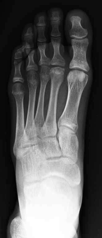
Radiograph of Freiberg Disease in the Right Foot. This frontal x-ray reveals avascular necrosis localized to the head of the 2nd metatarsal. Structural changes, including cortical thickening and subchondral collapse, are visible at the metatarsophalangeal joint.
Contributed by Kevin Carter, DO
(Click Image to Enlarge)
Radiograph of a Classic Jones Fracture. The image shows a clear cortical disruption at the base of the 5th metatarsal, specifically at the junction between the metaphysis and diaphysis.
Contributed by Mark A. Dreyer, DPM, FACFAS
(Click Image to Enlarge)
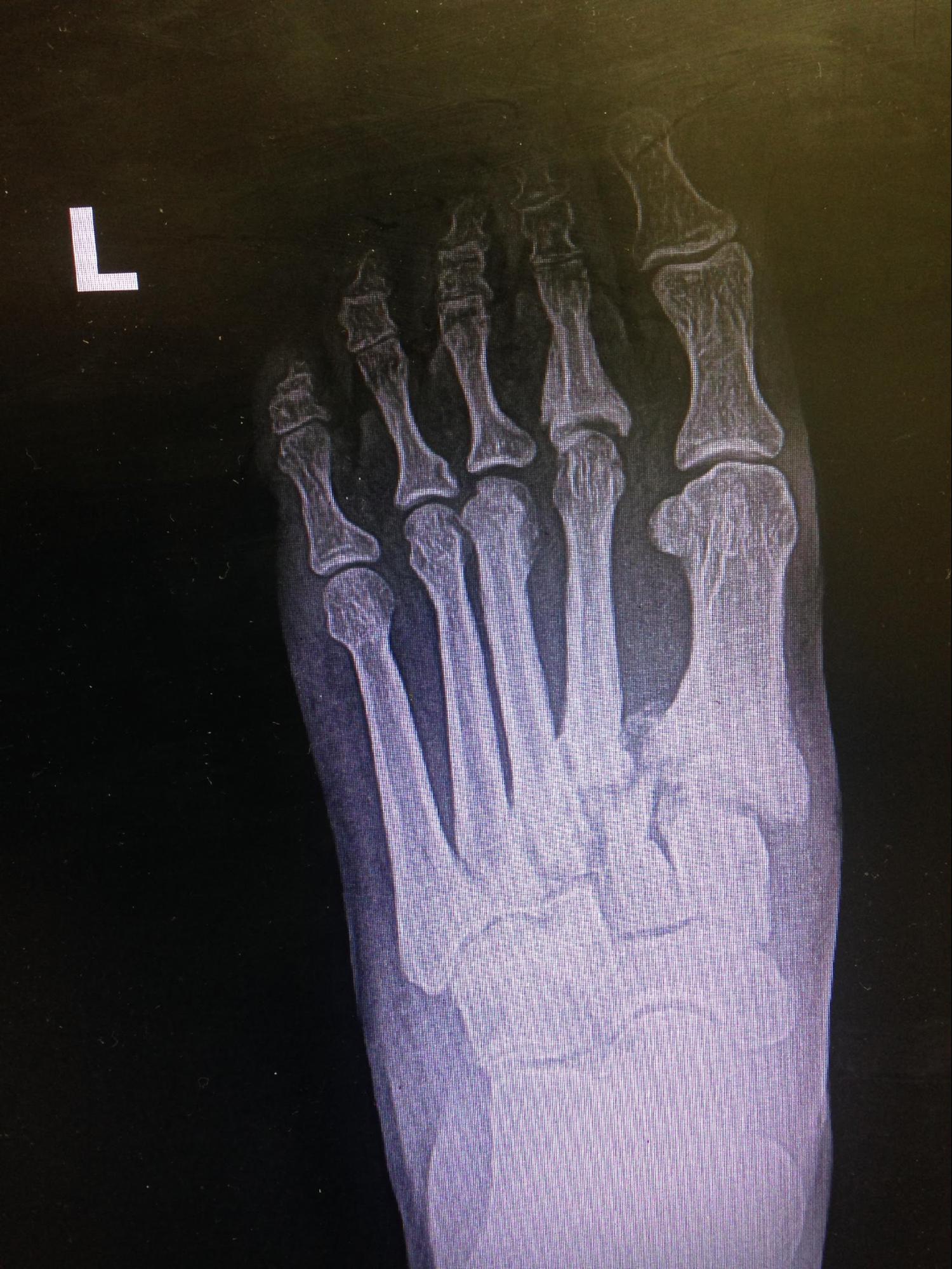
Lisfranc Fracture-Dislocation of the Left Foot. This anteroposterior radiograph reveals a significant disruption of the tarsometatarsal joint complex, specifically showing lateral displacement of the 2nd through 5th metatarsals. A medial tarsometatarsal fracture is evident, indicating a high-energy injury that has compromised the structural integrity of the Lisfranc ligament and the midfoot arch.
Contributed by MA Dreyer, DPM, FACFAS
References
Chen W, Cheng J, Sun R, Zhang Z, Zhu Y, Ipaktchi K, Zhang Y. Prevalence and variation of sesamoid bones in the hand: a multi-center radiographic study. International journal of clinical and experimental medicine. 2015:8(7):11721-6 [PubMed PMID: 26380010]
Chan CW, Rudins A. Foot biomechanics during walking and running. Mayo Clinic proceedings. 1994 May:69(5):448-61 [PubMed PMID: 8170197]
Mróz I, Kielczewski S, Pawlicki D, Kurzydło W, Bachul P, Konarska M, Bereza T, Walocha K, Kaythampillai LN, Depukat P, Pasternak A, Bonczar T, Chmielewski P, Mizia E, Skrzat J, Mazur M, Warchoł Ł, Tomaszewski K. Blood vessels of the shin - anterior tibial artery - anatomy and embryology - own studies and review of the literature. Folia medica Cracoviensia. 2016:56(1):33-47 [PubMed PMID: 27513837]
Gros J, Tabin CJ. Vertebrate limb bud formation is initiated by localized epithelial-to-mesenchymal transition. Science (New York, N.Y.). 2014 Mar 14:343(6176):1253-6. doi: 10.1126/science.1248228. Epub [PubMed PMID: 24626928]
Level 3 (low-level) evidenceLezak B, Summers S. Anatomy, Bony Pelvis and Lower Limb: Leg Anterior Compartment. StatPearls. 2026 Jan:(): [PubMed PMID: 30969547]
Lowe D, Grambart S, Cheng CI, Adelman V, Adelman R. Screw characteristics and intramedullary fixation for Jones fractures and associated outcomes: A systematic review and meta-analysis. The Journal of foot and ankle surgery : official publication of the American College of Foot and Ankle Surgeons. 2026 Apr 22:():. pii: S1067-2516(26)00111-0. doi: 10.1053/j.jfas.2026.04.014. Epub 2026 Apr 22 [PubMed PMID: 42031019]
Level 1 (high-level) evidenceVan der Meij D, Weaver B, Ebrahimi A, Bejarano-Pineda L, Waryasz G, Kloen P, Kwon JY, Ashkani-Esfahani S. Risk Factors for Union Deformities in Fifth Metatarsal Fractures: A Scoping Review. Foot & ankle specialist. 2026 Mar 23:():19386400261426853. doi: 10.1177/19386400261426853. Epub 2026 Mar 23 [PubMed PMID: 41871111]
Level 2 (mid-level) evidenceDobrotă RD, Pogărășteanu M, Barbilian AG, Moga M. Clinical and Biomechanical Determinants of Fixation Failure in Fifth Metatarsal Fractures: Implications for Surgical Decision-Making. Journal of clinical medicine. 2026 Feb 24:15(5):. doi: 10.3390/jcm15051680. Epub 2026 Feb 24 [PubMed PMID: 41827098]
Dobrotă RD, Pogărășteanu M, Ferechide D, Lebada IC, Moga M. Comparative Biomechanical Evaluation of Bicortical Screw Versus Plate Fixation in Jones Fractures of the Fifth Metatarsal Using 3D-Printed Models. Journal of clinical medicine. 2025 Oct 21:14(20):. doi: 10.3390/jcm14207449. Epub 2025 Oct 21 [PubMed PMID: 41156318]
Level 2 (mid-level) evidenceSegal KR, Schmitt P, Molino J, Galambas A, Liu J, Blankenhorn B, Hsu RY. Trends in Surgical Management of Lisfranc Injuries: An Analysis of American Board of Orthopaedic Surgery Part II Candidates. Foot & ankle orthopaedics. 2026 Apr:11(2):24730114261433594. doi: 10.1177/24730114261433594. Epub 2026 Apr 18 [PubMed PMID: 42027403]
Hammad A, Ahmad Y, Abdelnour J. Lisfranc Injuries: Latest Updates on Diagnostics and Management. Translational sports medicine. 2026:2026():3933956. doi: 10.1155/tsm2/3933956. Epub 2026 Jan 7 [PubMed PMID: 41522288]
Carmont MR, Rees RJ, Blundell CM. Current concepts review: Freiberg's disease. Foot & ankle international. 2009 Feb:30(2):167-76 [PubMed PMID: 19254514]
Bernstein DT, Mitchell RJ, McCulloch PC, Harris JD, Varner KE. Treatment of Proximal Fifth Metatarsal Fractures and Refractures With Plantar Plating in Elite Athletes. Foot & ankle international. 2018 Dec:39(12):1410-1415. doi: 10.1177/1071100718791835. Epub 2018 Aug 6 [PubMed PMID: 30079768]
Singer G, Cichocki M, Schalamon J, Eberl R, Höllwarth ME. A study of metatarsal fractures in children. The Journal of bone and joint surgery. American volume. 2008 Apr:90(4):772-6. doi: 10.2106/JBJS.G.00547. Epub [PubMed PMID: 18381315]
Level 2 (mid-level) evidencePorter DA. Fifth Metatarsal Jones Fractures in the Athlete. Foot & ankle international. 2018 Feb:39(2):250-258. doi: 10.1177/1071100717741856. Epub 2017 Dec 11 [PubMed PMID: 29228800]
Yates J, Feeley I, Sasikumar S, Rattan G, Hannigan A, Sheehan E. Jones fracture of the fifth metatarsal: Is operative intervention justified? A systematic review of the literature and meta-analysis of results. Foot (Edinburgh, Scotland). 2015 Dec:25(4):251-7. doi: 10.1016/j.foot.2015.08.001. Epub 2015 Aug 11 [PubMed PMID: 26481787]
Level 1 (high-level) evidenceLau S, Guest C, Hall M, Tacey M, Joseph S, Oppy A. Functional Outcomes Post Lisfranc Injury-Transarticular Screws, Dorsal Bridge Plating or Combination Treatment? Journal of orthopaedic trauma. 2017 Aug:31(8):447-452. doi: 10.1097/BOT.0000000000000848. Epub [PubMed PMID: 28731965]
Buda M, Kink S, Stavenuiter R, Hagemeijer CN, Chien B, Hosseini A, Johnson AH, Guss D, DiGiovanni CW. Reoperation Rate Differences Between Open Reduction Internal Fixation and Primary Arthrodesis of Lisfranc Injuries. Foot & ankle international. 2018 Sep:39(9):1089-1096. doi: 10.1177/1071100718774005. Epub 2018 May 29 [PubMed PMID: 29812959]
Magill HHP, Hajibandeh S, Bennett J, Campbell N, Mehta J. Open Reduction and Internal Fixation Versus Primary Arthrodesis for the Treatment of Acute Lisfranc Injuries: A Systematic Review and Meta-analysis. The Journal of foot and ankle surgery : official publication of the American College of Foot and Ankle Surgeons. 2019 Mar:58(2):328-332. doi: 10.1053/j.jfas.2018.08.061. Epub [PubMed PMID: 30850102]
Level 1 (high-level) evidence