 Tools
Tools
 Anatomy, Shoulder and Upper Limb, Clavicle
Introduction
The clavicle is a sigmoid-shaped long bone with a convex surface along its medial end when observed from a cephalad position. The clavicle connects the axial and appendicular skeletons and, along with the scapula, forms the pectoral girdle.[1] Although not as large as other supporting structures in the body, clavicular attachments permit substantial upper extremity function and range of motion and protect posterior neurovascular structures. Each part of this long bone serves a distinct purpose related to its attachments and contributes to the overall physiology of the pectoral girdle.
Medially, the clavicle articulates with the manubrial portion of the sternum, forming the sternoclavicular joint. Surrounded by a fibrous capsule, this joint contains a fibrocartilaginous intraarticular disc between the clavicle and the sternum. Superiorly, the interclavicular ligament connects the ipsilateral and contralateral clavicles and provides additional stability.[2]
Laterally, the clavicle articulates with the acromion, forming the acromioclavicular joint. The surrounding area provides attachment for the shoulder joint capsule. Like the sternoclavicular joint, the acromioclavicular joint is lined by fibrocartilage and contains an intraarticular disc. The acromioclavicular, coracoclavicular, and coracoacromial ligaments support this joint.[3]
The shaft of the clavicle is clinically divided into 2 parts: the medial 2/3 and the lateral 1/3. These regions are used to identify muscle attachment sites.
The medial 2/3 contains an attachment site for the sternocleidomastoid (SCM) on its superior aspect and an attachment for the subclavius muscle along the subclavian groove on its inferior surface. The anterior surface provides attachment for the pectoralis major muscle, and the posterior surface provides attachment for the sternohyoid muscle. The costal tuberosity, which receives the insertion of the costoclavicular ligament and supports the sternoclavicular joint, is also located on the inferior surface.[4] The lateral 1/3 provides attachment for the deltoid and trapezius muscles anteriorly and posteriorly, respectively. Inferiorly, the conoid and trapezoid components of the coracoclavicular ligament stabilize the clavicle and coracoid process of the scapula (see Image. Bony Features and Attachments of the Clavicle).
The clavicle is one of the most commonly fractured bones in the human body. Clavicular fractures can result from direct impact or force transmitted by a fall onto an outstretched hand. Treatment is determined individually due to injury-specific factors. Surgical intervention may be indicated, depending on the extent of fracture displacement.
Structure and Function
Register For Free And Read The Full Article
 Search engine and full access to all medical articles
- 10 free questions in your specialty
- Free CME/CE Activities
- Free daily question in your email
- Save favorite articles to your dashboard
- Emails offering discounts
Learn more about a Subscription to StatPearls Point-of-Care
Structure and Function
Although small, the clavicle supports optimal upper extremity function and protects the limb by dispersing forces transmitted through direct contact. Clavicular positioning maintains sufficient distance between the upper extremity and thorax, allowing unimpeded shoulder range of motion. Strut-like mechanics allow the scapula to glide smoothly along the posterior thoracic wall, which is critical for full upper extremity motion.[5] The anatomical location also protects the brachial plexus, subclavian artery, and subclavian vein, disruption of which substantially increases morbidity.[6]
Embryology
The clavicle is the first bone to begin ossification during embryologic development and is derived primarily from the lateral mesoderm. The medial and lateral ends undergo endochondral ossification, unlike the shaft.[7] Endochondral ossification begins with chondrocyte formation of a cartilaginous model that subsequently undergoes mineralization and ossification. In contrast, the shaft forms through intramembranous ossification, producing woven bone directly without a cartilaginous precursor. Subsequent remodeling produces lamellar bone in both processes. Despite its early ossification, the clavicle is one of the last to complete the process, and growth plates may not close until age 20 to 25 years.[8][9]
Blood Supply and Lymphatics
The clavicle has a relatively small or poorly defined medullary cavity compared with typical long bones. Previous studies have demonstrated a periosteal arterial blood supply but no central nutrient artery. The suprascapular, thoracoacromial, and internal thoracic arteries supply the clavicle.[10]
Nerves
The primary sensory innervation of the clavicle remains controversial. Anesthesiology studies following clavicular fractures suggest contributions from the supraclavicular, subclavian, long thoracic, and suprascapular nerves, in isolation or in combination.[11] A common anatomical variation is a perforating branch of the supraclavicular nerve that traverses the superior surface of the clavicle. Postmortem studies have demonstrated passage of this nerve through bony tunnels or grooves, creating susceptibility to injury and potentially explaining entrapment neuropathy following clavicular fracture.[12]
Muscles
The clavicle has multiple muscular and ligamentous attachments of anatomical importance. The anterior deltoid originates from the anterior aspect of the superior surface and assists in shoulder flexion, whereas the trapezius inserts onto the posterior aspect. The trapezius primarily stabilizes the scapula.[13] The subclavius occupies the subclavian groove on the inferior surface and depresses the shoulder while pulling the clavicle anteroinferiorly. Laterally, the coracoclavicular ligament supports the clavicle from the underlying coracoid process. The conoid ligament, which forms the medial component of the coracoclavicular ligament, inserts onto the conoid tubercle, whereas the trapezoid ligament, which forms the lateral component, inserts onto the trapezoid line.
Anteriorly, the clavicular head of the pectoralis major originates from the medial clavicle and contributes to flexion, horizontal adduction, and internal rotation of the humerus. Posteriorly, the trapezius inserts onto the posterosuperior clavicle. The clavicular head of the SCM occupies a similar location along the medial 1/3 of the clavicle. Unilateral SCM contraction rotates the head to the contralateral side and laterally flexes the head ipsilaterally. Bilateral SCM contraction produces head flexion. The sternohyoid originates from the inferior aspect of the posterior surface of the clavicle, the manubrium, and the posterior portion of the sternoclavicular ligament. Sternohyoid contraction depresses the hyoid bone.
Physiologic Variants
The clavicle exhibits several distinct features compared with other long bones. Thickness and length vary by sex, with men having longer and thicker clavicles than women. Men also exhibit greater clavicular curvature than women. Cadaveric studies have also shown that left clavicles are substantially longer than contralateral clavicles.[14] A rare but clinically relevant genetic disorder, cleidocranial dysplasia, can present with bilateral absence or partial absence of the clavicles. Other features include dental abnormalities, delayed fontanel closure, and failure of cranial suture fusion.[15]
Surgical Considerations
Clavicular fractures are among the most common fractures and typically involve the middle 1/3 of the bone. Most stable medial and lateral fractures can be managed nonoperatively, whereas midshaft fractures may exhibit greater displacement and a higher incidence of malunion or nonunion. Surgical intervention may be warranted, depending on fracture displacement, fragment shortening, and whether neurovascular compromise is present. Pediatric clavicular injuries typically involve the physes and, given the substantial healing potential of the immature skeleton, is often managed nonoperatively.[16]
Open reduction and internal fixation with plates and screws, as well as intramedullary nailing, have been used to manage these fractures.[17] Operative management has been shown to improve short-term functional outcomes, though long-term functional prognosis is comparable to that following nonoperative management. Surgical management has also been associated with increased patient satisfaction, earlier return to physical activity, and improved cost-effectiveness compared with nonoperative treatment. Still, decisions regarding operative intervention should be individualized according to current recommendations.[18]
Clinical Significance
The midclavicular line is a key surface landmark that serves as a reference point for several anatomical structures. The left midclavicular line approximates the location of the cardiac apex beat, while its right counterpart aids in assessing liver size. The intersection of the right midclavicular line and the transpyloric plane approximates the location of the gallbladder fundus, though localization accuracy varies among examiners.[19]
Clavicular fractures account for approximately 10% of all fractures and are the most common acute clavicular injury. The degree of comminution, displacement, and shortening determines the need for surgical intervention. Superior displacement of the medial fragment in midshaft fractures may result from SCM tension and contribute to further instability. Clavicular fractures typically result from trauma, with direct lateral impact to the shoulder accounting for 87% of cases. Clavicular fractures may also result from a fall onto an outstretched hand or direct medial impact to the clavicle.[20]
Acromioclavicular joint dislocation is common in contact sports and accounts for 9% of all traumatic shoulder girdle injuries.[21] Radiographic assessment classifies acromioclavicular joint injuries into 6 types. Injury severity increases progressively from types I through VI and depends on the degree of separation at the acromioclavicular articulation.[22]
Type I and II injuries are managed nonoperatively. Type I injuries manifest as acromioclavicular joint tenderness without instability. Type II injuries exhibit horizontal instability only because of disruption of the acromioclavicular joint, with an increase in coracoclavicular distance of less than 25% compared with the contralateral side.
Type III injuries are often managed nonoperatively, although this approach remains somewhat controversial. For example, a survey of 28 Major League Baseball team orthopedic surgeons found that 72% (20/28) preferred nonoperative treatment despite advances in surgical techniques.[23] Interestingly, this finding closely mirrored the classic report by McFarland et al, in which 69% of team clinicians favored nonoperative management of type III acromioclavicular separations.[24] Notably, surgical repair of acute, complete acromioclavicular joint separation was strongly favored 30 years earlier. A survey by Powers and Bach of 163 orthopedic program chairpersons in the US found that 92% advocated surgical treatment.[25]
Types IV through VI injuries are typically managed surgically. Type IV injuries consist of posterior displacement of the lateral clavicle through the trapezial fascia. Type V injuries represent an increase in coracoclavicular distance greater than 100% compared with the contralateral side. Type VI injuries are characterized by inferior dislocation of the lateral clavicle into the subacromial or subcoracoid position.[26]
Acromioclavicular joint osteoarthritis has multiple etiologies, including degenerative, posttraumatic, septic, and inflammatory causes. As the most common disorder of the acromioclavicular joint, this condition can impair activities of daily living substantially, particularly overhead activities. Management may include anti-inflammatory medication use, intraarticular injections, and physical therapy. Persistent symptoms may warrant acromioclavicular joint resection in selected patients.[27]
Sternoclavicular joint injuries can also occur but are less common. Anterior dislocations can result from anterolateral loading of the distal clavicle, whereas posterior dislocations occur with posterolateral loading. A much less common mechanism of posterior sternoclavicular joint dislocation involves substantial posteriorly directed force applied to the medial end of the clavicle. Women with ligamentous laxity have a higher incidence of sternoclavicular joint injuries, which may also be associated with trapezius nerve palsy.[28]
Media
(Click Image to Enlarge)
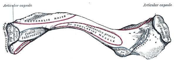
Bony Features and Attachments of the Clavicle. This inferior view of the clavicle shows the bone's structural landmarks from the sternal end to the acromial end. The image depicts the articular capsule, costal tuberosity, subclavian groove, articulating cartilage of the 1st rib, articulation with the sternum, deltoid attachment, conoid tuberosity, acromion of the scapula, and trapezoid ligament.
Henry Vandyke Carter, Public Domain, via Wikimedia Commons
References
Javed O, Maldonado KA, Ashmyan R. Anatomy, Shoulder and Upper Limb, Muscles. StatPearls. 2026 Jan:(): [PubMed PMID: 29494017]
Tubbs RS, Loukas M, Slappey JB, McEvoy WC, Linganna S, Shoja MM, Oakes WJ. Surgical and clinical anatomy of the interclavicular ligament. Surgical and radiologic anatomy : SRA. 2007 Jul:29(5):357-60 [PubMed PMID: 17563831]
La Rose J, Tiwari V. Anatomy, Shoulder and Upper Limb, Acromioclavicular Joint. StatPearls. 2026 Jan:(): [PubMed PMID: 29763033]
Tubbs RS, Shah NA, Sullivan BP, Marchase ND, Cömert A, Acar HI, Tekdemir I, Loukas M, Shoja MM. The costoclavicular ligament revisited: a functional and anatomical study. Romanian journal of morphology and embryology = Revue roumaine de morphologie et embryologie. 2009:50(3):475-9 [PubMed PMID: 19690777]
Hsu JE, Hulet DA, McDonald C, Whitson A, Russ SM, Matsen FA 3rd. The contribution of the scapula to active shoulder motion and self-assessed function in three hundred and fifty two patients prior to elective shoulder surgery. International orthopaedics. 2018 Nov:42(11):2645-2651. doi: 10.1007/s00264-018-4027-3. Epub 2018 Jul 9 [PubMed PMID: 29987556]
Okwumabua E, Thompson JH. Anatomy, Shoulder and Upper Limb, Axillary Nerve. StatPearls. 2026 Jan:(): [PubMed PMID: 29630264]
Ogata S, Uhthoff HK. The early development and ossification of the human clavicle--an embryologic study. Acta orthopaedica Scandinavica. 1990 Aug:61(4):330-4 [PubMed PMID: 2402985]
Vieth V, Schulz R, Brinkmeier P, Dvorak J, Schmeling A. Age estimation in U-20 football players using 3.0 tesla MRI of the clavicle. Forensic science international. 2014 Aug:241():118-22. doi: 10.1016/j.forsciint.2014.05.008. Epub 2014 May 21 [PubMed PMID: 24908196]
Schmidt S, Ottow C, Pfeiffer H, Heindel W, Vieth V, Schmeling A, Schulz R. Magnetic resonance imaging-based evaluation of ossification of the medial clavicular epiphysis in forensic age assessment. International journal of legal medicine. 2017 Nov:131(6):1665-1673. doi: 10.1007/s00414-017-1676-5. Epub 2017 Sep 9 [PubMed PMID: 28889331]
Knudsen FW, Andersen M, Krag C. The arterial supply of the clavicle. Surgical and radiologic anatomy : SRA. 1989:11(3):211-4 [PubMed PMID: 2588097]
Tran DQ, Tiyaprasertkul W, González AP. Analgesia for clavicular fracture and surgery: a call for evidence. Regional anesthesia and pain medicine. 2013 Nov-Dec:38(6):539-43. doi: 10.1097/AAP.0000000000000012. Epub [PubMed PMID: 24121609]
Natsis K, Totlis T, Chorti A, Karanassos M, Didagelos M, Lazaridis N. Tunnels and grooves for supraclavicular nerves within the clavicle: review of the literature and clinical impact. Surgical and radiologic anatomy : SRA. 2016 Aug:38(6):687-91. doi: 10.1007/s00276-015-1602-9. Epub 2015 Dec 24 [PubMed PMID: 26702936]
Bordoni B, Varacallo MA. Neuroanatomy, Cranial Nerve 11 (Accessory). StatPearls. 2026 Jan:(): [PubMed PMID: 29939544]
Bernat A, Huysmans T, Van Glabbeek F, Sijbers J, Gielen J, Van Tongel A. The anatomy of the clavicle: a three-dimensional cadaveric study. Clinical anatomy (New York, N.Y.). 2014 Jul:27(5):712-23. doi: 10.1002/ca.22288. Epub 2013 Oct 21 [PubMed PMID: 24142486]
Level 2 (mid-level) evidenceModgil R, Arora KS, Sharma A, Mohapatra S, Pareek S. Cleidocranial Dysplasia: Presentation of Clinical and Radiological Features of a Rare Syndromic Entity. Mymensingh medical journal : MMJ. 2018 Apr:27(2):424-428 [PubMed PMID: 29769514]
van der Meijden OA, Gaskill TR, Millett PJ. Treatment of clavicle fractures: current concepts review. Journal of shoulder and elbow surgery. 2012 Mar:21(3):423-9. doi: 10.1016/j.jse.2011.08.053. Epub 2011 Nov 6 [PubMed PMID: 22063756]
Vlček M, Niedoba M, Jakubička J, Pech J, Kalvach J. [Surgical treatment of midshaft clavicular fractures using intramedullary nail]. Rozhledy v chirurgii : mesicnik Ceskoslovenske chirurgicke spolecnosti. 2018 Spring:97(4):176-188 [PubMed PMID: 29726264]
Hoogervorst P, van Schie P, van den Bekerom MP. Midshaft clavicle fractures: Current concepts. EFORT open reviews. 2018 Jun:3(6):374-380. doi: 10.1302/2058-5241.3.170033. Epub 2018 Jun 20 [PubMed PMID: 30034818]
Naylor CD, McCormack DG, Sullivan SN. The midclavicular line: a wandering landmark. CMAJ : Canadian Medical Association journal = journal de l'Association medicale canadienne. 1987 Jan 1:136(1):48-50 [PubMed PMID: 2947672]
Le MQT, Myers K, May T, Hosseinzadeh S. Clavicle Fracture. StatPearls. 2026 Jan:(): [PubMed PMID: 29939669]
Mazzocca AD, Arciero RA, Bicos J. Evaluation and treatment of acromioclavicular joint injuries. The American journal of sports medicine. 2007 Feb:35(2):316-29 [PubMed PMID: 17251175]
Eschler A, Rösler K, Rotter R, Gradl G, Mittlmeier T, Gierer P. Acromioclavicular joint dislocations: radiological correlation between Rockwood classification system and injury patterns in human cadaver species. Archives of orthopaedic and trauma surgery. 2014 Sep:134(9):1193-8. doi: 10.1007/s00402-014-2045-1. Epub 2014 Jul 4 [PubMed PMID: 24993589]
Liu JN, Garcia GH, Weeks KD, Joseph J, Limpisvasti O, McFarland EG, Dines JS. Treatment of Grade III Acromioclavicular Separations in Professional Baseball Pitchers: A Survey of Major League Baseball Team Physicians. American journal of orthopedics (Belle Mead, N.J.). 2018 Jul:47(7):. doi: 10.12788/ajo.2018.0051. Epub [PubMed PMID: 30075044]
Level 3 (low-level) evidenceMcFarland EG, Blivin SJ, Doehring CB, Curl LA, Silberstein C. Treatment of grade III acromioclavicular separations in professional throwing athletes: results of a survey. American journal of orthopedics (Belle Mead, N.J.). 1997 Nov:26(11):771-4 [PubMed PMID: 9402211]
Level 3 (low-level) evidencePowers JA, Bach PJ. Acromioclavicular separations. Closed or open treatment? Clinical orthopaedics and related research. 1974 Oct:(104):213-23 [PubMed PMID: 4411824]
Stucken C, Cohen SB. Management of acromioclavicular joint injuries. The Orthopedic clinics of North America. 2015 Jan:46(1):57-66. doi: 10.1016/j.ocl.2014.09.003. Epub 2014 Oct 11 [PubMed PMID: 25435035]
Menge TJ, Boykin RE, Bushnell BD, Byram IR. Acromioclavicular osteoarthritis: a common cause of shoulder pain. Southern medical journal. 2014 May:107(5):324-9. doi: 10.1097/SMJ.0000000000000101. Epub [PubMed PMID: 24937735]
Kiel J, Ponnarasu S, Kaiser K. Sternoclavicular Joint Injury. StatPearls. 2026 Jan:(): [PubMed PMID: 29939671]