 Tools
Tools
 Anatomy, Smooth Muscle
Introduction
Smooth muscle is found throughout the body, where it serves a variety of functions. Smooth muscle in the stomach and intestines helps with digestion and nutrient absorption. Smooth muscle throughout the urinary system helps eliminate toxins and maintain electrolyte balance. Smooth muscle throughout the arteries and veins plays a vital role in regulating blood pressure and tissue oxygenation. Without these vital functions, the body would not be able to maintain basic homeostasis.
Smooth muscle differs from skeletal muscle in a variety of ways, perhaps most importantly in its ability to contract and be controlled involuntarily. The nervous system can use smooth muscle to tightly regulate many of the body's subsystems without conscious control. A person does not need to think about blood pressure for it to adapt to the increased oxygen demands of exercise. Instead, the nervous system uses hormones, neurotransmitters, and other receptors to control smooth muscle activity.
Smooth muscle also plays an important role in the disease process throughout the body. The use of bronchodilators to relax airway smooth muscle is an important and life-saving treatment in patients with asthma.[1] Likewise, medications such as metoclopramide can stimulate and promote gastric emptying by increasing smooth muscle signaling. Perhaps one of the most well-known therapeutic applications involving smooth muscle is the use of nitrates in the treatment of ischemic heart disease,[2] where nitrates, in combination with angiotensin-converting enzyme inhibitors, can reduce mortality.[3] The substantial impact of smooth muscle throughout the body makes this tissue an important topic for clinicians to understand, as many treatments rely on modifying the signaling pathways that regulate it.
Structure and Function
Register For Free And Read The Full Article
 Search engine and full access to all medical articles
- 10 free questions in your specialty
- Free CME/CE Activities
- Free daily question in your email
- Save favorite articles to your dashboard
- Emails offering discounts
Learn more about a Subscription to StatPearls Point-of-Care
Structure and Function
Smooth muscle differs from skeletal muscle in function. Unlike skeletal muscle, smooth muscle can maintain tone for extended periods and often contracts involuntarily. At the cellular level, smooth muscle is an involuntary, nonstriated muscle. Smooth muscle consists of thick and thin filaments that are not arranged into sarcomeres, giving it a nonstriated pattern. On microscopic examination, smooth muscle appears homogeneous. Smooth muscle cytoplasm contains a large amount of actin and myosin. Actin and myosin are the main proteins involved in muscle contraction. Actin filaments attach to dense bodies spread throughout the cell. Dense bodies can be observed under an electron microscope and appear dark. Another important structure is the calcium-containing sarcoplasmic reticulum, which helps maintain contraction. The shape of smooth muscle is described as fusiform, meaning round in the center and tapered at each end. Smooth muscle can contract and relax but has greater elasticity than striated muscle. This elasticity is important in organs such as the urinary bladder, where contractile tone must be maintained.
Actin and myosin form continuous chains within the smooth muscle cell that are anchored at the dense bodies. The intermediate and thin filaments formed by actin and myosin chains can then stretch to dense bodies on adjacent smooth muscle cells, forming a meshlike network encircling many smooth muscle cells. Through adherens junctions or connexins, smooth muscle cells contract uniformly in what has been described as a spiral, corkscrew-like fashion.
The function of smooth muscle can be extended to a much larger scale, encompassing the organ systems that this tissue helps regulate. The functions of smooth muscle in each organ system are an incredibly broad topic and are beyond the overall scope of this article. For simplicity, the basic functions of smooth muscle in the organ systems are listed below.
- Gastrointestinal tract: Propulsion of the food bolus
- Cardiovascular: Regulation of blood flow and pressure via vascular resistance
- Renal: Regulation of urine flow
- Genital: Contractions during pregnancy, propulsion of sperm
- Respiratory tract: Regulation of bronchiole diameter
- Integument: Elevation of hair through the arrector pili muscle
- Sensory: Dilation and constriction of the pupil as well as changing lens shape
Embryology
Smooth muscle is derived from both mesoderm and neural crest cells. Smooth muscle is found in many different tissues throughout the body. One unique feature of neural crest cells is migration during embryonic development. For this reason, numerous tissues throughout the body are derived from neural crest cells. Neural crest cells play an important role in the development of smooth muscle throughout the body, particularly in regulating blood vessels.
Vascular smooth muscle cells arise from multiple origins, which is clinically significant because this may contribute to the site-specific localization of vascular diseases. For example, atherosclerosis and aortic aneurysms often present at specific vascular locations. Historically, clinicians attributed this localization to hemodynamics and underlying vessel structure. However, increasing evidence suggests that smooth muscle cell embryonic lineage may play a role in determining the location and presentation of vascular disease.[4] Smooth muscle cell development is also an important factor in the development of the endothelial network. Vascular smooth muscle cells, sometimes called mural cells, are important for vascular development and stability. Mural cells wrap around larger vessels and are heavily relied upon in the regulation of blood flow, endothelial network growth, and vessel stability. However, little is known about the effects of their developmental origins or the signaling processes that underlie vessel development. The development of vascular smooth muscle cells is an important target for vascular tissue engineering and therapeutic revascularization.[5]
Blood Supply and Lymphatics
Because smooth muscle’s widespread throughout the body, blood supply and lymphatic drainage vary by region. Almost every artery in the body supplies blood to smooth muscle, whether in the arterial wall or in organs, such as the gastrointestinal tract. Recognizing how smooth muscle affects blood supply is more clinically important. For example, within the cardiovascular system, smooth muscle helps regulate blood flow by controlling vessel diameter. As previously discussed, vascular pathologies of smooth muscle can have devastating effects on the body and lead to significant pathology. Atherosclerosis, once thought to be only a function of hemodynamics and vessel structure, has more recently been linked to smooth muscle development.[4] Results from studies have shown that continuous activation of vascular smooth muscle can lead to pulmonary hypertension.[6] Within the lungs, pathologic activation of smooth muscle can lead to asthma. Asthma occurs when smooth muscle constriction leads to airway obstruction. Results from recent studies have shown that the smooth muscle layer may thicken before the onset of asthma, suggesting a possible genetic association.[7]
Nerves
Similar to the blood supply, the innervation of smooth muscle varies widely by location and function. Vascular smooth muscle is primarily innervated by the sympathetic nervous system. α1 and α2 receptors cause vasoconstriction by contracting vascular smooth muscle cells, leading to systemic hypertension. β2 receptors also respond to sympathetic stimulation but produce a vasodilatory effect, leading to systemic hypotension. However, parasympathetic stimulation also plays an important role in the contraction of smooth muscle cells. Results from studies performed as early as 1925 demonstrated the effect of parasympathetic innervation on the gastrointestinal tract.[8] More recently, study results have shown that the sympathetic, parasympathetic, and enteric nervous systems work together to regulate smooth muscle contraction.[9] Sympathetic stimulation of smooth muscle is received by contributions from spinal levels T1 to L2. Each of these contributions enters the sympathetic trunk, which routes autonomic nervous supply to organs and tissues throughout the body. The parasympathetic nervous system functions through 3 parts: the cranial nerves, the vagus nerve, and the pelvic splanchnic nerves. Each nerve in the parasympathetic system regulates a specific portion of the body. For instance, the vagus nerve innervates the gastrointestinal tract from the esophagus to the proximal portion of the large intestine, and also sends branches to the heart, larynx, trachea, bronchi, liver, and pancreas. The sympathetic and parasympathetic nervous systems are collectively referred to as the autonomic nervous system. The complex nature of the autonomic nervous system enables tight, unconscious control of digestion, respiratory rate, urination, heart rate, blood pressure, and many other critical body functions.
Ultimately, innervation by the autonomic nervous system leads to calcium release in smooth muscle. Smooth muscle contraction is dependent on calcium influx. Calcium is increased within the smooth muscle cell through 2 different processes. First, depolarization, hormones, or neurotransmitters cause calcium to enter the cell through L-type calcium channels located in the caveolae of the membrane. Intracellular calcium then stimulates the release of calcium from the sarcoplasmic reticulum via ryanodine receptors and inositol 1,4,5-trisphosphate; this process is referred to as calcium-induced calcium release.[10] Unlike skeletal muscle, smooth muscle calcium release from the sarcoplasmic reticulum is not physically coupled to the ryanodine receptor. Once calcium has entered the cell, it is free to bind calmodulin, which then becomes activated. Calmodulin then activates the enzyme myosin light chain kinase. Myosin light chain kinase then phosphorylates a regulatory light chain on myosin. Once phosphorylation has occurred, a conformational change takes place in the myosin head. This change increases myosin ATPase activity, which promotes interaction between the myosin head and actin. Cross-bridge cycling then occurs, generating tension. The tension generated is relative to the calcium concentration within the cell. ATPase activity is much lower in smooth muscle than in skeletal muscle. Lower ATPase activity leads to the much slower cycling speed of smooth muscle. However, a longer contraction period can lead to a greater force of contraction in smooth muscle. Smooth muscle contraction is further enhanced by connexins. Connexins enable intercellular communication by allowing calcium and other molecules to flow between neighboring smooth muscle cells. This intercellular communication enables rapid signaling between cells and a smooth contraction pattern.
Steps involved in smooth muscle cell contraction:
- Depolarization of membrane or hormone/neurotransmitter activation
- Opening L-type voltage-gated calcium channels
- Calcium-induced calcium release from the sarcoplasmic reticulum
- Increased intracellular calcium
- Calmodulin binds calcium
- Myosin light chain kinase activation
- Phosphorylation of myosin light chain
- Increased myosin ATPase activity
- Myosin-P binds actin
- Cross-bridge cycling leads to muscle tone
Dephosphorylation of myosin light chains terminates smooth muscle contraction. Unlike skeletal muscle, smooth muscle is phosphorylated during its activation. This process creates a potential difficulty because simply reducing calcium levels does not produce muscle relaxation. Myosin light chain phosphatase is responsible for dephosphorylation of the myosin light chains, ultimately leading to smooth muscle relaxation.
Muscles
Smooth muscle can be found in all the organ systems below:
- Gastrointestinal tract
- Cardiovascular: Blood vessels and lymphatic vessels
- Renal: Urinary bladder
- Genital: Male and female reproductive tracts
- Respiratory tract
- Integument: Erector pili of the skin
- Sensory: Ciliary muscle and iris of the eye
Physiologic Variants
Smooth muscle consists of 2 types: single-unit and multiunit. Single-unit smooth muscle consists of multiple cells connected through connexins that can be stimulated in a synchronous pattern from only 1 synaptic input. Connexins enable cell-to-cell communication among single-unit smooth muscle cells. Intercellular communication allows ions and molecules to diffuse between cells, giving rise to calcium waves. This property allows synchronous contraction.[11] Multiunit smooth muscle differs from single-unit smooth muscle in that each smooth muscle cell receives its own synaptic input. This arrangement allows multiunit smooth muscle to be controlled much more finely. Multiunit smooth muscle is found in the airways of the lungs, large arteries, and ciliary muscles of the eye.
Surgical Considerations
Monitoring a patient's vital signs during a surgical procedure is paramount to procedural success, and the stressors of surgery can substantially impact the autonomic nervous system, which regulates smooth muscle contraction. Surgical procedures can even be targeted at modifying the function of smooth muscle, such as vagotomy. Overstimulation of the vagus nerve has been speculated to be a possible cause of peptic ulcer disease, and vagotomy is a classic surgical procedure that aims to treat this disorder by transecting the vagus nerve at the level of the stomach and thereby reducing stimulation. However, this procedure has recently fallen out of favor because of advancements in medical therapy for peptic ulcer disease but may still show some benefit in certain patients.[12] Another example is the treatment of certain neuroendocrine tumors, such as adrenal pheochromocytoma, which can cause cardiovascular complications during a surgical procedure by releasing excess catecholamines. Proper treatment requires in-depth knowledge of how α- and β-blockade affect smooth muscle and the downstream effects of these changes on bodily functions.[13] Because of smooth muscle’s regulatory effects, a sufficient understanding of smooth muscle contraction and its impact on body systems is paramount when preparing for and performing any surgical procedure.
Clinical Significance
Estimated health care costs associated with asthma reached $81.9 billion in the US in 2013.[14] This substantial health care burden reflects, in part, the clinical impact of smooth muscle contraction. Smooth muscle is an integral part of the human body; its function is essential for life, and it can be found in almost every organ system. In the cardiovascular system, smooth muscle in blood vessels helps maintain blood pressure and flow. In the lungs, smooth muscle opens and closes airways. In the gastrointestinal tract, smooth muscle plays a role in motility and nutrient absorption, and it also serves a purpose in almost every other organ system. The wide distribution of smooth muscle throughout the body and its many unique properties make an in-depth understanding of its anatomy, physiology, function, and disease applications imperative for clinicians.
From a functional perspective, smooth muscle physiology is responsible for maintaining and preserving every vital sign. Regardless of whether a patient presents with an acute emergency or a chronic disease, smooth muscle likely plays some role in disease development. In an acute setting, many potentially life-saving therapies directly target smooth muscle. In these settings, a firm foundation in smooth muscle physiology can help healthcare professionals save lives. An even broader understanding of smooth muscle can help clinicians improve their patients' quality of life. As part of the biopsychosocial model, clinicians should also consider psychosocial factors that may be overlooked in smooth muscle diseases. For example, a patient diagnosed with neurogenic bladder disease may become socially isolated to avoid embarrassment associated with the condition. When approaching smooth muscle dysfunction, healthcare professionals should appreciate the many facets of how the disease may affect their patients.
As with all aspects of medicine, ongoing research will likely change our understanding of smooth muscle and its effects on disease. Current research into smooth muscle has shown promise for future implications, such as restoring endothelial tissue, which could lead to new ways to encourage revascularization. Even small changes in understanding could have a substantial impact on the treatment and mortality of cardiovascular disease in the future.[4] While smooth muscle remains an exceptionally complex topic, a solid understanding of its impact on health care, even at the most basic level, gives healthcare professionals tools to improve health care outcomes now and into the future.
Other Issues
Smooth muscle anatomy, physiology, and function remain broad and incompletely understood topics despite substantial funding and research efforts directed toward understanding them. As more time and effort are devoted to understanding smooth muscle, clinicians’ ability to treat the pathophysiology associated with its dysfunction will broaden. Clinicians should continue to learn and study the impact of smooth muscle. As discussed, future methods may involve stimulating tissue regrowth using smooth muscle–modulating factors. Future advances in medical treatment may focus on modifying smooth muscle function.
Media
(Click Image to Enlarge)
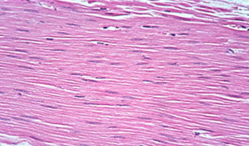
Smooth Muscle.
Contributed by William Gossman collection
(Click Image to Enlarge)
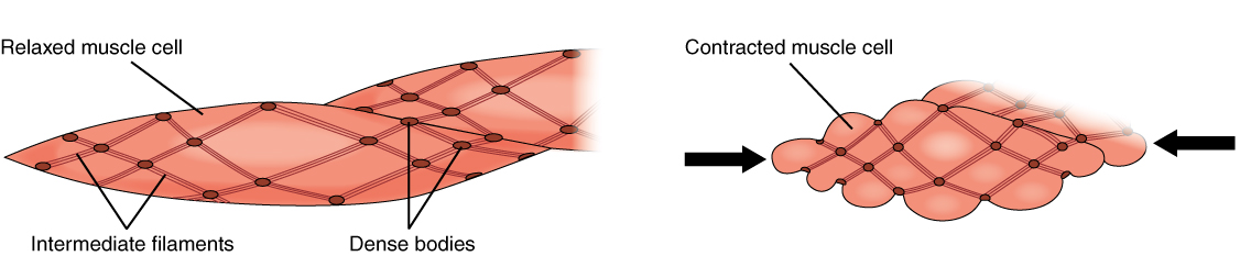
Smooth Muscle Contraction By OpenStax - https://cnx.org/contents/FPtK1zmh@8.25:fEI3C8Ot@10/Preface, CC BY 4.0, https://commons.wikimedia.org/w/index.php?curid=30015054
References
Williams DM, Rubin BK. Clinical Pharmacology of Bronchodilator Medications. Respiratory care. 2018 Jun:63(6):641-654. doi: 10.4187/respcare.06051. Epub [PubMed PMID: 29794201]
Giuseppe C, Paul J, Hans-Ulrich I. Use of nitrates in ischemic heart disease. Expert opinion on pharmacotherapy. 2015:16(11):1567-72. doi: 10.1517/14656566.2015.1052742. Epub 2015 Jun 1 [PubMed PMID: 26027641]
Level 3 (low-level) evidence. GISSI-3: effects of lisinopril and transdermal glyceryl trinitrate singly and together on 6-week mortality and ventricular function after acute myocardial infarction. Gruppo Italiano per lo Studio della Sopravvivenza nell'infarto Miocardico. Lancet (London, England). 1994 May 7:343(8906):1115-22 [PubMed PMID: 7910229]
Level 1 (high-level) evidenceSinha S, Iyer D, Granata A. Embryonic origins of human vascular smooth muscle cells: implications for in vitro modeling and clinical application. Cellular and molecular life sciences : CMLS. 2014 Jun:71(12):2271-88. doi: 10.1007/s00018-013-1554-3. Epub 2014 Jan 18 [PubMed PMID: 24442477]
Level 3 (low-level) evidenceBargehr J, Low L, Cheung C, Bernard WG, Iyer D, Bennett MR, Gambardella L, Sinha S. Embryological Origin of Human Smooth Muscle Cells Influences Their Ability to Support Endothelial Network Formation. Stem cells translational medicine. 2016 Jul:5(7):946-59. doi: 10.5966/sctm.2015-0282. Epub 2016 May 18 [PubMed PMID: 27194743]
Khalil RA. Regulation of Vascular Smooth Muscle Function. 2010:(): [PubMed PMID: 21634065]
James AL, Noble PB, Drew SA, Mauad T, Bai TR, Abramson MJ, McKay KO, Green FHY, Elliot JG. Airway smooth muscle proliferation and inflammation in asthma. Journal of applied physiology (Bethesda, Md. : 1985). 2018 Oct 1:125(4):1090-1096. doi: 10.1152/japplphysiol.00342.2018. Epub 2018 Jul 19 [PubMed PMID: 30024335]
Veach HO. Studies on the innervation of smooth muscle: III. Splanchnic Effects on the Lower End of the Oesophagus and Stomach of the Cat. The Journal of physiology. 1925 Oct 31:60(5-6):457-78 [PubMed PMID: 16993768]
Lazebnik LB, Lychkova AE. Interaction between different parts of the autonomic nervous system in the regulation of smooth muscles in the femoral artery and trachea. Bulletin of experimental biology and medicine. 2006 Jan:141(1):5-8 [PubMed PMID: 16929950]
Level 3 (low-level) evidenceJackson WF, Boerman EM. Voltage-gated Ca(2+) channel activity modulates smooth muscle cell calcium waves in hamster cremaster arterioles. American journal of physiology. Heart and circulatory physiology. 2018 Oct 1:315(4):H871-H878. doi: 10.1152/ajpheart.00292.2018. Epub 2018 Jun 29 [PubMed PMID: 29957015]
Pogoda K, Kameritsch P, Mannell H, Pohl U. Connexins in the control of vasomotor function. Acta physiologica (Oxford, England). 2019 Jan:225(1):e13108. doi: 10.1111/apha.13108. Epub 2018 Jul 22 [PubMed PMID: 29858558]
Lagoo J, Pappas TN, Perez A. A relic or still relevant: the narrowing role for vagotomy in the treatment of peptic ulcer disease. American journal of surgery. 2014 Jan:207(1):120-6. doi: 10.1016/j.amjsurg.2013.02.012. Epub 2013 Oct 16 [PubMed PMID: 24139666]
Ramachandran R, Rewari V. Current perioperative management of pheochromocytomas. Indian journal of urology : IJU : journal of the Urological Society of India. 2017 Jan-Mar:33(1):19-25. doi: 10.4103/0970-1591.194781. Epub [PubMed PMID: 28197025]
Nurmagambetov T, Kuwahara R, Garbe P. The Economic Burden of Asthma in the United States, 2008-2013. Annals of the American Thoracic Society. 2018 Mar:15(3):348-356. doi: 10.1513/AnnalsATS.201703-259OC. Epub [PubMed PMID: 29323930]