 Tools
Tools
 Anatomy, Bony Pelvis and Lower Limb, Hip
Introduction
The articulation of the femoral head with the acetabulum forms the hip joint. This articulation connects the axial skeleton to the lower extremities and transmits forces encountered during daily activities from the axial skeleton to the lower extremities. The hip joint’s ability to balance forces throughout its full range of motion provides the stability required to perform everyday tasks such as standing upright, maintaining a smooth, balanced gait, rising from a chair, and lifting a weight from a squatting position. [1]
The hip joint is a spheroidal, ball-and-socket synovial joint stabilized by bony and ligamentous restraints. The osseous anatomy of the femoroacetabular articulation contributes to the hip’s inherent stability. The pelvis is composed of 3 parts: the ilium, ischium, and pubis. These innominate bones come together at the triradiate cartilage to form the cup-shaped socket known as the acetabulum. At 15 to 17 years of age, the triradiate cartilage begins to ossify, and the cartilage completely fuses by 20 to 25 years of age. Acetabular development is a complex process that involves both endochondral growth from the triradiate cartilage and intramembranous growth from primary and secondary ossification centers of the innominate bones.[2] The acetabulum covers approximately 40% of the femoral head throughout hip motion. Additional stability is conferred by the labrum, a rim composed of circumferential collagen fibers that surrounds the acetabulum, deepens the hip socket, and limits extreme motion. The acetabular labrum contributes approximately 22% of the articulating surface of the hip and increases the volume of the acetabulum by 33%.[1]
The hip joint capsule is formed by 3 major ligaments: the iliofemoral, pubofemoral, and ischiofemoral ligaments. The capsular ligaments run in a spiral fashion, limiting hip extension, and are surrounded by thick longitudinal fibers that provide additional stability in the lateral plane. The capsule is thicker anterosuperiorly, where the predominant weight-bearing stresses occur, and thinner posteroinferiorly.[1] The ligamentum teres fans out from the fovea of the femoral head and attaches to nearly the full length of the acetabular ligament.
Structure and Function
Register For Free And Read The Full Article
 Search engine and full access to all medical articles
- 10 free questions in your specialty
- Free CME/CE Activities
- Free daily question in your email
- Save favorite articles to your dashboard
- Emails offering discounts
Learn more about a Subscription to StatPearls Point-of-Care
Structure and Function
The hip acts as a multiaxial ball-and-socket joint that supports the upper body during stance and gait. The balance and stability provided by the hip joint allow motion while supporting forces encountered during daily activities. The congruity of the femoral head with the acetabulum allows the rotational motion required to perform these tasks without any detectable translational motion, which would destabilize the joint and increase the risk of dislocation. The inherent stability provided by the osseous anatomy of the joint, combined with the stabilizing forces of the fibrous capsule and neuromuscular anatomy, defines the absolute limits of motion of the hip joint before bony impingement occurs.
- Flexion: 120°
- Extension: 10°
- Abduction: 45°
- Adduction:25°
- Internal rotation:15°
- External rotation: 35°
Ischiofemoral ligament: The ischiofemoral ligament attaches to the posterior surface of the acetabular rim and labrum and courses circumferentially around the joint to its insertion on the anterior aspect of the femur. The ischiofemoral ligament limits internal rotation and hip adduction with flexion.
Iliofemoral ligament (Y Ligament of Bigelow): The iliofemoral ligament is triangular and attaches along the intertrochanteric line of the femur and converges into its attachment on the anterior inferior iliac spine. The iliofemoral ligament is the strongest ligament in the body. The iliofemoral ligament limits extension and external rotation of the hip and helps maintain a static erect posture with minimal muscular activity. Biomechanical analysis has concluded that the iliofemoral ligament is the strongest of the 3-ligament complex.[1] Consequently, the hip can withstand the greatest force in anterior translation before failure. These data support the greater incidence of posterior hip dislocations (90%) relative to anterior dislocations.[3][4]
Pubofemoral ligament: Located on the anterior aspect of the hip joint, this ligament extends from the anterior portion of the pubic ramus to the anterior surface of the intertrochanteric fossa, often blending with the inferior fibers of the iliofemoral ligament. The pubofemoral ligament limits hip abduction and extension.
Zona orbicularis (annular ligament): Not visible externally, it encircles the femoral neck like a buttonhole and acts as a biomechanical locking ring wrapped around the femoral neck. The zona orbicularis forms a locking ring around the femur which resists distraction forces on the hip.
Ligamentum teres: Located deep in the hip, it has a pyramidal shape with a broad origin from nearly the entire transverse acetabular ligament attaching to the ischial and pubic bases by two bundles, with the posterior bundle being stronger than the anterior bundle. The ligamentum teres’ function prior to puberty has been well-described, as it provides a secondary blood supply to the head of the femur. However, its function in adulthood is a subject of debate. In a recent meta-analysis of several cadaveric studies, O’Donnell et al concluded that the ligamentum teres acts as a secondary stabilizer to supplement the work of the capsular ligaments.[5]
Acetabular labrum: The acetabular labrum is a fibrocartilaginous rim, composed of circumferential collagen fibers, that spans the entirety of the acetabulum and is continuous with the transverse acetabular ligament. The labrum contributes approximately 22% of the articulating surface of the hip and increases the volume of the acetabulum by 33%. The acetabular labrum limits the extreme range of motion and deepens the acetabulum, helping dissipate large forces across the hip during athletic strides. Additionally, the acetabular labrum provides a sealing rim around the joint, increasing hydrostatic pressure to facilitate synovial lubrication and resist joint distraction.[1]
A thick layer of highly organized type 2 collagen fibers and hydrophilic glycosaminoglycans makes up the hyaline cartilage that covers the articular surfaces of the joint. The hyaline cartilage acts synergistically with the subchondral bone to absorb shock and appropriately distribute the high forces generated across the joint. The hydrophilic glycosaminoglycans trap water in the substance of the cartilage, resulting in additional stress-shielding properties. The cartilage is thickest at the ventrocranial surface of the acetabulum and ventrolateral surface of the femoral head, with cartilage density decreasing concentrically from these points.[1] The thickest regions coincide with the areas that receive the greatest amount of force when the joint is loaded.
Embryology
Primary ossification of the ischium, ilium, pubis, and femoral shaft occurs in utero. Secondary ossification of the femur occurs at 2 ossification centers:
1. The proximal femoral epiphysis appears at age 4 to 8 months.2. The trochanter appears at age 4 years.
The proximal femoral epiphyses fuse by 18 years of age, and the trochanteric apophyses fuse between 16 and 18 years of age. The proximal femoral epiphyses contribute significantly to metaphyseal growth of the femoral neck, whereas the trochanteric apophyses contribute largely to appositional growth of the greater trochanter.
Blood Supply and Lymphatics
From age 0 to 4 years, the femoral head receives significant blood supply from the medial femoral circumflex artery (MFCA), the lateral femoral circumflex artery (LFCA), and the artery of the ligamentum teres. From age 4 to 8 years, the MFCA provides the majority of the blood supply, with supplementary contributions from the LFCA and artery of the ligamentum teres. After 8 years of age, the MFCA predominates, with negligible contribution from the LFCA and artery of the ligamentum teres.[6]
The MFCA most commonly originates from the profunda femoris. However, results from cadaveric studies showed that the MFCA may also originate from the common femoral artery.[6] The MFCA has 5 consistent branches: superficial, ascending, acetabular, descending, and deep. The deep branch runs toward the intertrochanteric crest between the pectineus medially and the iliopsoas tendon laterally, along the inferior border of the obturator externus. Posteriorly, the deep branch runs deep to the quadratus femoris and can be identified in the space between the quadratus femoris and the inferior gemellus. Identification of these posterior landmarks is critical because the vessel is at risk of iatrogenic injury during hip arthroplasty via a posterior approach. The main division of the deep branch crosses posterior to the tendon of the obturator externus and anterior to the tendons of the superior gemellus, obturator internus, and inferior gemellus. The main division perforates the capsule just cranial to the superior gemellus tendon and distal to the piriformis tendon and divides into 2 to 4 terminal branches. These branches maintain a posterosuperior course along the neck of the femur before perforating the femoral head approximately 2 to 4 mm from the cartilage-bone junction. Posteroinferior terminal branches may also contribute to the blood supply. The exposed nature of the subsynovial branches of the MFCA increases the risk of avascular necrosis of the femoral head in patients with displaced femoral neck fractures.
The consistency of contributions from the inferior gluteal artery to femoral head blood supply is a subject of debate. In a recent cadaveric study, Grose et al examined the contribution of the inferior gluteal artery to the extracapsular anastomosis supplying the femoral head and demonstrated that the deep branch of the MFCA receives a significant and consistent inflow from vessels derived from the inferior gluteal artery.[7] Findings from this study support the belief that the inferior gluteal artery is capable of providing a compensatory blood supply after an injury to the deep branch of the MFCA.
The acetabular branch of the obturator artery provides the blood supply to the acetabular fossa while coursing through the acetabular notch. The pelvic surface of the acetabulum receives blood supply from pubic branches of the obturator artery. The superior and posteroinferior regions of the acetabulum are supplied by an anastomosis of deep branches of the superior and inferior gluteal arteries. Lymphatic drainage from the anterior aspect of the hip drains to the deep inguinal nodes, while drainage from the medial and posterior aspects passes into the internal iliac nodes.
Nerves
Obturator nerve: The obturator nerve originates from nerve roots L2 through L4 and exits through the obturator canal before splitting into an anterior division that runs anterior to the obturator externus and a posterior division that runs posterior to the obturator externus. The obturator nerve supplies sensory innervation to the inferomedial thigh via the cutaneous branch of the obturator nerve and motor innervation to the gracilis (anterior division), adductor longus (anterior division), adductor brevis (anterior and posterior divisions), and adductor magnus (posterior division). Iatrogenic injury can occur from retractors placed behind the transverse acetabular ligament when using a medial approach to the hip, an approach commonly used in pediatric patients.
Genitofemoral nerve: The genitofemoral nerve originates from nerve roots L1 through L2. The nerve pierces the psoas muscle and continues down the anteromedial surface of the psoas before dividing into femoral and genital branches. The femoral branch provides sensory innervation to the proximal anterior thigh over the femoral triangle. The genital branch provides sensory innervation to the scrotum and labia. The genitofemoral nerve has no motor function.
Lateral femoral cutaneous nerve: The lateral femoral cutaneous nerve originates from nerve roots L2 through L3. The nerve crosses inferior to the anterior superior iliac spine and provides sensory innervation to the lateral thigh. The lateral femoral cutaneous nerve has no motor function. Iatrogenic injury can occur during hip arthroplasty using an anterior approach or when placing the anterior portal for hip arthroscopy.
Femoral nerve: The femoral nerve originates from nerve roots L2 through L4. The femoral nerve lies between the psoas major and iliacus and branches in the femoral triangle. The femoral nerve provides sensory innervation to the anteromedial thigh via anterior cutaneous branches and motor innervation to the psoas, pectineus, sartorius, and quadriceps (rectus femoris, vastus lateralis, vastus intermedius, and vastus medialis).
Sciatic nerve: The sciatic nerve originates from the sacral plexus and projects through the greater sciatic foramen, descending down the posterior thigh deep to the hamstrings and superficial to the adductor magnus. The sciatic nerve has 2 distinct divisions: the tibial and common peroneal divisions. The tibial division originates from nerve roots L4 through S3 and provides motor innervation to the biceps femoris (long head), semitendinosus, and semimembranosus. The tibial division provides no sensory innervation in the thigh. The common peroneal division originates from nerve roots L4 through L2 and provides motor innervation to the biceps femoris (short head). Anatomical variations have been noted in the relationship between nerve divisions and the piriformis muscle. In most people, both divisions of the sciatic nerve pass beneath the piriformis. Two distinct anatomical variations have been described: the common fibular division passes through the piriformis or over the piriformis. The sciatic nerve is at risk for iatrogenic injury during hip arthroplasty using a posterior approach. Injury can be avoided by reflecting the piriformis.
Posterior femoral cutaneous nerve: The posterior femoral cutaneous nerve originates from nerve roots S1 through S3 and passes through the greater sciatic foramen medial to the sciatic nerve. The posterior femoral cutaneous nerve provides sensory innervation to the posterior thigh and has no motor function.
Muscles
Smooth gait is achieved through a series of concentric and eccentric muscular contractions, both voluntary and involuntary. A complex neuromuscular loop receives proprioceptive feedback from both the position of the body and intrinsic muscular properties, such as muscle spindle fiber and sarcomere length, and maintains the proper position of the femoral head within the acetabulum. Understanding the synchronization of muscle contractions that facilitate balanced gait is critical when evaluating pathology of the hip’s articular surface.
Hip Flexors
Psoas major:
- Origin: T12 through L5 vertebrae
- Insertion: Lesser trochanter
- Innervation: Femoral nerve
Psoas minor (present in 50% of the population):
- Origin: T12 through L1 vertebrae
- Insertion: Iliopubic eminence
- Innervation: L1 ventral ramus
Pectineus (flexes and adducts thigh):
- Origin: Pectineal line of pubis
- Insertion: Pectineal line of femur
- Innervation: Femoral nerve
Iliacus:
- Origin: Iliac fossa and sacral ala
- Insertion: Lesser trochanter
- Innervation: Femoral nerve
Hip Extensors and External Rotators
Gluteus maximus:
- Origin: Ilium, dorsal sacrum
- Insertion: Iliotibial band, gluteal tuberosity
- Innervation: Inferior gluteal nerve
Obturator externus:
- Origin: Ischiopubic rami, obturator membrane
- Insertion: Trochanteric fossa
- Innervation: Obturator nerve
Short External Rotators
Piriformis:
- Origin: Anterior sacrum
- Insertion: Superior greater trochanter
- Innervation: Nerve to piriformis (S2, posterior division of lumbosacral plexus)
Superior gemellus:
- Origin: Ischial spine
- Insertion: Medial greater trochanter
- Innervation: Nerve to obturator internus (L5 through S2, anterior division of the lumbosacral plexus)
Obturator internus:
- Origin: Ischiopubic rami, obturator membrane
- Insertion: Medial greater trochanter
- Innervation: Nerve to obturator internus (L5 through S2, anterior division of lumbosacral plexus)
Inferior gemellus:
- Origin: Ischial tuberosity
- Insertion: Medial greater trochanter
- Innervation: Nerve to quadratus femoris (L4 through S1, anterior division of lumbosacral plexus)
Quadratus femoris:
- Origin: Ischial tuberosity
- Insertion: Intertrochanteric crest
- Innervation: Nerve to quadratus femoris (L4 through S1, anterior division of lumbosacral plexus)
Hip Abductors
Tensor fascia latae:
- Origin: Iliac crest, anterior superior iliac spine
- Insertion: Iliotibial band/proximal tibia
- Innervation: Superior gluteal nerve
Gluteus medius:
- Origin: Ilium between anterior and posterior gluteal lines
- Insertion: Greater trochanter
- Innervation: Superior gluteal nerve
Gluteus minimus:
- Origin: Ilium between anterior and posterior gluteal lines
- Insertion: Greater trochanter
- Innervation: Superior gluteal nerve
Hip Adductors
Adductor magnus:
- Origin: Pubic ramus, ischial tuberosity
- Insertion: Linea aspera, adductor tubercle
- Innervation: Obturator nerve, sciatic nerve
Adductor longus:
- Origin: Body of pubis
- Insertion: Linea aspera
- Innervation: Obturator nerve
Adductor brevis:
- Origin: Body and inferior pubic ramus
- Insertion: Pectineal line, linea aspera
- Innervation: Obturator nerve
Gracilis:
- Origin: Body and inferior pubic ramus
- Insertion: Proximal medial tibia (pes anserinus)
- Innervation: Obturator nerve
Surgical Considerations
Total hip arthroplasty (THA) is one of the most successful orthopedic procedures performed today, with an estimated 300,000 procedures performed each year in the US. THA is a surgical procedure that replaces the diseased articular surface with synthetic components to restore native hip anatomy, resulting in pain relief and improved joint kinematics. Performing THA requires a nuanced understanding of hip anatomy and biomechanics.
THA can be performed through multiple approaches, with approach selection dictated by surgeon preference, prior incisions, obesity, risk of dislocation, implant selection, and degree of deformity. Standard approaches include direct anterior (Smith-Peterson), anterolateral (Watson-Jones), direct lateral (Hardinge), posterolateral (Kocher-Langenbach), and posterior (Moore or Southern). Results from a recent study comparing the relatively new direct anterior approach with the conventional posterior approach suggested that the direct anterior approach may confer functional advantages early in recovery, although randomized trials are needed to validate these findings.[8]
Clinical Significance
Hip osteoarthritis is one of the most common causes of debilitating pain in the general population. A recent study estimates a rise in mean prevalence from 4.0% in the 1970s to 8.6% in the 2000s, with men having a higher prevalence before age 50 and women after age 50. Hip osteoarthritis is treated in a stepwise manner, using nonoperative treatment options, including weight loss, activity modification, physical therapy, assisted ambulatory devices (eg, cane, walker), nonsteroidal anti-inflammatory drugs (NSAIDs), and corticosteroid injections. The use of needle lavage, acupuncture, glucosamine and chondroitin supplementation, viscoelastic joint injections, growth factor injections, and platelet-rich plasma injections remains controversial. When nonoperative treatment modalities have been exhausted, total hip arthroplasty is the operative treatment of choice.
Media
(Click Image to Enlarge)
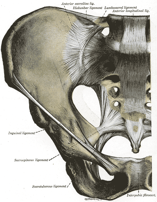
Anterior Articulations, Pelvis. The illustration depicts the sacroiliac, iliolumbar, lumbosacral, longitudinal, inguinal, sacrospinous, sacrotuberous ligaments, intrapubic fibrocartilage, and hip.
Henry Vandyke Carter, Public Domain, via Wikimedia Commons
(Click Image to Enlarge)
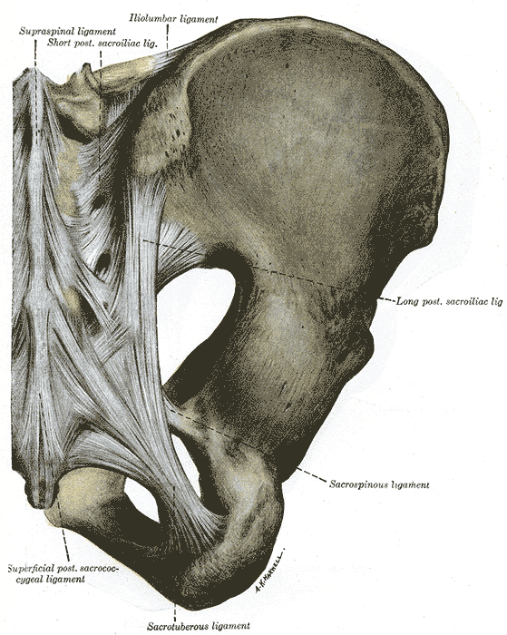
Posterior Articulations, Pelvis. The illustrated image portrays the supraspinal, short posterior, sacroiliac, iliolumbar, sacrospinous, sacrotuberous, superficial posterior sacrococcygeal ligaments, and hip.
Henry Vandyke Carter, Public Domain, via Wikimedia Commons
(Click Image to Enlarge)
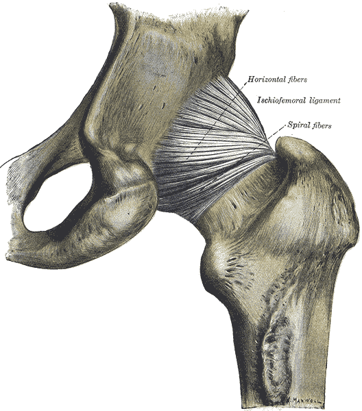
Hip Joint, Posterior View.
Henry Vandyke Carter, Public Domain, via Wikimedia Commons
References
Bowman KF Jr, Fox J, Sekiya JK. A clinically relevant review of hip biomechanics. Arthroscopy : the journal of arthroscopic & related surgery : official publication of the Arthroscopy Association of North America and the International Arthroscopy Association. 2010 Aug:26(8):1118-29. doi: 10.1016/j.arthro.2010.01.027. Epub [PubMed PMID: 20678712]
Parvaresh KC, Pennock AT, Bomar JD, Wenger DR, Upasani VV. Analysis of Acetabular Ossification From the Triradiate Cartilage and Secondary Centers. Journal of pediatric orthopedics. 2018 Mar:38(3):e145-e150. doi: 10.1097/BPO.0000000000001120. Epub [PubMed PMID: 29309383]
Graber M, Marino DV, Johnson DE. Anterior Hip Dislocation. StatPearls. 2026 Jan:(): [PubMed PMID: 29939591]
Mandell JC, Marshall RA, Weaver MJ, Harris MB, Sodickson AD, Khurana B. Traumatic Hip Dislocation: What the Orthopedic Surgeon Wants to Know. Radiographics : a review publication of the Radiological Society of North America, Inc. 2017 Nov-Dec:37(7):2181-2201. doi: 10.1148/rg.2017170012. Epub [PubMed PMID: 29131775]
O'Donnell JM, Devitt BM, Arora M. The role of the ligamentum teres in the adult hip: redundant or relevant? A review. Journal of hip preservation surgery. 2018 Jan:5(1):15-22. doi: 10.1093/jhps/hnx046. Epub 2018 Jan 10 [PubMed PMID: 29423246]
Gautier E, Ganz K, Krügel N, Gill T, Ganz R. Anatomy of the medial femoral circumflex artery and its surgical implications. The Journal of bone and joint surgery. British volume. 2000 Jul:82(5):679-83 [PubMed PMID: 10963165]
Grose AW, Gardner MJ, Sussmann PS, Helfet DL, Lorich DG. The surgical anatomy of the blood supply to the femoral head: description of the anastomosis between the medial femoral circumflex and inferior gluteal arteries at the hip. The Journal of bone and joint surgery. British volume. 2008 Oct:90(10):1298-303. doi: 10.1302/0301-620X.90B10.20983. Epub [PubMed PMID: 18827238]
Rodriguez JA, Deshmukh AJ, Rathod PA, Greiz ML, Deshmane PP, Hepinstall MS, Ranawat AS. Does the direct anterior approach in THA offer faster rehabilitation and comparable safety to the posterior approach? Clinical orthopaedics and related research. 2014 Feb:472(2):455-63. doi: 10.1007/s11999-013-3231-0. Epub [PubMed PMID: 23963704]