 Tools
Tools
 Anatomy, Abdomen and Pelvis: Anterolateral Abdominal Wall
Introduction
The abdominal wall is a complex organ with many functions that contribute to patients’ quality of life. The anatomical core of the anterolateral abdominal wall is mainly composed of 4 paired, symmetrical muscles. Classic descriptions present the anterolateral abdominal wall in separate layers from superficial to deep in the following order:
- Skin
- Subcutaneous tissues (subdivided into the superficial Camper fascia and the deeper Scarpa fascia)
- External oblique muscle
- Internal oblique muscle
- Transversus abdominis muscle
- Transversalis fascia
- Parietal peritoneum
Each component makes a unique contribution to the abdominal wall and will be described further in this review.
Structure and Function
Register For Free And Read The Full Article
 Search engine and full access to all medical articles
- 10 free questions in your specialty
- Free CME/CE Activities
- Free daily question in your email
- Save favorite articles to your dashboard
- Emails offering discounts
Learn more about a Subscription to StatPearls Point-of-Care
Structure and Function
The main functions of the anterolateral abdominal muscles include stabilizing the vertebral column, moving the trunk, and tensing the abdominal wall. The function of the anterolateral wall as a whole includes protection of the abdominal viscera, maintenance of anatomical position, assistance in forceful expiration, and involvement in any activity that increases intra-abdominal pressure.
Embryology
Embryonic differentiation is based on 3 germ layers: the outermost protective layer, termed the ectoderm; the middle layer, termed the mesoderm; and the innermost layer, termed the endoderm. The mesoderm is further divided into 2 layers: the splanchnic layer, which gives rise to the abdominal viscera, and the somatic layer, which gives rise to the abdominal wall.
Blood Supply and Lymphatics
Arterial Supply
The arterial supply to the abdominal wall is derived from the following:
Six most inferior intercostal arteries and lumbar arteries: These vessels course from lateral to medial between the transversus abdominis and internal oblique muscles, along with the intercostal, iliohypogastric, and ilioinguinal nerves. The branches pierce the lateral border of the rectus sheath and freely communicate with the epigastric arteries.
Superior epigastric arteries: These arteries are terminal branches of the internal thoracic artery, also known as the internal mammary artery,bilaterally. They descend within the rectus sheath, posterior to the rectus muscle but anterior to the posterior rectus sheath, to form an anastomosis with the inferior epigastric artery.
Inferior epigastric arteries: These arteries are branches of the external iliac artery just before the artery crosses the inguinal ligament. They course superiorly in the preperitoneal space, between the transversalis fascia and parietal peritoneum, to meet the superior epigastric vessels.
Deep circumflex iliac arteries: These arteries arise from the external iliac artery laterally, just distal to the inferior epigastric artery branching, and contribute blood to the abdominal wall via an ascending branch.
Venous Drainage
The venous drainage of the abdominal wall superior to the umbilicus is through the internal thoracic, intercostal, and long thoracic veins. These veins ultimately drain into the superior vena cava. Inferior to the umbilicus, the venous drainage is through the superficial epigastric, circumflex iliac, and pudendal veins. These veins drain to the groin at the saphenofemoral junction and ultimately reach the inferior vena cava. An important embryological consideration of the venous drainage is the ligamentum teres. The ligamentum teres contains the remnant of the umbilical vein, which is a connection between the abdominal wall veins and the left portal vein branch. In the setting of portal hypertension, this vein may recanalize, leading to dilation of the abdominal wall veins, termed caput medusae.
Lymphatic Drainage
The lymphatic drainage of the abdominal wall parallels the venous drainage. The abdominal wall lymphatics superior to the umbilicus drain to the axillary lymph node basin, and those inferior to the umbilicus drain to the inguinal lymph node basin. Additionally, the embryological origin of the ligamentum teres allows gastrointestinal tract cancers to metastasize to the periumbilical region of the abdominal wall, forming Sister Mary Joseph nodules.
Nerves
The sensory and motor innervation to the abdominal wall is primarily derived from the anterior rami of T7 through T12. These nerves course in an inferior and medial direction between the transversus abdominis and internal oblique muscles. The anterior ramus of T12, along with the first lumbar nerve, contributes to the common trunk of the ilioinguinal and iliohypogastric nerves. The ilioinguinal nerve is found within the inguinal canal and provides sensation to the ipsilateral medial thigh and scrotum. The iliohypogastric nerves provide sensation to the anterior abdominal wall in the suprapubic region.
Muscles
Layers of the Anterolateral Abdominal Wall
Subcutaneous tissues: Camper fascia can be thought of as superficial subcutaneous fat; this layer contains little collagen and therefore provides little strength. Scarpa fascia is contiguous with the tensor fascia lata of the thigh. The Scarpa fascia lies deeper than the fatty layer and contains more collagen; many surgeons approximate this layer during abdominal wall closures.
External oblique: The external oblique is the most superficial, largest, and thickest of the 3 anterolateral abdominal wall muscles. This muscle originates from the lower 7 ribs and runs obliquely from superolateral to inferomedial (hands-in-pockets orientation) to insert on the anterior half of the iliac crest. The most inferior extension folds posteriorly and superiorly to form the inguinal ligament. At the midclavicular line, the muscle belly ends, but the aponeurosis extends medially to the linea alba, contributing to the anterior rectus sheath.
Internal oblique: The internal oblique is the middle layer of the 3 anterolateral abdominal wall muscles, originating from the iliopsoas fascia, lateral iliac crest, and thoracolumbar fascia. Its fibers course opposite to the external oblique, from inferolateral to superomedial, to insert on the cartilage of the lower 5 ribs. Inferiorly, the fibers take a more transverse course and insert onto the pubic tubercle. At this inferior portion, the internal oblique aponeurosis joins the aponeurosis of the transversus abdominis muscle to create the conjoined tendon. Like the external oblique, this muscle gives off a medial extension of the aponeurosis to contribute to the rectus sheath. Above the semicircular line of Douglas, approximately just inferior to the level of the umbilicus, the internal oblique aponeurosis splits around the rectus abdominis muscle to contribute to the anterior and posterior rectus sheaths. Below this line, the aponeurosis contributes only to the anterior rectus sheath. Inferiorly, this muscle gives off fibers that encircle the spermatic cord, termed the cremasteric fibers.
Transversus abdominis: The transversus abdominis is the thinnest and deepest of the 3 anterolateral abdominal wall muscles. This muscle originates from the lower 6 costal cartilages, lumbar vertebrae, iliac crests, and iliopsoas fascia. The fibers are oriented transversely and contribute to the medial aspect of the rectus sheath. Above the semicircular line of Douglas, the aponeurosis courses posterior to the rectus muscles to contribute to the posterior rectus sheath. Inferiorly, the aponeurosis contributes only to the anterior rectus sheath.
Transversalis fascia: The endoabdominal fascia lines the entire abdominal cavity. The endoabdominal fascia has specific names by anatomic location, including the diaphragmatic, obturator, and iliopsoas fascias, among others. The transversalis fascia is just deep to the transversus abdominis and posterior rectus sheaths. The transversalis fascia is an important layer because its violation defines an abdominal wall hernia.
Parietal peritoneum: The parietal peritoneum is a thin layer of connective tissue deep to the transversalis fascia. The deep surface contains a single cell layer of squamous mesothelium.[1]
Physiologic Variants
Anatomical variations are common in the literature.[2] Classic anatomy is described in just over 50% of observed specimens. Common variations include different sensory distribution of nerves, presence or absence of accessory internal oblique or pyramidalis muscles, and different composition of the anterior and posterior rectus sheaths. Embryologic malformations may result in ventral abdominal wall defects at birth. These malformations include ectopia cordis, bladder exstrophy, omphalocele, and gastroschisis.[3]
Surgical Considerations
Detailed knowledge of the abdominal wall is necessary when performing abdominal wall reconstruction. Techniques such as component separation require meticulous dissection of the different abdominal wall layers and preservation of the vascular supply. Surgeons must also avoid injury to the innervation of the abdominal wall because denervation can contribute to loss of function and abdominal wall weakness.[4][5]
Clinical Significance
Knowledge of abdominal wall innervation and its anatomic location is important when considering postoperative analgesia. Clinicians use several techniques to deliver local anesthetic to the nerves of the abdominal wall to improve analgesia after abdominopelvic surgical procedures. A popular example is termed the transversus abdominis plane block (TAP block).[6] This established technique involves administering a local anesthetic to the sensory innervation of the abdominal wall, situated in a plane between the transversus abdominis and internal oblique muscles. Typically, 2 techniques are used: one involves ultrasonography guidance, and the other uses direct visualization. Both methods have yielded statistically significant improvements in postoperative pain control.
Other Issues
Expanding knowledge of the abdominal wall will likely lead to new techniques for abdominal wall reconstruction and postoperative analgesia. Every clinician, particularly surgeons, should have a thorough understanding of this complex anatomy.
Media
(Click Image to Enlarge)
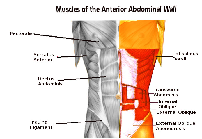
Abdominal Wall Muscles.
Contributed by S Dulebohn, MD
References
Stumpf M, Conze J, Prescher A, Junge K, Krones CJ, Klinge U, Schumpelick V. The lateral incisional hernia: anatomical considerations for a standardized retromuscular sublay repair. Hernia : the journal of hernias and abdominal wall surgery. 2009 Jun:13(3):293-7. doi: 10.1007/s10029-009-0479-0. Epub 2009 Feb 12 [PubMed PMID: 19214648]
Monkhouse WS, Khalique A. Variations in the composition of the human rectus sheath: a study of the anterior abdominal wall. Journal of anatomy. 1986 Apr:145():61-6 [PubMed PMID: 2962970]
Sadler TW. The embryologic origin of ventral body wall defects. Seminars in pediatric surgery. 2010 Aug:19(3):209-14. doi: 10.1053/j.sempedsurg.2010.03.006. Epub [PubMed PMID: 20610194]
Ramirez OM, Ruas E, Dellon AL. "Components separation" method for closure of abdominal-wall defects: an anatomic and clinical study. Plastic and reconstructive surgery. 1990 Sep:86(3):519-26 [PubMed PMID: 2143588]
Level 3 (low-level) evidenceDiBello JN Jr, Moore JH Jr. Sliding myofascial flap of the rectus abdominus muscles for the closure of recurrent ventral hernias. Plastic and reconstructive surgery. 1996 Sep:98(3):464-9 [PubMed PMID: 8700983]
Tsai HC, Yoshida T, Chuang TY, Yang SF, Chang CC, Yao HY, Tai YT, Lin JA, Chen KY. Transversus Abdominis Plane Block: An Updated Review of Anatomy and Techniques. BioMed research international. 2017:2017():8284363. doi: 10.1155/2017/8284363. Epub 2017 Oct 31 [PubMed PMID: 29226150]