 Tools
Tools
 Juvenile Xanthogranuloma
Introduction
Juvenile xanthogranuloma is a relatively common condition and is the most common form of non-Langerhans cell histiocytic disorder of childhood.[1][2] Lesions are estimated to appear in approximately 75% of cases within the first year of life, with 15% to 20% of patients presenting with lesions at birth. Juvenile xanthogranuloma is rare in adults, with a peak incidence in the late twenties to thirties, although rare cases have been described in older individuals.[3] The majority of adult patients have solitary lesions.[4] The true incidence, however, may be underestimated, as many lesions, especially those that are solitary and small (up to 90% of patients), may go unrecognized.
Typically, the clinical presentation consists of solitary or multiple firm papules or nodules with a yellow-orange to brown coloration.[5] The most common locations include the face, neck, and upper torso.[4] Oral lesions are rare and often occur as a yellow nodule on the lateral aspects of the tongue. Oral lesions can also arise on the gingiva, buccal mucosa, and midline hard palate and may ulcerate and bleed. Lesions of the oral mucosa may appear verrucous, umbilicated, pedunculated, or fibroma-like. Cutaneous lesions are typically asymptomatic and most undergo spontaneous involution over several years.
Although rare, ocular involvement is the most common extracutaneous site, followed by the lungs. Ocular juvenile xanthogranuloma is nearly always unilateral and develops in less than 0.5% of patients. Approximately 40% of patients with ocular juvenile xanthogranuloma, however, have multiple cutaneous lesions at the time of diagnosis. Ocular disease typically occurs before age 2 and commonly involves the iris. Hyphema and glaucoma are serious complications that can result in blindness and necessitate early referral to an ophthalmologist for evaluation and possible treatment. Bone, visceral, and or central nervous system (CNS) involvement (eg, diabetes insipidus) is unusual. Disease-related deaths are rare.[1]
Unlike other xanthomatous diseases, juvenile xanthogranuloma is not associated with metabolic or lipid disorders.[4]
Etiology
Register For Free And Read The Full Article
 Search engine and full access to all medical articles
- 10 free questions in your specialty
- Free CME/CE Activities
- Free daily question in your email
- Save favorite articles to your dashboard
- Emails offering discounts
Learn more about a Subscription to StatPearls Point-of-Care
Etiology
Juvenile xanthogranuloma is presumed arise in response to an unknown stimulus, possibly infectious or physical, triggering a granulomatous histiocytic reaction.[4] However, the true etiology of juvenile xanthogranuloma is currently unknown, and conservative management is generally advised.[6]
Juvenile xanthogranuloma has been associated with juvenile chronic myelogenous leukemia.[7] Juvenile myelomonocytic leukemia is also observed in patients with neurofibromatosis, with an estimated incidence of 1:2000 to 1:5000. Rarely, it is observed in patients with both juvenile xanthogranuloma and neurofibromatosis.[6] However, when this does occur, these children have a 20- to 32-fold increased risk of juvenile myelomonocytic leukemia when compared to patients with neurofibromatosis alone.[8] There have been many cases of neurofibromatosis type 1 (NF-1) presenting with juvenile xanthogranuloma, with possible correlation with lymphoproliferative diseases, and their prevalence is currently being studied.[9]
Other diseases associated with juvenile xanthogranuloma include urticaria pigmentosa, insulin-dependent diabetes mellitus, aquagenic pruritis, and cytomegalovirus infection.[7]
Epidemiology
Juvenile xanthogranuloma most commonly occurs in early childhood, with 15% to 20% of patients having lesions at birth. In approximately 75% of cases, lesions appear during the first year of life.[1] Approximately 10% of cases may appear in adulthood and are known as adult xanthogranuloma.[1] In children, the disease most often affects males; however, in adults, there is no sex prevalence.[4]
Histopathology
Juvenile xanthogranuloma most commonly occurs in the skin and has a characteristic morphology. Lesions show well-demarcated, dense infiltration of numerous histiocytes within the superficial dermis, often accompanied by blunting of the rete ridges and scattered lymphocytes, plasma cells, and eosinophils (see Image. Juvenile Xanthogranuloma).[1] Mononuclear cells may appear spindled or elongated.[7] Foam cells, foreign body giant cells, and Touton giant cells may be observed in the mature stage.[1] The characteristic multinucleated giant cell (Touton cell), observed in 85% of cases, exhibits a garland-like arrangement (see Image. Classic Multinucleated Giant (Touton) Cell of Juvenile Xanthogranuloma).[4][1] Late-stage lesions show increased fibroblasts and replacement of the infiltrate by fibrosis.[1]
In ocular involvement, tumor cells typically have abundant cytoplasm and fewer Touton cells compared to cutaneous juvenile xanthogranuloma.[1]
Immunohistochemically, juvenile xanthogranuloma lesions classically stain positive for CD68 (Ki-M1P), factor XIIIa, vimentin, and CD4.[1]
History and Physical
Juvenile xanthogranuloma generally occurs in 2 forms. The first form consists of multiple dome-shaped papules measuring 2 to 5 mm that appear pink to reddish-brown and subsequently turn yellow.[10] The second form comprises 1 or more large nodules that measure 1 to 2 cm.[11] Other variants and presentations include giant juvenile xanthogranuloma, atrophic plaque, cutaneous horn, and subcutaneous mass.[11]
Serious complications can arise when systemic juvenile xanthogranuloma develops. Systemic juvenile xanthogranuloma develops in 4% of children, with an overall mortality of 5% to 10%.[8] Fatalities are typically due to progressive central nervous system (CNS) disease or liver failure.[8] Extracutaneous juvenile xanthogranuloma most commonly affects the eye, and ocular involvement has been observed in 0.3% to 10% of children with cutaneous juvenile xanthogranuloma.[1] Most patients with eye lesions are younger than 1 year.[4] The iris is most commonly involved, and complications can arise that lead to spontaneous intraocular hemorrhages, glaucoma, and blinding eye disease.[1] Notably, spontaneous hyphema, with or without an increase in intraocular pressure, is a common sign of juvenile iris xanthogranuloma.[12]
Other CNS complications include seizures, increased intracranial pressure, diabetes insipidus, and developmental delay.[8] These extracutaneous lesions may also be observed in the deeper soft tissues, spleen, and lung.[8]
Evaluation
Dermoscopy is a noninvasive technique used as an aid in the diagnosis of juvenile xanthogranuloma. This technique shows a characteristic setting sun appearance, characterized by a red-yellow center and a discrete erythematous halo.[13] Definitive diagnosis can be confirmed through complete surgical excision and histopathologic examination.[2]
Treatment / Management
Curative skin excisions may be performed at easily accessible sites; however, most lesions in childhood spontaneously involute and require no therapy.[8]
Management of non-cutaneous (internal) lesions is more difficult and may involve any combination of surgery, chemotherapy, radiotherapy, and immunosuppression.[4] In general, chemotherapy and radiotherapy are options for patients with symptomatic, unresectable, or incompletely resected extracutaneous disease.[6] (B3)
Patients with concurrent NF-1 should receive frequent follow-up with a complete blood count to monitor for juvenile chronic myelogenous leukemia.[8]
Eye examination is recommended for high-risk patients. Topical, intralesional, and subconjunctival corticosteroids may be administered for ocular involvement. In cases of complications such as glaucoma or hyphema, surgery or systemic steroids may be required.[8]
Differential Diagnosis
The differential of juvenile xanthogranuloma includes both benign and malignant entities, depending on the clinical presentation and site of involvement.
Langerhans cell histiocytosis is a key differential diagnosis of juvenile xanthogranuloma, as both conditions may contain eosinophils. Distinguishing morphologic features of Langerhans cell histiocytosis include grooved, vesicular nuclei with a characteristic coffee bean appearance.[14] Additionally, immunohistochemistry shows that Langerhans cell histiocytosis cells are positive for CD1a and S-100, whereas juvenile xanthogranuloma cells are negative for these markers.[14] CD1a is a relatively specific marker for Langerhans cell histiocytosis and is not expressed in non-Langerhans cell histiocytosis diseases.[1]
Reticulohistiocytoma, another non-Langerhans cell histiocytosis histiocytic disorder, has a histologic appearance similar to juvenile xanthogranuloma.[14] This lesion demonstrates large, mono- or multinucleated histiocytes, a lymphocytic infiltrate, and dermal fibrosis.[14] Although reticulohistiocytoma is positive for CD68, it is negative for vimentin, unlike juvenile xanthogranuloma.[14]
Although juvenile xanthogranuloma may be found in uncommon locations, such as the vulva, it is important to remember that other entities, such as embryonal rhabdomyosarcoma, may have a similar histologic appearance and should be ruled out. [14] The morphologic appearance of monomorphic small round cells with a cambium layer and necrosis may help direct a malignant diagnosis in such cases.[14]
Other conditions to consider in the differential diagnosis include additional xanthomatous lesions, mastocytomas, dermatofibromas, and Spitz nevus.[6]
Prognosis
The prognosis of juvenile xanthogranuloma is generally excellent, with most lesions undergoing spontaneous involution over the course of 3 to 6 years. Lesions that are of cosmetic concern or pose a risk to vital functions can be treated.[8]
Complications
There are often minimal to no complications of juvenile xanthogranuloma due to its self-limiting nature. Occasionally, lesions are removed for cosmetic concerns. Ocular lesions often require intervention to avoid potential complications of blindness, glaucoma, or hyphema.
Deterrence and Patient Education
Deterrence and patient education in juvenile xanthogranuloma focus on reassuring families about the typically benign, self-limited nature of the condition while promoting vigilance for uncommon but clinically significant complications. Parents should be informed that most cutaneous lesions involute spontaneously over several years without scarring, and that no specific lifestyle modifications are required. However, clinicians must emphasise the importance of monitoring for extracutaneous involvement, particularly ocular disease, which, although rare, can lead to hyphema, secondary glaucoma, and vision loss if not promptly detected. Families should be counselled to seek early ophthalmologic evaluation in infants with multiple, large, or periocular lesions, and to remain attentive to symptoms such as photophobia, tearing, or visual changes. Education should also include recognition of atypical features such as rapid lesion enlargement, ulceration, or systemic symptoms, which warrant reevaluation to exclude alternative diagnoses, including Langerhans cell histiocytosis or malignancy. In cases with numerous lesions, patients and caregivers should understand the association with NF-1 and the need for periodic multidisciplinary follow-up. Clear communication, written information, and scheduled review appointments help reduce anxiety and reinforce adherence to monitoring recommendations. Ultimately, well-structured patient education empowers families, ensures timely detection of complications, and supports optimal long-term outcomes.
Pearls and Other Issues
Isolated intracranial lesions of juvenile xanthogranuloma have been associated with a more favorable prognosis than other forms of systemic disease. These lesions have been reported to harbor a BRAF V600E mutation, similar to that observed in Langerhans cell histiocytosis and Erdheim-Chester disease. However, further study with a larger patient population is necessary to interpret these findings.[15]
Enhancing Healthcare Team Outcomes
The management of solitary lesions of juvenile xanthogranuloma is often straightforward with routine observation, as they are benign and often resolve spontaneously. Surgical excision may be considered for symptomatic or cosmetically concerning lesions, but most skin-only cases have a favorable prognosis.
Multiple lesions or those involving vital structures require an interprofessional approach. While skin lesions can be excised, lesions elsewhere on the body may compress or obstruct vital tissues. Patients with skin-only lesions generally have favorable outcomes, whereas those with CNS involvement may experience surgical complications. Collaborative care is essential to optimize outcomes and minimize complications.[16][17][18]
Media
(Click Image to Enlarge)
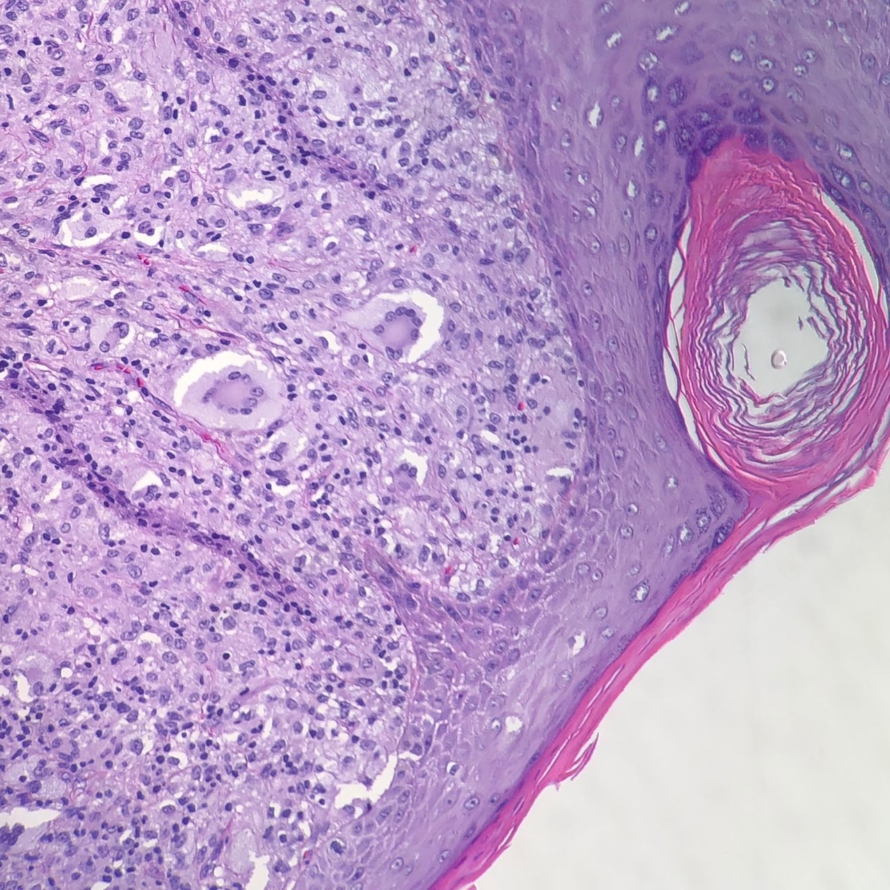
Juvenile Xanthogranuloma.
Contributed by J Collie, DO
(Click Image to Enlarge)
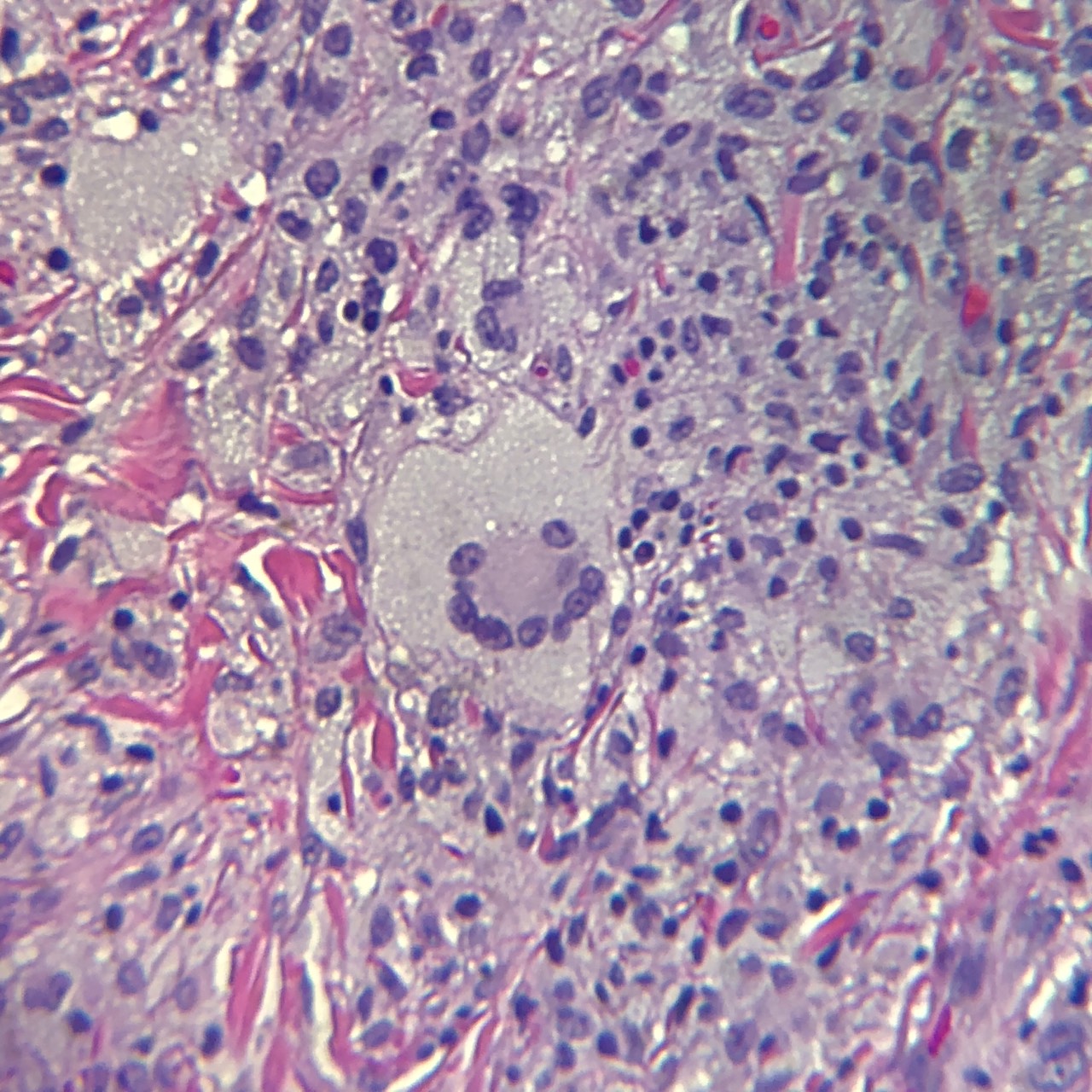
Classic Multinucleated Giant (Touton) Cell of Juvenile Xanthogranuloma.
Contributed by J Collie, DO
References
Pantalon A, Ștefănache T, Danciu M, Zurac S, Chiseliță D. Iris juvenile xanthogranuloma in an infant - spontaneous hyphema and secondary glaucoma. Romanian journal of ophthalmology. 2017 Jul-Sep:61(3):229-236 [PubMed PMID: 29450403]
Oliveira TE, Tarlé RG, Mesquita LAF. Dermoscopy in the diagnosis of juvenile xanthogranuloma. Anais brasileiros de dermatologia. 2018 Jan-Feb:93(1):138-140. doi: 10.1590/abd1806-4841.20186849. Epub [PubMed PMID: 29641718]
. What Is a Juvenile Xanthogranuloma? Pediatric dermatology. 2022 Jan:39(1):119-120. doi: 10.1111/pde.14920. Epub [PubMed PMID: 35106819]
Szczerkowska-Dobosz A, Kozicka D, Purzycka-Bohdan D, Biernat W, Stawczyk M, Nowicki R. Juvenile xanthogranuloma: a rare benign histiocytic disorder. Postepy dermatologii i alergologii. 2014 Jun:31(3):197-200. doi: 10.5114/pdia.2014.40918. Epub 2014 Jun 13 [PubMed PMID: 25097495]
Level 3 (low-level) evidenceZou T, Wei A, Ma H, Lian H, Liu Y, Wang D, Zhao Y, Cui L, Li Z, Zhang R, Wang T. Systemic juvenile xanthogranuloma: A systematic review. Pediatric blood & cancer. 2023 May:70(5):e30232. doi: 10.1002/pbc.30232. Epub 2023 Feb 13 [PubMed PMID: 36779547]
Level 1 (high-level) evidenceMurphy JT, Soeken T, Megison S, Perez E. Juvenile xanthogranuloma: diverse presentations of noncutaneous disease. Journal of pediatric hematology/oncology. 2014 Nov:36(8):641-5. doi: 10.1097/MPH.0000000000000027. Epub [PubMed PMID: 24065046]
Level 3 (low-level) evidenceFreyer DR, Kennedy R, Bostrom BC, Kohut G, Dehner LP. Juvenile xanthogranuloma: forms of systemic disease and their clinical implications. The Journal of pediatrics. 1996 Aug:129(2):227-37 [PubMed PMID: 8765620]
Level 3 (low-level) evidenceHaroche J, Abla O. Uncommon histiocytic disorders: Rosai-Dorfman, juvenile xanthogranuloma, and Erdheim-Chester disease. Hematology. American Society of Hematology. Education Program. 2015:2015():571-8. doi: 10.1182/asheducation-2015.1.571. Epub [PubMed PMID: 26637774]
Miraglia E, Laghi A, Moramarco A, Giustini S. Juvenile xanthogranuloma in neurofibromatosis type 1. Prevalence and possible correlation with lymphoproliferative diseases: experience of a single center and review of the literature. La Clinica terapeutica. 2022 Jul-Aug:173(4):353-355. doi: 10.7417/CT.2022.2445. Epub [PubMed PMID: 35857053]
Garcia-Sirvent L, Espineira-Sicre J, Ruiz-Sanchez J, Cuesta-Montero L. Multiple juvenile xanthogranuloma. Dermatology online journal. 2024 Mar 15:30(1):. doi: 10.5070/D330163291. Epub 2024 Mar 15 [PubMed PMID: 38762861]
Vignault C, Bourgeault É, Gagné É, Bujold J. A Rare Case of Solitary Giant Congenital Juvenile Xanthogranuloma: A Case Report. Journal of cutaneous medicine and surgery. 2017 May/Jun:21(3):267-269. doi: 10.1177/1203475417690718. Epub 2017 Feb 1 [PubMed PMID: 28300446]
Level 3 (low-level) evidenceMeyer P, Graeff E, Kohler C, Munier F, Bruder E. Juvenile xanthogranuloma involving concurrent iris and skin: Clinical, pathological and molecular pathological evaluations. American journal of ophthalmology case reports. 2018 Mar:9():10-13. doi: 10.1016/j.ajoc.2017.09.004. Epub 2017 Sep 23 [PubMed PMID: 29468209]
Level 3 (low-level) evidencePeruilh-Bagolini L, Silva-Astorga M, Hernández San Martín MJ, Manoli MS, Papageorgiou C, Apalla Z, Lallas A. Dermoscopy of Juvenile Xanthogranuloma. Dermatology (Basel, Switzerland). 2021:237(6):946-951. doi: 10.1159/000510265. Epub 2020 Oct 19 [PubMed PMID: 33075787]
Gupta B, Yadav S, Khurana N, Sharma M. Juvenile Xanthogranuloma in Vulva of a 10-Year-Old Child. Journal of clinical and diagnostic research : JCDR. 2016 Nov:10(11):ED21-ED22. doi: 10.7860/JCDR/2016/22266.8930. Epub 2016 Nov 1 [PubMed PMID: 28050386]
Techavichit P, Sosothikul D, Chaichana T, Teerapakpinyo C, Thorner PS, Shuangshoti S. BRAF V600E mutation in pediatric intracranial and cranial juvenile xanthogranuloma. Human pathology. 2017 Nov:69():118-122. doi: 10.1016/j.humpath.2017.04.026. Epub 2017 May 10 [PubMed PMID: 28504206]
Mori H, Nakamichi Y, Takahashi K. Multiple Juvenile Xanthogranuloma of the Eyelids. Ocular oncology and pathology. 2018 Sep:4(2):73-78. doi: 10.1159/000478101. Epub 2017 Aug 12 [PubMed PMID: 30320084]
Höck M, Zelger B, Schweigmann G, Brunner B, Zelger B, Kropshofer G, Kiechl-Kohlendorfer U. The various clinical spectra of juvenile xanthogranuloma: imaging for two case reports and review of the literature. BMC pediatrics. 2019 Apr 24:19(1):128. doi: 10.1186/s12887-019-1490-y. Epub 2019 Apr 24 [PubMed PMID: 31018833]
Level 3 (low-level) evidenceShenoy A, Singhal SS, Shinde S. Solitary Intraspinal Juvenile Xanthogranuloma in an Infant. Asian journal of neurosurgery. 2018 Jan-Mar:13(1):172-175. doi: 10.4103/1793-5482.180896. Epub [PubMed PMID: 29492155]