 Tools
Tools
 Anatomy, Abdomen and Pelvis: Uterus Round Ligament
Introduction
The round ligament of the uterus is fibromuscular connective tissue. This ligament resembles a round band of rope. One end of the round ligament is attached to the superior and lateral aspect of the uterus. This uterine attachment occurs at the cornu of the uterus. From the cornu of the uterus, the round ligament crosses the pelvis through the deep inguinal ring, traverses the inguinal canal, and then enters the labia majora, where it terminates as its fibers blend into the mons pubis. Two round ligaments are present, one on each side of the uterus. Each round ligament is about 10 to 12 cm long. The round ligament is covered by folded peritoneum and comprises the superior margin of the broad ligament on each side of the uterus. The medial side of the broad ligament is attached to the lateral side of the uterus on each side. The broad ligament fans out to the pelvic sidewall. The broad ligament contains the round ligament, the fallopian tube, arteries, veins, lymphatics, nerve fibers, and loose connective tissue (see Image. Uterine Tubal Anatomy and Ligaments).
Structure and Function
Register For Free And Read The Full Article
 Search engine and full access to all medical articles
- 10 free questions in your specialty
- Free CME/CE Activities
- Free daily question in your email
- Save favorite articles to your dashboard
- Emails offering discounts
Learn more about a Subscription to StatPearls Point-of-Care
Structure and Function
The uterus is a pear-shaped muscular organ of the female reproductive system. The uterus is divided into the fundus, lower uterine segment, and cervix uteri. The uterus hosts and nourishes the embryo and fetus until delivery.
- The fallopian tubes are 2 tubular structures attached to the uterus on each side. The fimbriated ends of the fallopian tubes float freely adjacent to the ovaries on each side of the pelvis. The round ligament of the uterus originates at the uterine cornu and blends with the tissue of the mons pubis and labia majora.
- The cardinal ligament is the inferior demarcation at the base of the broad ligament. The 2 cardinal ligaments support the uterus and cervix uteri. The broad ligament is the peritoneal fold around the round ligament, parametrial connective tissue, arteries, veins, lymphatics, and nerves.
- The uterosacral ligaments are attached anteriorly to the cervix uteri. The uterosacral ligaments are attached posteriorly to the sacral vertebrae. The ovaries rest in the ovarian fossa in the lateral part of the pelvic cavity next to the iliac vessels and contain germ cells.
- The ovarian ligaments are attached to the posterolateral aspect of the uterus. The infundibulopelvic ligaments are peritoneal reflections of the broad ligaments. The bladder is located anterior to the uterus, and the ureters insert into the trigone.
- The rectum is located between the vagina and the sacrum. The pelvic diaphragm supports all the viscera. The round ligament helps maintain the anteverted position of the uterus during pregnancy, whereas the cardinal ligaments support the uterus.
Embryology
The round ligament of the uterus develops from the gubernaculum. In a fetus, the gubernaculum is undifferentiated mesenchymal tissue attached to ovarian tissue in a female fetus and testicular tissue in a male fetus. During the early stages of urogenital development in the female fetus, the gubernaculum develops as a connective tissue band. This ligamentous connective tissue band is attached to the ovary on one side and the labia majora on the other side. During its course from the ovary, the gubernaculum runs to the cornu of the uterus and adheres to the uterus. The gubernaculum then continues toward the labia majora.
In adults, the gubernaculum develops into the following 2 parts:
- The round ligament of the uterus: The part between the cornu of the uterus and the labia majora. This ligament is also called the ligamentum teres uteri and is longer than the ovarian ligament.
- The ovarian ligament: The ovarian ligament is the part between the cornu of the uterus and the ovary.
Blood Supply and Lymphatics
The round ligament receives blood from the Sampson artery, also called the artery of the round ligament. The common iliac artery divides into the external and internal iliac arteries. The internal iliac artery branches into the uterine artery, vaginal artery, superior vesical artery, obturator artery, inferior gluteal artery, internal pudendal artery, and obliterated umbilical artery. Sampson artery, a branch of the uterine artery, runs along the length of the round ligament. Ovarian arteries originate directly from the aorta. The left ovarian vein drains into the left renal vein. The right ovarian vein drains directly into the inferior vena cava. Uterine lymphatics drain to the pelvic and para-aortic nodes. Cervical lymphatics drain to the parametrial, obturator, pelvic, and para-aortic nodes. Ovarian lymphatics drain to the pelvic and para-aortic nodes.
Clinical Significance
In rare cases, the gubernaculum may fail to adhere to the uterus. Failed gubernacular adherence may cause the ovaries to descend through the inguinal canal into the labia majora. This ovarian malposition may resemble the testes.[1][2][3]
During the reproductive years, pelvic endometriosis may infiltrate the round ligaments. Endometriosis is defined as the presence of ectopic endometrial glands and stroma outside the uterus. Endometriosis may lead to severe dysmenorrhea, dyspareunia, chronic pelvic pain, and infertility. Pelvic endometriosis can affect the uterosacral ligaments, rectum, vagina, cul-de-sac, and urinary bladder. Deep, extensive pelvic endometriosis may infiltrate the round ligaments of the uterus. Magnetic resonance imaging (MRI) is helpful for diagnosing round ligament lesions. If medical therapy fails, surgical treatment remains the best therapeutic option for advanced endometriosis.
Round ligament varices (RLV) are conditions in which veins become tortuous and twisted. RLV develop during pregnancy. Venous blood from the round ligament and the inguinal canal drains into the inferior epigastric vein. During pregnancy, blood volume and cardiac output increase. An enlarged gravid uterus increases venous pressure, leading to venous stagnation. Increased progesterone levels contribute to venous dilation and smooth muscle relaxation. Increased venous return and engorgement of veins and tributaries progress to RLV. The patient may present with bilateral asymptomatic inguinal swellings during pregnancy. Ultrasonography may help diagnose and differentiate RLV from an inguinal hernia. Multiple dilated vessels without bowel contents may be visualized on ultrasonography. Conservative treatment with close monitoring is recommended throughout pregnancy. After pregnancy, the hormonal effects of progesterone and the pressure of the gravid uterus are relieved, leading to spontaneous resolution during the postpartum recovery period.
Round Ligament Pain
The round ligaments of the uterus increase in diameter and length during pregnancy. During the second trimester, the round ligaments may cause cramping and pain due to stretching and contractions. At times, this pain may be sharp and can cause pulling sensations. Round ligament pain is considered physiologic in normal pregnancy. Usually, the pain from the round ligaments is temporary and resolves spontaneously with rest. Round ligament pain is not considered a symptom of disease. If pain persists, immediate follow-up is recommended to rule out other causes of pain during pregnancy. Hormone levels decrease sharply after delivery. After the postpartum period, the uterine size is reduced to the normal nonpregnant state. The hypertrophic round ligaments of the uterus also shrink to the normal nonpregnant state. Following postpartum recovery, round ligament pain may occur, but pain from sudden movements of pelvic organs is unlikely.In a nonpregnant woman, round ligament pain may occur, but is unlikely. In a nonpregnant woman, the round ligament is a firm and flexible structure. Sudden movements of pelvic organs may not cause any symptoms.
Conservative Management
Warm compresses to the area may relieve pain. Acetaminophen is safe to take during pregnancy. Acetaminophen is the most prescribed analgesic during pregnancy.Rest during pregnancy is one of the best remedies for round ligament pain. Sudden active movements are discouraged to prevent worsening of round ligament pain. Prolonged standing and heavy lifting should be avoided to prevent worsening of round ligament pain.
Daily stretching exercises may be helpful. Bending and flexion of the hip joint may reduce the pulling sensations and discomfort from stretching. Pregnant patients are encouraged to identify pain triggers and to avoid those triggers.
Media
(Click Image to Enlarge)
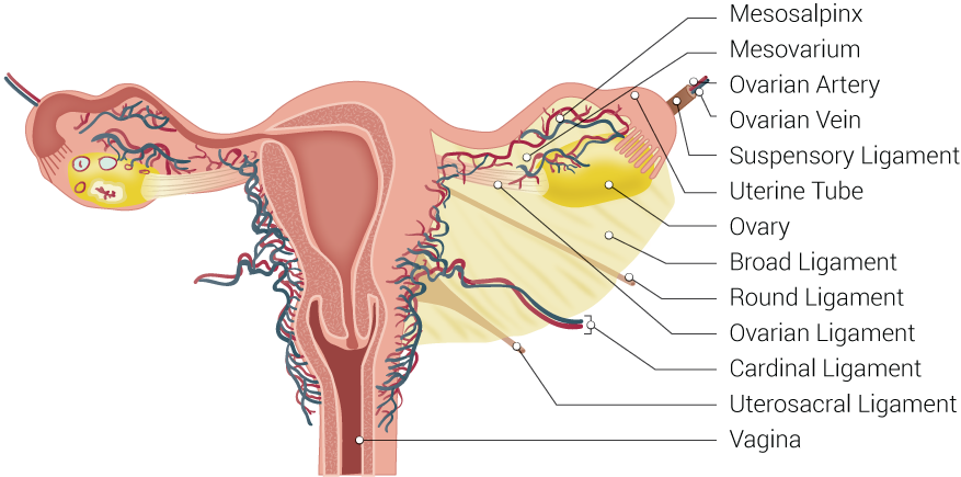
Uterine Tubal Anatomy and Ligaments. Shown in this illustration are anatomical structures surrounding the uterus and fallopian or uterine tubes, including the mesosalpinx, mesovarium, ovarian artery, ovarian vein, suspensory ligament, uterine tube, ovary, broad ligament, round ligament, ovarian ligament, cardinal ligament, uterosacral ligament, and vagina.
Illustrated by B Palmer
References
Kaniewska M, Gołofit P, Heubner M, Maake C, Kubik-Huch RA. Suspensory Ligaments of the Female Genital Organs: MRI Evaluation with Intraoperative Correlation. Radiographics : a review publication of the Radiological Society of North America, Inc. 2018 Nov-Dec:38(7):2195-2211. doi: 10.1148/rg.2018180089. Epub [PubMed PMID: 30422765]
Simó Alari F, Caveriviere P, Gutierrez I, Gillon C. Laparoscopic excision of round ligament endometrioma. BMJ case reports. 2018 Jun 29:2018():. pii: bcr-2017-223613. doi: 10.1136/bcr-2017-223613. Epub 2018 Jun 29 [PubMed PMID: 29960956]
Level 3 (low-level) evidenceBellier A, Cavalié G, Marnas G, Chaffanjon P. The round ligament of the uterus: Questioning its distal insertion. Morphologie : bulletin de l'Association des anatomistes. 2018 Jun:102(337):55-60. doi: 10.1016/j.morpho.2018.04.001. Epub 2018 May 3 [PubMed PMID: 29731327]