 Tools
Tools
 Cerebrovascular Disease
Introduction
A stroke or cerebrovascular accident is an acute compromise of the cerebral perfusion or vasculature. Approximately 85% of strokes are ischemic, and the remaining are hemorrhagic.[1] In this discussion, we mainly confine ourselves to ischemic strokes. Over the past several decades, the incidence of stroke and mortality has been decreasing.[2]
Stroke is the leading cause of adult disability worldwide; therefore, it is critical to recognize and treat this condition rapidly to prevent or minimize morbidity and mortality. There are many risk factors for stroke, with hypertension being the leading risk factor for an ischemic stroke. In younger populations, there are numerous causes of stroke, including clotting disorders, carotid dissection, and illicit drug abuse. In the acute setting, a quick history and examination should be performed as "time is brain". As acute stroke management is evolving rapidly, clinicians must consider patients for intravenous tissue plasminogen activator up to 4.5 hours and mechanical thrombectomy for up to 24 hours.[3][4]
Etiology
Register For Free And Read The Full Article
 Search engine and full access to all medical articles
- 10 free questions in your specialty
- Free CME/CE Activities
- Free daily question in your email
- Save favorite articles to your dashboard
- Emails offering discounts
Learn more about a Subscription to StatPearls Point-of-Care
Etiology
Ischemic etiologies can further be divided into embolic, thrombotic, and lacunar. In general, the common risk factors for stroke include hypertension, diabetes, smoking, obesity, atrial fibrillation, and drug use. Of all the risk factors, hypertension is the most common modifiable risk factor for stroke.[1]
Chronic, uncontrolled hypertension causes small vessel strokes mainly in the internal capsule, thalamus, pons, and cerebellum.[5] Lifestyle measures such as weight loss, salt restriction, and increased intake of fruits and vegetables (such as the Mediterranean diet) are helpful in lowering blood pressure.[6] Every 10 mm Hg reduction in blood pressure is associated with a one-third reduction in stroke risk in primary prevention.[6]
One-third of adults in the United States have elevated low-density lipoprotein levels, which can lead to plaque formation in the intracerebral vasculature. Eventually, due to the excessive plaque build-up, thrombotic strokes occur. In the older population, the risk of cardioembolic stroke increases mainly due to atrial fibrillation.[7] Lacunar strokes contribute to about 20% of all ischemic strokes and result from occlusion of the small penetrating branches of the middle cerebral artery, vertebral or basilar artery, or the lenticulostriate vessels. Typical causes of lacunar strokes include microemboli, fibrinoid necrosis secondary to vasculitis or hypertension, amyloid angiopathy, and hyaline arteriosclerosis.
Epidemiology
Stroke is the fifth leading cause of death in the United States. The incidence of stroke is around 800,000 people annually, and stroke is the leading cause of disability.[8] The incidence of stroke has declined, but the morbidity has increased. Due to longer life expectancy, the lifetime risk of stroke is higher in women. Globally, at least 5 million people die from strokes, and millions of others remain disabled.
Pathophysiology
A stroke results from ischemia in a brain region. The Na+/K+ adenosine triphosphatease pumps fail mainly due to poor adenosine triphosphate production and failure of the aerobic mechanism. Ischemia leads to cellular depolarization, resulting in calcium influx, elevated lactic acid, acidosis, and free radicals. Cell death increases glutamate levels and triggers a cascade of chemical reactions (excitotoxicity).[9]
History and Physical
The most important piece of historical information that the clinician should obtain is the time of symptoms onset or the time last seen normal. This is critical because it determines eligibility for recombinant tissue plasminogen activator (rtPA) or endovascular intervention for stroke.[10] Other important information to obtain is risk factors for arteriosclerosis and cardiovascular disease, diabetes, smoking, atrial fibrillation, drug abuse, migraine, seizures, infection, trauma, or pregnancy.
The stroke exam is a multi-person, coordinated, rapid exam. While the staff obtain vitals, attach telemetry, and obtain intravenous access, the clinician performs a rapid neurological evaluation. The National Institutes of Health Stroke Scale (NIHSS) is routinely used to get the baseline evaluation. The exam has to be rapid as “time is brain.” Clinicians must examine the following items:
- The level of consciousness (alert and responsive, arouses to noxious stimuli, comatose…)
- Language (fluency, naming, comprehension, repetition)
- Dysarthria (slurring), which may be picked up in the history
- Motor (subtle arm weakness can be picked up by performing a pronator drift)
- Visual field deficits
- Eye movement abnormalities (in general, if a gaze preference is present, the eyes deviate towards the side of the lesion)
- Facial paralysis (asking the patient to smile)
- Ataxia (finger to nose)
With a good history and physical exam, the stroke can be localized. There are various stroke syndromes.
Anterior Cerebral Artery Infarction
There is a significant collateral blood supply in the anterior cerebral artery (ACA) territory. Pure ACA strokes are rare. The ACA distribution mainly involves the Broca area, the primary motor and sensory cortices, and the prefrontal cortex. Patients present with motor aphasia, personality issues, and contralateral leg weakness and numbness; hands and face are usually spared.
Middle Cerebral Artery Infarction
The middle cerebral artery includes the main trunk (M1), and it divides into 2 M2 branches. The M1 (horizontal branch) supplies the basal ganglia, and the M2 (Sylvian branches) supplies part of the parietal, frontal, and temporal lobes. As MCA supplies a wide territory; therefore it is extremely important to rule out MCA occlusion. The MCA syndrome causes contralateral arm and facial numbness and weakness, and gaze deviation towards the affected side. Aphasia in the left-sided lesions and neglect in the right-sided lesions.
Posterior Cerebral Artery Infarction
The posterior cerebral artery (PCA) mainly supplies the occipital lobe, thalamus, and some portion of the temporal lobe. The classic presentation of PCA stroke is homonymous hemianopsia. Apart from this hypersomnolence, cognitive issues, and hemisensory loss can be seen when the deep PCA is involved. Sometimes there is bilateral infarction of distal PCAs producing cortical blindness, and the patient is unaware of the blindness and denies it; this is called Anton-Babinski syndrome.[11]
Cerebellar Infarction
Patients with cerebellar strokes present with ataxia, dysarthria, nausea, vomiting, and vertigo. Lacunar strokes are due to occlusion of small perforating vessels and can be pure motor, pure sensory, or ataxic hemiparetic strokes. In general, these strokes don't impair memory, cognition, level of consciousness, or speech.
The stroke can be quantified by the NIHSS scale, which includes the following:
- Visual function
- Level of consciousness
- Sensation and neglect
- Motor function
- Cerebellar function
- Language
A high score suggests proximal vessel occlusion.
Evaluation
The initial workup of a patient with stroke involves stabilizing the airway, breathing, and circulation (ABC). This is followed by a rapid, concise history and exam, such as the NIHSS, which is administered simultaneously while the patient receives intravenous access, telemetry is initiated, and labs are drawn. The patient should then get a stat noncontrast head computed tomogram (CT) or a combination of head CT, CT angiography, and perfusion imaging.
"Time is brain," therefore, time is of the essence. Ideally, rtPA should be prepared as imaging is occurring, and as soon as the noncontrasted head CT can be visualized, and a bleed is excluded, rtPA should be administered after discussing the risks and benefits, and excluding rtPA contraindications. Time is critical, as only patients who receive all the required studies within 4.5 hours qualify for potentially lifesaving thrombolysis. After intravenous rtPA, the CT angiography should be reviewed to determine whether the patient also qualifies for endovascular therapy.
The earliest CT sign of stroke is:
- A hyperdense vessel sign, representing direct visualization of an intravascular thrombus or embolus, most commonly involving the middle cerebral artery, is seen as the hyperdense middle cerebral artery sign or middle cerebral artery dot sign.
Interpretation of CT perfusion scan:
- The infarct core demonstrates a matched decrease in cerebral blood volume (CBV) and a prolonged mean transit time (MTT), reflecting irreversibly infarcted tissue with absent or severely reduced perfusion.
- The ischemic penumbra demonstrates prolonged MTT with preserved CBV, reflecting hypoperfused but potentially salvageable brain tissue.
Interpretation of MRI scan:
- Early hyperacute: Increased diffusion-weighted image signal and reduced apparent diffusion coefficient values
- Late hyperacute (>6 hours): T2 signal in fluid-attenuated inversion recovery image
In recent years, there have been significant advancements in acute stroke care. Results from multiple stroke trials in 2015 showed that endovascular thrombectomy in the first 6 hours is much better than standard medical care in patients with large vessel occlusion in the arteries of the proximal anterior circulation. These benefits sustained irrespective of geographical location and patient characteristics.[12]
Again in 2018, a significant paradigm shift happened in stroke care. The DAWN trial demonstrated significant benefits of endovascular thrombectomy in patients with large-vessel occlusion in the proximal anterior circulation. This trial extended the stroke window up to 24 hours in selected patients using perfusion imaging. As a result, more patients can be treated, even for up to 24 hours.[3]
Following these monumental shifts, the 2019 American Heart Association/American Stroke Association guidelines for the early management of acute ischemic stroke formalized these advancements and introduced critical updates regarding thrombolytic selection and extended treatment windows. A pivotal change was the strong endorsement of advanced multimodal neuroimaging, specifically diffusion-weighted MRI–fluid-attenuated inversion recovery (DWI-FLAIR) mismatch or CT perfusion, to identify salvageable penumbra and to justify intravenous alteplase administration for patients with an unknown time of symptom onset, such as wake-up strokes.
Furthermore, the 2019 guidelines officially recognized tenecteplase (TNK) as a viable alternative to standard rtPA. Specifically, a single-bolus 0.25 mg/kg dose of TNK is now recommended for patients without contraindications who are also eligible for mechanical thrombectomy, largely due to its high fibrin specificity and convenient administration that expedites door-in-door-out transfer times. These updates underscore a definitive transition from strict time-based protocols to highly individualized, tissue-based viability assessments.
All patients should be treated with an antiplatelet agent and a statin and be admitted for full stroke evaluation. Hypertension is often seen in acute stroke. This should not be aggressively treated. A baseline electrocardiogram is recommended. The following labs would be indicated when a diagnosis of stroke is entertained:
- Basic metabolic panel
- Complete blood count
- Cardiac markers
- Coagulation profile: prothrombin time, international normalized ratio, and activated partial thromboplastin time
- Lipid Panel
- Hemoglobin A1C
A transthoracic echocardiogram, telemetry monitoring, and neck vessel imaging are necessary to elucidate the etiology of stroke.
Treatment / Management
Patients with acute ischemic stroke who meet the criteria for rtPA and do not have any contraindications should receive intravenous rtPA. Patients with large-vessel occlusions should be evaluated for possible endovascular intervention. All patients suspected of having an acute ischemic stroke should be admitted for a full neurological workup.
Neurology consultation should be obtained. The workup of acute ischemic stroke includes a search for a source of thrombus, which includes carotid artery evaluation by ultrasound, computed tomography angiography, MR angiography, or conventional angiography. A transthoracic echocardiogram is obtained to ascertain for low ejection fraction, the cardiac source of the clot, or patent foramen ovale.
Electrocardiogram and telemetry are obtained to ascertain for rhythms predisposing to stroke, such as atrial fibrillation. Labs, such as a fasting lipid panel and hemoglobin A1C, are obtained to assess modifiable risk factors for stroke. Other labs, such as a hypercoagulable panel in young individuals or B12 and syphilis testing in selected patients, are also obtained. Antiplatelet and statins remain the mainstay of medical management of stroke. For ischemic stroke, intravenous rtPA within 4.5 hours of stroke onset is the standard of care.
The following patients are excluded from receiving intravenous rtPA if they present between 3 and 4.5 hours:
- Age >80 years
- Severe stroke (NIHSS >25)
- History of diabetes and prior stroke
- Taking an oral anticoagulant regardless of the international normalized ratio (INR) [13]
Contraindications for intravenous rtPA include the following:
- Unclear time and/or unwitnessed symptom onset
- Time last known to be at baseline state is >3 or 4.5 hours
- Current intracranial or subarachnoid hemorrhage
- Severe head trauma or ischemic stroke within 3 months
- Cranial or spinal surgery within 3 months
- Gastrointestinal malignancy or bleed within 21 days
- Active internal bleeding
- Coagulopathy (platelets <100,000/mm, INR >1.7, activated partial thromboplastin time >40 s, or prothrombin time >15 s)
- Receiving a dose of low-molecular-weight heparin within 24 hours
- Infective endocarditis
- Direct thrombin inhibitors or direct factor Xa inhibitors
- Glycoprotein IIb/IIIa receptor inhibitors
- Intra-axial intracranial neoplasm
- Current severe uncontrolled hypertension (not maintained less than or equal to 185/110 mm Hg) [13]
The indications for mechanical thrombectomy with a stent retriever in a patient aged
older than 18 include:
- Minimal prestroke disability
- Causative occlusion of the internal carotid artery or proximal MCANIHSS of ≥6
- Reassuring noncontrast head CT [Alberta Stroke Program Early Computed Tomography Score (ASPECT) score of ≥6]
- If the patient can be treated within 6 hours of the last known normal [13]
Building upon these established timelines, the 2019 American Heart Association/American Stroke Association guidelines dramatically modernized our approach by shifting focus from strict time clocks to tissue-based viability. Crucially, the guidelines extended the window for mechanical thrombectomy to 24 hours from last known normal for patients who met specific clinical and imaging mismatch criteria (based on the DAWN and DEFUSE 3 trials). For intravenous thrombolysis, the guidelines officially endorsed the use of advanced imaging, such as MRI demonstrating a DWI-FLAIR mismatch, to identify salvageable tissue and justify administering intravenous alteplase in patients with an unknown time of onset or wake-up strokes.
Furthermore, TNK, administered as a single 0.25 mg/kg bolus, was introduced as a safe, highly fibrin-specific alternative to alteplase, particularly for patients without contraindications who are eligible for mechanical thrombectomy. Finally, for patients presenting with minor, non-disabling strokes (NIHSS ≤ 3) or high-risk transient ischemic attacks who do not receive thrombolytics, the 2019 updates strongly recommend initiating dual antiplatelet therapy (aspirin and clopidogrel) within 24 hours and continuing for 21 days to prevent early recurrence.
For the above, see:
- Thomalla G, et al. (WAKE-UP Investigators). MRI-Guided Thrombolysis for Stroke with Unknown Time of Onset. N Engl J Med. 2018. (Established the use of MRI DWI-FLAIR mismatch for treating wake-up strokes with alteplase).
- Ma H, et al. (EXTEND Investigators). Thrombolysis Guided by Perfusion Imaging up to 9 Hours after Onset of Stroke. N Engl J Med. 2019. (Supported extending the IV thrombolysis window using CT or MRI perfusion imaging).
- Johnston SC, et al. (POINT Investigators). Clopidogrel and Aspirin in Acute Ischemic Stroke and High-Risk TIA. N Engl J Med. 2018. (The primary evidence for using 21 days of dual antiplatelet therapy in minor stroke/high-risk TIA).
- Menon BK, et al. (AcT Trial Investigators). Intravenous tenecteplase compared with alteplase for acute ischaemic stroke in Canada (AcT): a pragmatic, multicentre, open-label, registry-linked, randomised, controlled, non-inferiority trial. Lancet. 2022. (A critical modern study confirming TNK is an excellent, non-inferior alternative to alteplase for all-comers, further cementing its use beyond just thrombectomy-eligible patients).
A notable potential complication after fibrinolytic therapy is hemorrhagic transformation. Hemorrhagic transformation is classified as hemorrhagic infarction and parenchymal hematoma, each with 2 subsets. Predictive factors for the occurrence of this complication include increased infarction area, gray matter location, atrial fibrillation, cerebral embolism, acute hyperglycemia, low platelet count, and poor collateral circulation.[14]
Aspirin is recommended within 24 to 48 hours after stroke onset; its administration can be delayed for 24 hours in patients treated with intravenous rtPA. Mono/dual antiplatelet therapy is not a contraindication for receiving rtPA. Determining the optimal timing for initiation of anticoagulation in patients with atrial fibrillation following an acute ischemic stroke remains a complex clinical challenge. Usually, it depends on various factors, such as stroke size and other comorbidities. Usually, if the stroke is small to moderate and there is no hemorrhage, we start anticoagulation at 7 to 14 days.[15](B3)
Sometimes there are patients with small hemorrhagic transformation after an acute stroke, and in this scenario, it is better to wait for anticoagulation for a couple of weeks. This delay is not associated with an increased risk of stroke recurrence.[16] For patients with significant disabilities, physical therapy and occupational therapy consults should be obtained.(B3)
Similarly, if swallowing and speech are of concern, then speech/swallow consults should be obtained. All patients should have follow-ups arranged with their primary care clinicians and neurology at appropriate post-discharge intervals. For symptomatic and significant carotid artery stenosis, referrals to vascular or neurological surgery should be sought.
The patient's comorbidity has to be addressed, and one should avoid hyperthermia. Oxygen supplementation is recommended when the oxygen saturation is less than 94%. Both hypo- and hyperglycemia need to be addressed as they can affect the outcome.
Significant cerebral edema can occur after a stroke, and thus, a CT scan should be performed in patients who have altered mentation. Mannitol can be used, but there is no evidence to support routine corticosteroid use. Patient positioning, hyperosmolar therapy, hyperventilation, and barbiturate coma may be used to reduce intracranial edema.
Seizures occur in about 15% of patients within the first few days of the stroke. Those who develop chronic seizures need treatment. Understanding that palliative care is also a critical component of stroke management is important. Some patients have a severe stroke and are incapacitated. Thus, clinicians need to discuss end-of-life care, palliation, and do-not-resuscitate orders with the family.
Differential Diagnosis
The differential diagnosis is broad and can include stroke mimics such as:
- Transient ischemic attack
- Hypoglycemia
- Hyponatremia
- Hemiplegic migraine
- Encephalitis
- Brain abscess
- Cerebral neoplasm
- Syncope
- Conversion disorder [14]
Prognosis
Stroke continues to carry high morbidity and mortality. One-year survival for stroke varies from 60% to 80%. For those who survive, the recovery is prolonged, and the risk of another stroke is high. Most patients remain disabled or have partial neurological deficits following a stroke that prevent them from being active in the workforce.
Complications
Complications after a stroke include worsening neurological status from the extension of the stroke or hemorrhagic conversion and a multitude of other complications associated with prolonged immobility and hospitalization. According to the results from a multicenter study, the incidence of complications after a stroke in the acute phase was:
- Recurrent stroke: 9%
- Epileptic seizure: 3%
- Urinary tract infection: 24%
- Pneumonia: 22%
- Pressure sores: 21%
- Deep venous thrombosis: 2%
- Pulmonary embolism: 1%
- Depression: 16%
- Anxiety: 14% [17]
Deterrence and Patient Education
Stroke prevention, whether primary or secondary, is the primary treatment goal. Patients at the highest risk should be identified early and counseled on lifestyle changes as well as control of comorbid conditions to prevent this devastating outcome. Conditions that increase a patient's risk for cerebrovascular accidents include uncontrolled diabetes mellitus, uncontrolled hypertension, nicotine abuse, and atrial fibrillation. These are the most common and highly prevalent diseases; however, other conditions are also linked to vascular pathologies, such as vasculitides and certain autoimmune diseases. Primary care and specialty clinicians must diligently identify these patients and provide focused counseling and aggressive therapy for underlying diseases to prevent this outcome.
The American Stroke Association has suggested the acronym ACT FAST to recognize the early symptoms of a stroke. They include:
- F (face): A droop or an uneven smile on a person’s face.
- A (arms): Arm numbness or weakness, elicited by asking the patient to lift the arms
- S (speech difficulty): Slurred speech or difficulty in understanding speech
- T (time): If any of the above features are present, even if transient, call the emergency helpline (911).
The additional symptoms of stroke to be watched for are:
- Sudden numbness
- Sudden confusion
- Sudden trouble seeing
- Sudden trouble walking
- Sudden severe headache
Pearls and Other Issues
Other issues to remember:
- A stroke generally presents with an acute focal neurologic deficit with no pain.
- The most important part of the history is the time of symptom onset.
- Rapid evaluation and treatment are paramount for the best outcomes.
- The use of a stroke rating scale, preferably the NIHSS, is recommended, and the AHA/ASA recommends that all clinicians who care for patients with stroke be certified in the NIHSS.
- Intravenous rtPA is approved for use up to 4.5 hours after symptom onset, but the earlier it is administered, the better the outcomes.
- At a minimum, a noncontrast head CT should be obtained to rule out hemorrhage before administering intravenous rtPA.
- The only lab needed before administering rtPA is a finger-stick serum glucose, unless a bleeding diathesis or clinical situation warrants more labs.
- Endovascular or interventional therapy should be considered in patients who have significant deficits (NIHSS >6) up to 6 hours following symptom onset if treatment can be initiated within 6 hours.
- The risk of intracranial hemorrhage in stroke mimics treated with intravenous rtPA is extremely low (~1%).
- For patients who do not qualify for rtPA, permissive hypertension is indicated for at least the first 24 hours, and blood pressure should not be lowered unless it exceeds 220/120 mm Hg.
- Before initiating rtPA, blood pressure should be gently lowered to a value below 185/110 mm Hg.
- After rtPA, blood pressure should be kept below 180/105 mm Hg for 24 hours.
- For patients who do not receive rtPA, an antiplatelet agent and statin should be administered.
Enhancing Healthcare Team Outcomes
Stroke continues to carry high morbidity and mortality. In addition, the cost to the healthcare system is enormous. Even though treatments for strokes exist, the outcomes are not satisfactory, and many people remain permanently disabled.
To improve outcomes, there is ample evidence that stroke management is best delivered by an interprofessional team that includes the emergency department clinician, nurse practitioner, neurologist, radiologist, and stroke team. The key is to first identify if the stroke is embolic or hemorrhagic and then institute thrombolytic treatment accordingly. For those who recover, physical, speech, and occupational therapy may be necessary.
Early identification of a stroke is crucial. The nursing staff must be trained to identify potential stroke victims and immediately contact the clinician for evaluation. Once the diagnosis is made, depending on the type, clinicians should work with the pharmacist to perform rapid screening for medication interactions and implement treatment promptly.
However, today the focus is on stroke prevention as it is more cost-effective. All clinicians are responsible for educating the public on methods to prevent strokes. The pharmacist should encourage blood pressure medication compliance since uncontrolled blood pressure is one of the risk factors for stroke. Clinicians, including nurses, should educate patients on quitting smoking, eating a healthy diet, exercising regularly, and maintaining a healthy body weight. Patients should be provided with supporting literature and educated about stroke and its complications.
For those who have developed a stroke, one may need to involve a social worker to ensure that the home is a safe place and that the patient has the support systems. A dietary consultation should be made to educate the patient on what to eat. In addition, some patients may need to enroll in rehabilitation to regain speech, muscle, and joint function. Only through an interprofessional team approach and open communication can this lead to improved outcomes and a better quality of life.
Outcomes
The outcomes for patients with mild embolic strokes are good, but those with hemorrhagic strokes tend to have poor outcomes.[18][19]
Media
(Click Image to Enlarge)
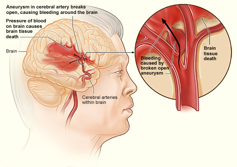
Hemorrhagic Stroke Process. The illustration shows how a hemorrhagic stroke can occur in the brain. An aneurysm in a cerebral artery breaks open, which causes bleeding in the brain. The pressure of the blood causes brain tissue death.
National Heart Lung and Blood Institute
(Click Video to Play)
Hemiballism, Involuntary Movement, Cerebrovascular Accident, Stroke, Subthalamic Nucleus
Contributed by RS Menon, MD
(Click Image to Enlarge)
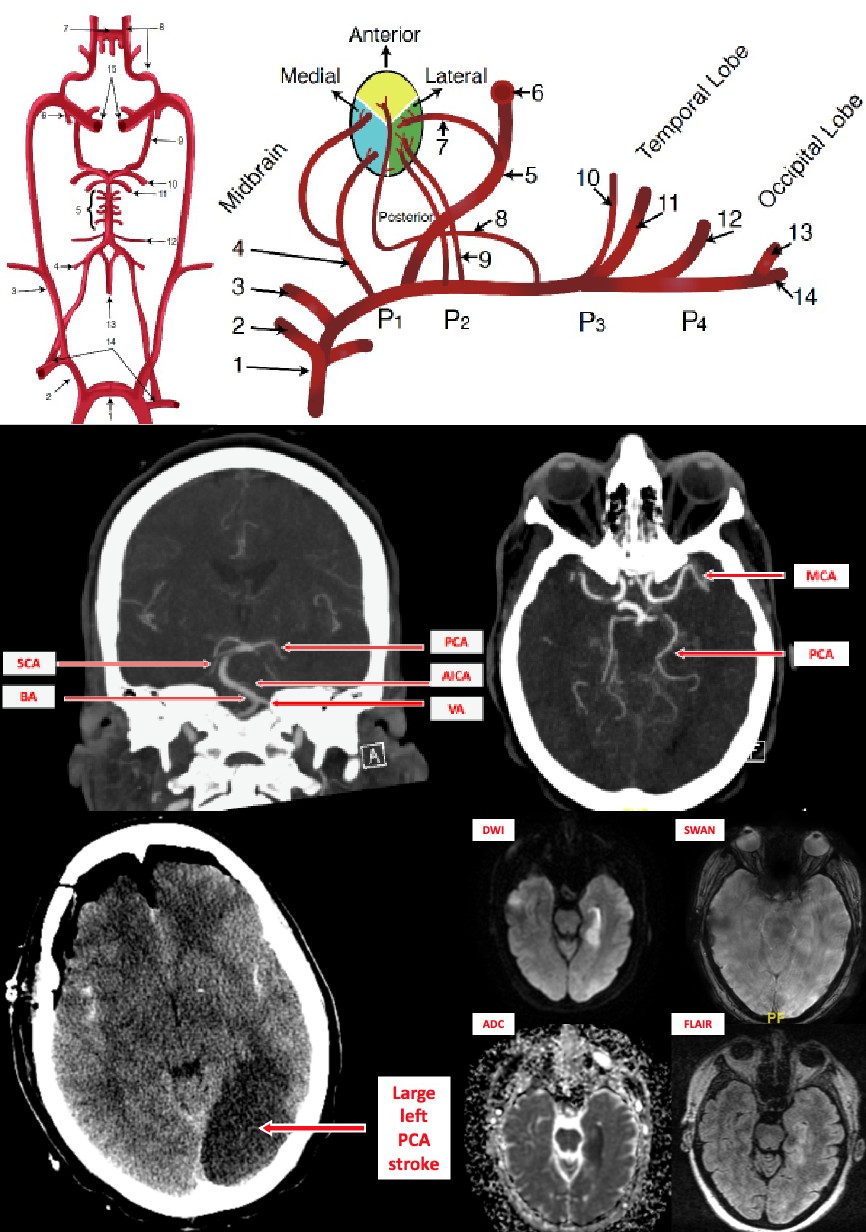
Stroke, Posterior Cerebral Artery.
Contributed by O KUYBU, MD
(Click Image to Enlarge)
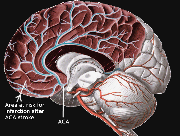
Anterior Cerebral Artery Stroke. Infarcts involving the territory of the anterior cerebral artery (ACA) account for approximately 20% of all ischemic strokes. The ACA itself often divides into 5 segments, usually labeled as A1 through A5, or as proximal (A1), ascending (A2, A3), and horizontal segments.
Contributed by S Bhimji, MD
(Click Image to Enlarge)
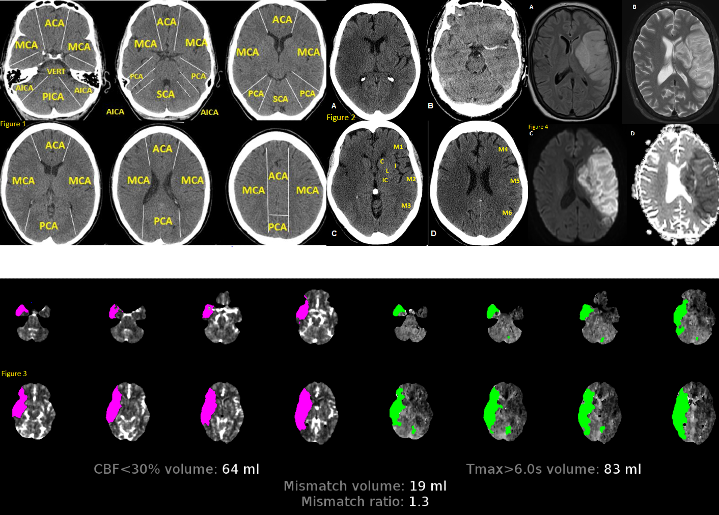
Anatomy of Brain Vascular Territories. Figure 1: ACA (anterior cerebral artery), MCA (middle cerebral artery), PCA (posterior cerebral artery), AICA (anterior inferior cerebellar artery), PICA (posterior inferior cerebellar artery), and SCA (superior cerebellar artery). Figure 2: Noncontrast CT shows loss of gray-white matter differentiation in the right MCA territory, consistent with acute large right MCA infarction. Figure 3: CT perfusion imaging reveals stroke; areas with increased mean transit time and time-to-peak/time-to-maximum, along with decreased cerebral blood volume and cerebral blood flow, indicate infarct core. Figure 4: MRI shows large left MCA infarction. It appears hyperintense on FLAIR (A) and T2 (B), with mass effect suggesting subacute infarction. Diffusion restriction is confirmed by hyperintensity on DWI (C) and hypointensity on ADC map (D).
Contributed by O Shafaat, MD
References
Writing Group Members, Mozaffarian D, Benjamin EJ, Go AS, Arnett DK, Blaha MJ, Cushman M, Das SR, de Ferranti S, Després JP, Fullerton HJ, Howard VJ, Huffman MD, Isasi CR, Jiménez MC, Judd SE, Kissela BM, Lichtman JH, Lisabeth LD, Liu S, Mackey RH, Magid DJ, McGuire DK, Mohler ER 3rd, Moy CS, Muntner P, Mussolino ME, Nasir K, Neumar RW, Nichol G, Palaniappan L, Pandey DK, Reeves MJ, Rodriguez CJ, Rosamond W, Sorlie PD, Stein J, Towfighi A, Turan TN, Virani SS, Woo D, Yeh RW, Turner MB, American Heart Association Statistics Committee, Stroke Statistics Subcommittee. Heart Disease and Stroke Statistics-2016 Update: A Report From the American Heart Association. Circulation. 2016 Jan 26:133(4):e38-360. doi: 10.1161/CIR.0000000000000350. Epub 2015 Dec 16 [PubMed PMID: 26673558]
Fang MC, Coca Perraillon M, Ghosh K, Cutler DM, Rosen AB. Trends in stroke rates, risk, and outcomes in the United States, 1988 to 2008. The American journal of medicine. 2014 Jul:127(7):608-15. doi: 10.1016/j.amjmed.2014.03.017. Epub 2014 Mar 25 [PubMed PMID: 24680794]
Nogueira RG, Jadhav AP, Haussen DC, Bonafe A, Budzik RF, Bhuva P, Yavagal DR, Ribo M, Cognard C, Hanel RA, Sila CA, Hassan AE, Millan M, Levy EI, Mitchell P, Chen M, English JD, Shah QA, Silver FL, Pereira VM, Mehta BP, Baxter BW, Abraham MG, Cardona P, Veznedaroglu E, Hellinger FR, Feng L, Kirmani JF, Lopes DK, Jankowitz BT, Frankel MR, Costalat V, Vora NA, Yoo AJ, Malik AM, Furlan AJ, Rubiera M, Aghaebrahim A, Olivot JM, Tekle WG, Shields R, Graves T, Lewis RJ, Smith WS, Liebeskind DS, Saver JL, Jovin TG, DAWN Trial Investigators. Thrombectomy 6 to 24 Hours after Stroke with a Mismatch between Deficit and Infarct. The New England journal of medicine. 2018 Jan 4:378(1):11-21. doi: 10.1056/NEJMoa1706442. Epub 2017 Nov 11 [PubMed PMID: 29129157]
Powers WJ, Rabinstein AA, Ackerson T, Adeoye OM, Bambakidis NC, Becker K, Biller J, Brown M, Demaerschalk BM, Hoh B, Jauch EC, Kidwell CS, Leslie-Mazwi TM, Ovbiagele B, Scott PA, Sheth KN, Southerland AM, Summers DV, Tirschwell DL. Guidelines for the Early Management of Patients With Acute Ischemic Stroke: 2019 Update to the 2018 Guidelines for the Early Management of Acute Ischemic Stroke: A Guideline for Healthcare Professionals From the American Heart Association/American Stroke Association. Stroke. 2019 Dec:50(12):e344-e418. doi: 10.1161/STR.0000000000000211. Epub 2019 Oct 30 [PubMed PMID: 31662037]
Shi Y, Wardlaw JM. Update on cerebral small vessel disease: a dynamic whole-brain disease. Stroke and vascular neurology. 2016 Sep:1(3):83-92. doi: 10.1136/svn-2016-000035. Epub 2016 Oct 25 [PubMed PMID: 28959468]
Lawes CM, Bennett DA, Feigin VL, Rodgers A. Blood pressure and stroke: an overview of published reviews. Stroke. 2004 Apr:35(4):1024 [PubMed PMID: 15053002]
Level 2 (mid-level) evidenceFerro JM. Cardioembolic stroke: an update. The Lancet. Neurology. 2003 Mar:2(3):177-88 [PubMed PMID: 12849239]
Krishnamurthi RV, Moran AE, Feigin VL, Barker-Collo S, Norrving B, Mensah GA, Taylor S, Naghavi M, Forouzanfar MH, Nguyen G, Johnson CO, Vos T, Murray CJ, Roth GA, GBD 2013 Stroke Panel Experts Group. Stroke Prevalence, Mortality and Disability-Adjusted Life Years in Adults Aged 20-64 Years in 1990-2013: Data from the Global Burden of Disease 2013 Study. Neuroepidemiology. 2015:45(3):190-202. doi: 10.1159/000441098. Epub 2015 Oct 28 [PubMed PMID: 26505983]
Xing C, Arai K, Lo EH, Hommel M. Pathophysiologic cascades in ischemic stroke. International journal of stroke : official journal of the International Stroke Society. 2012 Jul:7(5):378-85. doi: 10.1111/j.1747-4949.2012.00839.x. Epub [PubMed PMID: 22712739]
Yew KS, Cheng E. Acute stroke diagnosis. American family physician. 2009 Jul 1:80(1):33-40 [PubMed PMID: 19621844]
Das JM, Naqvi IA. Anton Syndrome. StatPearls. 2026 Jan:(): [PubMed PMID: 30844182]
Goyal M, Menon BK, van Zwam WH, Dippel DW, Mitchell PJ, Demchuk AM, Dávalos A, Majoie CB, van der Lugt A, de Miquel MA, Donnan GA, Roos YB, Bonafe A, Jahan R, Diener HC, van den Berg LA, Levy EI, Berkhemer OA, Pereira VM, Rempel J, Millán M, Davis SM, Roy D, Thornton J, Román LS, Ribó M, Beumer D, Stouch B, Brown S, Campbell BC, van Oostenbrugge RJ, Saver JL, Hill MD, Jovin TG, HERMES collaborators. Endovascular thrombectomy after large-vessel ischaemic stroke: a meta-analysis of individual patient data from five randomised trials. Lancet (London, England). 2016 Apr 23:387(10029):1723-31. doi: 10.1016/S0140-6736(16)00163-X. Epub 2016 Feb 18 [PubMed PMID: 26898852]
Level 1 (high-level) evidencePowers WJ, Rabinstein AA, Ackerson T, Adeoye OM, Bambakidis NC, Becker K, Biller J, Brown M, Demaerschalk BM, Hoh B, Jauch EC, Kidwell CS, Leslie-Mazwi TM, Ovbiagele B, Scott PA, Sheth KN, Southerland AM, Summers DV, Tirschwell DL, American Heart Association Stroke Council. 2018 Guidelines for the Early Management of Patients With Acute Ischemic Stroke: A Guideline for Healthcare Professionals From the American Heart Association/American Stroke Association. Stroke. 2018 Mar:49(3):e46-e110. doi: 10.1161/STR.0000000000000158. Epub 2018 Jan 24 [PubMed PMID: 29367334]
Tadi P, Lui F. Acute Stroke. StatPearls. 2026 Jan:(): [PubMed PMID: 30570990]
Paciaroni M, Agnelli G, Falocci N, Tsivgoulis G, Vadikolias K, Liantinioti C, Chondrogianni M, Bovi P, Carletti M, Cappellari M, Zedde M, Ntaios G, Karagkiozi E, Athanasakis G, Makaritsis K, Silvestrelli G, Lanari A, Ciccone A, Putaala J, Tomppo L, Tatlisumak T, Abdul-Rahim AH, Lees KR, Alberti A, Venti M, Acciarresi M, D'Amore C, Becattini C, Mosconi MG, Cimini LA, Soloperto R, Masotti L, Vannucchi V, Lorenzini G, Tassi R, Guideri F, Acampa M, Martini G, Sohn SI, Marcheselli S, Mumoli N, De Lodovici ML, Bono G, Furie KL, Tadi P, Yaghi S, Toni D, Letteri F, Tassinari T, Kargiotis O, Lotti EM, Flomin Y, Mancuso M, Maccarrone M, Giannini N, Bandini F, Pezzini A, Poli L, Padovani A, Scoditti U, Denti L, Consoli D, Galati F, Sacco S, Carolei A, Tiseo C, Gourbali V, Orlandi G, Giuntini M, Chiti A, Giorli E, Gialdini G, Corea F, Ageno W, Bellesini M, Colombo G, Monaco S, Maimone Baronello M, Karapanayiotides T, Caso V. Early Recurrence and Major Bleeding in Patients With Acute Ischemic Stroke and Atrial Fibrillation Treated With Non-Vitamin-K Oral Anticoagulants (RAF-NOACs) Study. Journal of the American Heart Association. 2017 Nov 29:6(12):. doi: 10.1161/JAHA.117.007034. Epub 2017 Nov 29 [PubMed PMID: 29220330]
Level 3 (low-level) evidencePaciaroni M, Bandini F, Agnelli G, Tsivgoulis G, Yaghi S, Furie KL, Tadi P, Becattini C, Zedde M, Abdul-Rahim AH, Lees KR, Alberti A, Venti M, Acciarresi M, D'Amore C, Mosconi MG, Cimini LA, Altavilla R, Volpi G, Bovi P, Carletti M, Rigatelli A, Cappellari M, Putaala J, Tomppo L, Tatlisumak T, Marcheselli S, Pezzini A, Poli L, Padovani A, Masotti L, Vannucchi V, Sohn SI, Lorenzini G, Tassi R, Guideri F, Acampa M, Martini G, Ntaios G, Athanasakis G, Makaritsis K, Karagkiozi E, Vadikolias K, Liantinioti C, Chondrogianni M, Mumoli N, Consoli D, Galati F, Sacco S, Carolei A, Tiseo C, Corea F, Ageno W, Bellesini M, Colombo G, Silvestrelli G, Ciccone A, Lanari A, Scoditti U, Denti L, Mancuso M, Maccarrone M, Ulivi L, Orlandi G, Giannini N, Gialdini G, Tassinari T, De Lodovici ML, Bono G, Rueckert C, Baldi A, D'Anna S, Toni D, Letteri F, Giuntini M, Lotti EM, Flomin Y, Pieroni A, Kargiotis O, Karapanayiotides T, Monaco S, Maimone Baronello M, Csiba L, Szabó L, Chiti A, Giorli E, Del Sette M, Imberti D, Zabzuni D, Doronin B, Volodina V, Michel P, Vanacker P, Barlinn K, Pallesen LP, Barlinn J, Deleu D, Melikyan G, Ibrahim F, Akhtar N, Gourbali V, Caso V. Hemorrhagic Transformation in Patients With Acute Ischemic Stroke and Atrial Fibrillation: Time to Initiation of Oral Anticoagulant Therapy and Outcomes. Journal of the American Heart Association. 2018 Nov 20:7(22):e010133. doi: 10.1161/JAHA.118.010133. Epub [PubMed PMID: 30571487]
Level 3 (low-level) evidenceLanghorne P, Stott DJ, Robertson L, MacDonald J, Jones L, McAlpine C, Dick F, Taylor GS, Murray G. Medical complications after stroke: a multicenter study. Stroke. 2000 Jun:31(6):1223-9 [PubMed PMID: 10835436]
Level 2 (mid-level) evidenceXu Y, Parikh NS, Jiao B, Willey JZ, Boehme AK, Elkind MSV. Decision Analysis Model for Prehospital Triage of Patients With Acute Stroke. Stroke. 2019 Apr:50(4):970-977. doi: 10.1161/STROKEAHA.118.023272. Epub [PubMed PMID: 30908159]
Rohde D, Gaynor E, Large M, Conway O, Bennett K, Williams DJ, Callaly E, Dolan E, Hickey A. Stroke survivor cognitive decline and psychological wellbeing of family caregivers five years post-stroke: a cross-sectional analysis. Topics in stroke rehabilitation. 2019 Apr:26(3):180-186. doi: 10.1080/10749357.2019.1590972. Epub 2019 Mar 23 [PubMed PMID: 30907273]
Level 2 (mid-level) evidence