 Tools
Tools
 Anatomy, Shoulder and Upper Limb, Arm Quadrangular Space
Introduction
The quadrangular (or quadrilateral) space is named for the shape of its anatomic boundaries (see Image. Quadrangular Space, Posterior View). The 4 borders include the teres minor (superior border), teres major (inferior border), long head of the triceps brachii (medial border), and surgical neck of the humerus (lateral border).[1] The posterior humeral circumflex artery (PHCA) and axillary nerve exit the axilla and pass through the quadrangular space to the posterolateral shoulder. The axillary nerve arises from the posterior cord of the brachial plexus and typically divides into muscular and cutaneous branches proximal to or within the quadrangular space.[2] The PHCA bifurcates into anterior and posterior branches within the space.[3]
The quadrangular space serves as an important anatomic landmark in multiple diagnostic and surgical procedures. Quadrangular space syndrome (QSS) is characterized by symptoms resulting from compression of the axillary nerve or PHCA and may arise from various etiologies. Understanding the anatomy and functional significance of the quadrangular space is essential for the evaluation and management of various upper-extremity conditions.
Structure and Function
Register For Free And Read The Full Article
 Search engine and full access to all medical articles
- 10 free questions in your specialty
- Free CME/CE Activities
- Free daily question in your email
- Save favorite articles to your dashboard
- Emails offering discounts
Learn more about a Subscription to StatPearls Point-of-Care
Structure and Function
Structure
The quadrangular space is defined by 4 anatomic borders. The superior border is formed by the teres minor, the inferior border by the teres major, the medial border by the long head of the triceps brachii, and the lateral border by the surgical neck of the humerus. The axillary nerve courses superior to the PHCA within the quadrangular space.[4]
Function
The quadrangular space serves as a passageway for the axillary nerve and PHCA as these structures exit the axilla and supply the posterolateral shoulder.[5] The axillary nerve is a terminal branch of the posterior cord of the brachial plexus and innervates the teres minor, deltoid, glenohumeral joint, and skin overlying the inferior deltoid region. The PHCA supplies approximately 64% of the blood flow to the humeral head.[6]
Blood Supply and Lymphatics
The axillary artery is divided into 3 parts by the pectoralis minor. The 1st part lies medial to the pectoralis minor and gives rise to 1 branch, the superior thoracic artery. The 2nd part lies posterior to the pectoralis minor and gives rise to 2 branches, the thoracoacromial and lateral thoracic arteries. The thoracoacromial trunk terminates into 4 branches: pectoral, deltoid, acromial, and clavicular. The 3rd part lies lateral to the pectoralis minor and gives rise to 3 branches: the PHCA, subscapular artery, and anterior humeral circumflex artery (AHCA).[7] The PHCA divides into anterior and posterior branches within the quadrangular space. These branches primarily perfuse the superior, inferior, and lateral aspects of the humeral head and surrounding musculature. Branches of the PHCA course around the surgical neck of the humerus and supply most of the proximal humerus.
The AHCA was previously considered the primary arterial supply to the proximal humerus. However, a study by Hettrich et al redefined current understanding through quantitative assessment of the relative contributions of the humeral circumflex arteries to proximal humeral perfusion. Approximately 64% of humeral head perfusion was found to derive from the PHCA, while the remaining 36% originated from the AHCA.[8] Clinical relevance stems from improved understanding of the relatively low rates of osteonecrosis associated with 3- and 4-part proximal humeral fractures.[9]
Nerves
The axillary nerve traverses the quadrangular space after arising from the posterior cord of the brachial plexus, crossing the subscapularis muscle and tendon anteroinferiorly, and passing posteriorly through the space. The axillary nerve courses superior to the PHCA within the quadrangular space and divides into anterior and posterior branches. The anterior branch innervates the anterior deltoid. The posterior branch innervates the posterior deltoid and the teres minor. The posterior branch also gives rise to the superior lateral brachial cutaneous nerve, which innervates the skin over the distal 2/3 of the posterior deltoid. Together, the anterior and posterior branches innervate the middle 3rd of the deltoid and the glenohumeral joint capsule.
The posterior division derivatives receive fibers from C5 to T1 and converge to form the posterior cord of the brachial plexus. The posterior cord gives rise to the upper and lower subscapular, thoracodorsal, axillary, and radial nerves. Spinal root contributions to these nerves are as follows:
- Upper subscapular nerve: C5
- Lower subscapular nerve: C6
- Thoracodorsal nerve: C6 to C8
- Axillary nerve: C5 and C6
- Radial nerve: C5 to T1
Variation in spinal root contributions is particularly relevant to the axillary and radial nerves, which follow the proximal-distal myotome distribution pattern. According to this principle, more rostral spinal nerve roots predominantly innervate proximal muscle groups, whereas more caudal roots innervate distal muscle groups. Consequently, axillary nerve myotomes in the shoulder region are primarily supplied by C5 and C6 fibers. In contrast, radial nerve myotomes involving the posterior arm, forearm, and hand receive contributions from C5, C6, and progressively more caudal roots through T1.[10]
Muscles
Muscles Bordering the Quadrangular Space
Three of the 4 structures delineating the quadrangular space include the teres minor, teres major, and long head of the triceps brachii. The teres minor originates from the lateral border of the scapula and inserts on the greater tubercle of the humerus. The teres minor functions primarily in external rotation of the arm at the glenohumeral joint. The teres major originates from the inferior angle of the scapula and inserts on the medial lip of the intertubercular groove of the humerus. Functions of the teres major include adduction, medial rotation, and extension of the arm.[11] The teres major also contributes to glenohumeral joint stability.[12] The long head of the triceps brachii originates from the infraglenoid tubercle of the scapula and inserts on the olecranon process of the ulna. Functions of the long head of the triceps brachii include extension and adduction of the arm at the glenohumeral joint and extension of the forearm at the elbow.[13]
Deltoid Neurovasculature
The neurovascular supply of the deltoid arises from structures traversing the quadrangular space. The axillary nerve provides motor innervation to the deltoid, while branches of the PHCA supply vascular perfusion to the muscle.
The deltoid originates from the lateral 3rd of the clavicle and the acromion and spine of the scapula, inserting on the deltoid tuberosity of the humerus. The deltoid is composed of 3 parts, each with distinct origins and functions. Collectively, the deltoid primarily abducts the arm, although each segment may function independently. The anterior (clavicular) portion medially rotates, abducts, and flexes the arm. The middle (acromial) portion serves as the primary contributor to arm abduction. The posterior (spinal) portion laterally rotates and extends the arm at the shoulder. Synergistic action between the clavicular and spinal portions also enables contribution to arm adduction.[14]
Physiologic Variants
Anatomical variants of the quadrangular space and its contents are clinically significant, as such anomalies may contribute to pathology and create diagnostic or surgical challenges. Variations may involve the muscles bordering the region. McClelland et al observed quadrangular space narrowing during external and internal rotation of the arm secondary to a fibrous sling connecting the long head of the triceps brachii to the teres major.[15] PHCA tortuosity has been implicated as a cause of QSS in the study by Mohandas Rao et al.[16] The PHCA may also originate from the subscapular artery rather than the 3rd part of the axillary artery, with potential variation in its course near the quadrangular space.
Axillary nerve branching patterns and course variants may also affect the quadrangular space. Beytell et al reported axillary nerve branching within the quadrangular space in 52% of 51 cadavers, while the remaining specimens demonstrated branching proximal to the space. This variability may influence surgical planning for the axillary approach. Pires et al documented an accessory subscapularis muscle passing anterior to the axillary nerve proximal to the quadrilateral space, a variant associated with potential development of QSS.[17]
Surgical Considerations
Quadrangular Space Syndrome
QSS is rare but demonstrates a known predilection for athletes and individuals in certain occupations. The condition is frequently misdiagnosed and requires a high index of suspicion in patients with a history of compressive symptoms and participation in activities involving repetitive overhead arm motion. At-risk populations include baseball pitchers, volleyball players, swimmers, electricians, and painters.[18]
QSS symptoms result from compression of the axillary nerve and PHCA. Reported etiologies include fibrous band formation, traumatic pathology, aneurysmal disease, paralabral cyst formation, the presence of intraarticular loose bodies, neoplastic disease, and hypertrophy of adjacent musculature.[19][20][21] Clinical presentation commonly includes shoulder pain and functional impairment.[22] Pain is typically localized to the posterolateral shoulder and exacerbated by abduction and external rotation.[23] Axillary nerve compression may present with altered sensation over the deltoid region, deltoid atrophy, and weakness during shoulder abduction.
QSS, although uncommon, is an important consideration in the differential diagnosis of shoulder pain. The condition may be confused with rotator cuff tears, adhesive capsulitis, thoracic outlet syndrome, cervical radiculopathy, and suprascapular nerve entrapment. Diagnostic modalities commonly include radiography, magnetic resonance imaging, computed tomography, ultrasound, arteriography, and electromyography.[24][25]
Nonoperative treatment
Most patients with QSS are managed nonoperatively, with conservative treatment often sufficient for both acute and chronic presentations. Clinical improvement typically occurs within 3 to 6 months. Conservative management includes nonsteroidal anti-inflammatory drug therapy and activity modification. Physical therapy protocols typically include glenohumeral joint mobilization, periscapular and rotator cuff strengthening, and posterior capsular stretching. Injection of 1% lidocaine into the quadrangular space may provide symptomatic relief and serve both diagnostic and therapeutic purposes. This intervention may be performed under ultrasound guidance.
Surgical management
Surgical management is indicated when QSS fails to resolve following conservative treatment. The operative technique depends on the underlying etiology. In vascular QSS (vQSS), the surgical strategy is determined by the extent of PHCA involvement (see Clinical Significance). PHCA aneurysms require resection. Arterial thrombus may be managed with PHCA ligation, with or without thrombolysis. Thromboembolectomy is indicated in cases of PHCA thrombus with digital embolization.[26]
Open decompression may benefit patients with fibrous adhesions or scar tissue causing axillary nerve impingement. Earlier surgical decompression should be considered in the presence of a space-occupying lesion or in cases with significant weakness and functional disability. Paralabral cysts, often associated with glenoid labral pathology, may require cyst decompression and arthroscopic labral repair.
Intraoperative identification and palpation of the axillary nerve and PHCA reduce the risk of iatrogenic injury to these structures. Following quadrangular space decompression, the axillary nerve and PHCA should be palpated while an assistant gently abducts and externally rotates the shoulder. This maneuver confirms unrestricted mobility of the neurovascular structures and preservation of a palpable PHCA pulse throughout the range of motion.[27]
Other Surgical Considerations
Open decompression of the quadrangular space is typically performed via a posterior approach, which facilitates access and dissection of the region. This technique carries an increased risk of injury to the deltoid muscle and its innervation, potentially causing chronic pain and weakness. Careful identification and protection of the axillary nerve and PHCA during dissection is essential.
The interscalene block is commonly used in shoulder surgery. However, this technique is frequently associated with ipsilateral phrenic nerve paralysis. Recent evidence suggests that a combined axillary and suprascapular nerve block may provide an effective alternative to the interscalene approach while preserving phrenic nerve function. Axillary nerve block using this technique involves the injection of an anesthetic within the quadrangular space.[28]
In cases of axillary nerve injury, the thoracodorsal nerve and the nerve to the medial head of the triceps brachii may be transferred to the axillary nerve to restore function. Access to the axillary nerve is achieved by opening the quadrangular space with the patient in the lateral decubitus position.[29]
Clinical Significance
Neurovascular structures within the quadrangular space are essential for upper-extremity function. Misdiagnosis of quadrangular space pathology may occur due to its complex clinical presentation.
Neurovascular Compression Within the Quadrangular Space
QSS occurs when neurovascular structures within the quadrangular space are mechanically compressed. Three types of QSS have been identified.
Neurogenic QSS (nQSS) occurs with compression of the axillary nerve within the quadrangular space. Clinical features of nQSS include quadrangular space tenderness, nondermatomal radicular pain, paresthesia typically localized to the posterior and lateral arm, deltoid fasciculations during abduction, and possible muscle atrophy and weakness secondary to denervation. VQSS occurs with compression of the PHCA within the quadrangular space. PHCA ischemia may result in pain, pallor, and diminished or absent distal pulses. Vascular compression may also lead to PHCA aneurysm formation and thrombus development, with potential distal digital embolization manifesting as cyanosis and cold hands and digits. Combined nQSS and vQSS may occur due to the close anatomical relationship between the PHCA and the axillary nerve within the quadrangular space.
Fibrous band formation within the quadrangular space is the most common cause of QSS. Muscular hypertrophy may also contribute to the development of QSS. Glenohumeral abduction and external rotation are commonly performed in sports involving repetitive overhead activity, including baseball, volleyball, and swimming. Similar biomechanical demands are encountered during occupational and daily activities such as window cleaning and ceiling light installation. Teres major hypertrophy and space-occupying lesions, including paralabral cysts (often associated with inferior labral tears), lipomas, axillary schwannomas, humeral osteochondromas, and fracture fragments, may also contribute to QSS.[30]
QSS is often diagnostically challenging. Radiography, magnetic resonance imaging, computed tomography, ultrasound, and electromyography are useful modalities for investigating underlying causes of treatment-refractory shoulder pain.[31]
Arm Trauma
Proximal humeral fractures may result in traumatic injury to neurovascular structures within or adjacent to the quadrangular space, potentially leading to complications, such as avascular necrosis of the humeral head. Clinical evaluation should include assessment of neurovascular integrity.
Media
(Click Image to Enlarge)
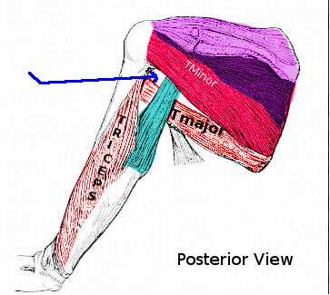
Quadrangular Space, Posterior View. This image shows the borders of the quadrangular space: teres major and minor, long head of the triceps brachii, and surgical neck of the humerus. The axillary nerve and posterior humeral circumflex artery pass through this space.
Contributed by S Bhimji, MD
References
Mishra N, Shirodkar K, Saran S, Pellakuru SR, Shah A, Raniga S, Shah AB, Iyengar KP, Botchu R. Exploring the quadrilateral space: Clinical anatomy, pathology, and imaging insights. Clinical imaging. 2025 May:121():110460. doi: 10.1016/j.clinimag.2025.110460. Epub 2025 Mar 18 [PubMed PMID: 40120476]
Beytell L, Mennen E, van Schoor AN, Keough N. The surgical anatomy of the axillary approach for nerve transfer procedures targeting the axillary nerve. Surgical and radiologic anatomy : SRA. 2023 Jul:45(7):865-873. doi: 10.1007/s00276-023-03168-x. Epub 2023 May 22 [PubMed PMID: 37212871]
Tang A, Varacallo MA. Anatomy, Head and Neck, Posterior Humeral Circumflex Artery. StatPearls. 2026 Jan:(): [PubMed PMID: 30855867]
Rothe C, Lund J, Jenstrup MT, Steen-Hansen C, Lundstrøm LH, Andreasen AM, Lange KHW. A randomized controlled trial evaluating the impact of selective axillary nerve block after arthroscopic subacromial decompression. BMC anesthesiology. 2020 Jan 31:20(1):33. doi: 10.1186/s12871-020-0952-y. Epub 2020 Jan 31 [PubMed PMID: 32005160]
Level 1 (high-level) evidenceFlynn LS, Wright TW, King JJ. Quadrilateral space syndrome: a review. Journal of shoulder and elbow surgery. 2018 May:27(5):950-956. doi: 10.1016/j.jse.2017.10.024. Epub 2017 Dec 20 [PubMed PMID: 29274905]
Mostafa E, Imonugo O, Varacallo MA. Anatomy, Shoulder and Upper Limb, Humerus. StatPearls. 2026 Jan:(): [PubMed PMID: 30521242]
Talbot NC, D'Antoni JV, Soileau LG, Storey NR, Fakoya A. Unusual Origin of the Posterior Circumflex Humeral Artery: A Case Report. Cureus. 2023 Mar:15(3):e36316. doi: 10.7759/cureus.36316. Epub 2023 Mar 17 [PubMed PMID: 37077595]
Level 3 (low-level) evidenceHettrich CM, Boraiah S, Dyke JP, Neviaser A, Helfet DL, Lorich DG. Quantitative assessment of the vascularity of the proximal part of the humerus. The Journal of bone and joint surgery. American volume. 2010 Apr:92(4):943-8. doi: 10.2106/JBJS.H.01144. Epub [PubMed PMID: 20360519]
Khmelnitskaya E, Lamont LE, Taylor SA, Lorich DG, Dines DM, Dines JS. Evaluation and management of proximal humerus fractures. Advances in orthopedics. 2012:2012():861598. doi: 10.1155/2012/861598. Epub 2012 Dec 18 [PubMed PMID: 23316376]
Level 3 (low-level) evidenceOkwumabua E, Black AC, Thompson JH. Anatomy, Shoulder and Upper Limb, Nerves. StatPearls. 2026 Jan:(): [PubMed PMID: 30252312]
Juneja P, Hubbard JB. Anatomy, Shoulder and Upper Limb, Teres Minor Muscle. StatPearls. 2026 Jan:(): [PubMed PMID: 30020696]
Barra-López ME, López-de-Celis C, Pérez-Bellmunt A, Puyalto-de-Pablo P, Sánchez-Fernández JJ, Lucha-López MO. The supporting role of the teres major muscle, an additional component in glenohumeral stability? An anatomical and radiological study. Medical hypotheses. 2020 Aug:141():109728. doi: 10.1016/j.mehy.2020.109728. Epub 2020 Apr 10 [PubMed PMID: 32298921]
Tiwana MS, Sinkler MA, Bordoni B. Anatomy, Shoulder and Upper Limb, Triceps Muscle. StatPearls. 2026 Jan:(): [PubMed PMID: 30725681]
Larionov A, Yotovski P, Link K, Filgueira L. Innervation of the clavicular part of the deltoid muscle by the lateral pectoral nerve. Clinical anatomy (New York, N.Y.). 2020 Nov:33(8):1152-1158. doi: 10.1002/ca.23555. Epub 2020 Jan 13 [PubMed PMID: 31894613]
McClelland D, Paxinos A. The anatomy of the quadrilateral space with reference to quadrilateral space syndrome. Journal of shoulder and elbow surgery. 2008 Jan-Feb:17(1):162-4 [PubMed PMID: 17993281]
Mohandas Rao KG, Somayaji SN, Ashwini LS, Ravindra S, Abhinitha P, Rao A, Sapna M, Jyothsna P. Variant course of posterior circumflex humeral artery associated with the abnormal origin of radial collateral artery: could it mimic the quadrangular space syndrome? Acta medica Iranica. 2012:50(8):572-6 [PubMed PMID: 23109033]
Pires LAS, Souza CFC, Teixeira AR, Leite TFO, Babinski MA, Chagas CAA. Accessory subscapularis muscle - A forgotten variation? Morphologie : bulletin de l'Association des anatomistes. 2017 Jun:101(333):101-104. doi: 10.1016/j.morpho.2017.04.003. Epub 2017 May 15 [PubMed PMID: 28522228]
Davis DD, Nickerson M, Varacallo MA. Swimmer's Shoulder. StatPearls. 2026 Jan:(): [PubMed PMID: 29262079]
Charmode S, Mehra S, Kushwaha S. Revisiting the Surgical Approaches to Decompression in Quadrilateral Space Syndrome: A Cadaveric Study. Cureus. 2022 Feb:14(2):e22619. doi: 10.7759/cureus.22619. Epub 2022 Feb 26 [PubMed PMID: 35371758]
Gahlot N, Singh M, Rajnish RK. Complete deltoid muscle paralysis due to post-traumatic para-labral cyst, an unusual cause of quadrilateral space syndrome. BMJ case reports. 2025 Dec 31:18(12):. pii: e269205. doi: 10.1136/bcr-2025-269205. Epub 2025 Dec 31 [PubMed PMID: 41475871]
Level 3 (low-level) evidenceAdke S, Driver ZV. Quadrilateral space syndrome secondary to a large loose body. BMJ case reports. 2025 Nov 12:18(11):. pii: e265962. doi: 10.1136/bcr-2025-265962. Epub 2025 Nov 12 [PubMed PMID: 41224363]
Level 3 (low-level) evidenceKemp TD, Kaye TR, Scali F. Quadrangular Space Syndrome: A Narrative Overview. Journal of chiropractic medicine. 2021 Mar:20(1):16-22. doi: 10.1016/j.jcm.2021.01.002. Epub 2021 May 12 [PubMed PMID: 34025301]
Level 3 (low-level) evidenceChen H, Narvaez VR. Ultrasound-guided quadrilateral space block for the diagnosis of quadrilateral syndrome. Case reports in orthopedics. 2015:2015():378627. doi: 10.1155/2015/378627. Epub 2015 Jan 20 [PubMed PMID: 25685573]
Level 3 (low-level) evidenceYing L, Han D, Ying L, Wang D, Wang Z, Tung TH, Zhou X, Zhang Q. MRI sign of venous varicosity aids in diagnosis of quadrilateral space syndrome: A comparative study. European journal of radiology. 2026 Mar:196():112716. doi: 10.1016/j.ejrad.2026.112716. Epub 2026 Feb 7 [PubMed PMID: 41671716]
Level 2 (mid-level) evidenceSmock CS, Buttinger JN, Luetmer MT, Beacom AS, Sanchez-Sotelo J, Meiling JB. Diagnostic muscle ultrasound and ultrasound-guided needle electromyography in quadrilateral space syndrome. Clinical neurophysiology : official journal of the International Federation of Clinical Neurophysiology. 2026 Jan:182():2111470. doi: 10.1016/j.clinph.2025.2111470. Epub 2025 Dec 4 [PubMed PMID: 41354012]
Brown SA, Doolittle DA, Bohanon CJ, Jayaraj A, Naidu SG, Huettl EA, Renfree KJ, Oderich GS, Bjarnason H, Gloviczki P, Wysokinski WE, McPhail IR. Quadrilateral space syndrome: the Mayo Clinic experience with a new classification system and case series. Mayo Clinic proceedings. 2015 Mar:90(3):382-94. doi: 10.1016/j.mayocp.2014.12.012. Epub 2015 Jan 31 [PubMed PMID: 25649966]
Level 2 (mid-level) evidenceHangge PT, Breen I, Albadawi H, Knuttinen MG, Naidu SG, Oklu R. Quadrilateral Space Syndrome: Diagnosis and Clinical Management. Journal of clinical medicine. 2018 Apr 21:7(4):. doi: 10.3390/jcm7040086. Epub 2018 Apr 21 [PubMed PMID: 29690525]
Saravanan R, Nivedita K, Karthik K. Selective Suprascapular and Axillary nerve (SSAX) block - A diaphragm sparing regional anesthetic technique for shoulder surgeries: A case series. Saudi journal of anaesthesia. 2022 Oct-Dec:16(4):457-459. doi: 10.4103/sja.sja_782_21. Epub 2022 Sep 3 [PubMed PMID: 36337404]
Level 2 (mid-level) evidenceJanes LE, Crowe C, Shah N, Sasson D, Ko JH. Alternative Nerve Transfer for Shoulder Function: Thoracodorsal and Medial Triceps to Anterior Axillary Nerve. Plastic and reconstructive surgery. Global open. 2022 Oct:10(10):e4614. doi: 10.1097/GOX.0000000000004614. Epub 2022 Oct 24 [PubMed PMID: 36299819]
Rollo J, Rigberg D, Gelabert H. Vascular Quadrilateral Space Syndrome in 3 Overhead Throwing Athletes: An Underdiagnosed Cause of Digital Ischemia. Annals of vascular surgery. 2017 Jul:42():63.e1-63.e6. doi: 10.1016/j.avsg.2016.10.051. Epub 2017 Mar 8 [PubMed PMID: 28284923]
Bredella MA, Tirman PF, Fritz RC, Wischer TK, Stork A, Genant HK. Denervation syndromes of the shoulder girdle: MR imaging with electrophysiologic correlation. Skeletal radiology. 1999 Oct:28(10):567-72 [PubMed PMID: 10550533]