 Tools
Tools
 Congenital Ptosis
Introduction
"Ptosis," in Greek, means "falling." Congenital ptosis is a condition in which abnormal drooping of the upper eyelid occurs since birth or within the first year of life.[1] It poses a significant functional and psychosocial impact on the child and is cosmetically alarming to both the child and the parents.[1] See Image. Congenital Ptosis.
Ptosis can be divided into 2 broad categories:
- True ptosis: True ptosis can be further divided depending on the timing since the development of ptosis into
- Congenital ptosis: Congenital maldevelopment of the levator palpebrae superioris muscle, resulting in drooping of the eyelid since birth or within the first year of life, is known as congenital ptosis.[2]
- Acquired ptosis: abnormal drooping of the eyelid after 1 year of age due to any cause. Acquired ptosis can be due to neurogenic, myogenic, aponeurotic, or mechanical causes.[2]
- Pseudo ptosis: Apparent drooping of the eyelid due to ocular and adnexal causes is known as pseudoptosis.[2]
Etiology
Register For Free And Read The Full Article
 Search engine and full access to all medical articles
- 10 free questions in your specialty
- Free CME/CE Activities
- Free daily question in your email
- Save favorite articles to your dashboard
- Emails offering discounts
Learn more about a Subscription to StatPearls Point-of-Care
Etiology
Congenital ptosis is associated with maldevelopment of the levator palpebrae superioris muscle.[1][3] The causes of congenital ptosis include:
- Simple congenital ptosis: Idiopathic in origin.
- Congenital ptosis along with superior rectus muscle weakness: often termed double-elevator palsy
- Marcus Gunn Jaw-winking ptosis (congenital synkinetic ptosis): the external pterygoid's motor innervation is misdirected to supply the ipsilateral levator muscle. With mastication, the ipsilateral eyelid elevates.[4]
- Blepharophimosis Syndrome: This syndrome comprises blepharophimosis, congenital ptosis, epicanthus inversus, and telecanthus.[5] See Image. Blepharophimosis Syndrome.
- Other less common causes of congenital ptosis include:
- Third cranial nerve palsy [6]
- Horner Syndrome: characterized by mild ptosis, miosis, anhidrosis, and ipsilateral heterochromia of the iris.[7]
- Secondary to birth trauma
- Periorbital tumors like plexiform neurofibromatosis, neuroblastoma, lymphoma, rhabdomyosarcoma, and neuroma leukemias can induce mechanical ptosis.
- Pseudotumor of the orbit- ptosis induced by inflammatory disease of the orbit, and secondarily affects the eyelids.
Epidemiology
In a study by Griepentrog et al, simple congenital ptosis was the most common form (81%), with a mean age at diagnosis of 3.7 years (0.1 to 16.7). Age and sex-adjusted incidence of simple congenital ptosis is 5.9 (4.6-7.2). A male preponderance was observed in patients with simple congenital ptosis (males 57%, females 43%). 11.7% of cases of simple congenital ptosis had a positive family history of ptosis. Bilateral involvement was seen in 4% of cases, and among all unilateral cases, 68% of patients had involvement of the left eye.[8]
Pathophysiology
There are 3 elevators of the upper eyelid:
- Levator Palpebrae Superioris
- It is the primary elevator of the upper eyelid.
- The oculomotor nerve supplies it.
- It originates from the lesser wing of the sphenoid.
- Along its path, it travels above the superior rectus muscle.
- It inserts into the upper eyelid skin, the upper tarsal plate's anterior surface, and the superior conjunctival fornix.
- Müller Muscle
- It is also known as the superior tarsal muscle.
- It is made of thin fibers of smooth muscle.
- The sympathetic nervous system innervates it.
- It is responsible for a 1.5 mm to 2 mm elevation of the upper eyelid.
- Frontalis Muscle
- It acts as an accessory upper eyelid elevator.
- It is innervated by cranial nerve VII.
- It elevates the brow and the upper eyelid.
- It is attached to the skin of the eyebrows.
- It joins the galea aponeurotica below the coronal suture.
Congenital ptosis is associated with levator muscle dysgenesis, in which muscle fibers are replaced by adipose and fibrous tissue. The muscle's elasticity is lost, and it is unable to contract or relax properly. In downgaze, the ptotic eyelid in congenital ptosis is higher because the levator is unable to relax sufficiently. In acquired ptosis caused by dehiscence of the levator aponeurosis, the upper eyelid is low in downgaze. There is a corresponding increased action of the frontalis muscle and Müller muscle when the levator palpebrae superioris is not functioning properly.[3] Blepharophimosis syndrome is a genetic disorder that is inherited as an autosomal dominant condition. The condition may occur sporadically or from de novo mutations in the FOXL2 gene.[5]
History and Physical
The History and Examination:
- age of onset and duration (congenital or acquired)
- any abnormal head position (eg, chin lift)
- associated symptoms indicating the underlying cause
- The progression of ptosis, as may be seen in conditions such as chronic progressive external ophthalmoplegia
- variability of ptosis, which may be seen in conditions such as myasthenia gravis
Presenting Complaints
Patients usually present with a complaint of visible drooping of 1 or both eyelids, cosmetic concerns, diminished vision, abnormal head posture, eyelid movement with mouth movement, heaviness in the eyes, and double vision in upgaze (if congenital ptosis is associated with superior rectus abnormality). See Image. Severe Congenital Ptosis.
History
- Trauma (traumatic ptosis)
- Medical conditions (eg, myasthenia gravis, myotonic dystrophy, chronic progressive external ophthalmoplegia, muscular dystrophies, hypertension, thyroid, and diabetes)
- Recurrent episodes (eg, in recurrent 3rd nerve palsy secondary to ischemia caused by diabetes and hypertension)
- Contact lens wear (poorly fitting contact lenses can lead to secondary blepharospasm giving rise to falsely small palpebral fissures)
- Previous history of amblyopia therapy or the use of spectacles
- Drug intake (eg, neostigmine)
- A recurrent stye, chalazion, vernal keratoconjunctivitis, giant papillary conjunctivitis, trachoma, eyelid tumor
Past Medical History
- Diabetes mellitus
- Hypertension
- Any neurological disorders
- Other associated congenital disabilities
- Thyroid disease
- Bleeding disorders (relevant if surgery is contemplated)
Past Surgical History
- Any strabismus surgery or ptosis correction surgery
- History of peribulbar block administration before any intraocular surgeries
Birth History
Delivery with the aid of forceps can be associated with an injury resulting in ptosis or facial palsy.
Family History
The presence of ptosis in family members should be ascertained, especially in cases of blepharophimosis syndrome.
Examination of a Case of Ptosis
- A study of previous photographs helps determine the duration, severity, or variation of the ptosis.
- Handshake to rule out myotonic dystrophy.
- Visual acuity should be tested in all ptosis patients to rule out any associated refractive error and amblyopia, which should be addressed.
- Facial symmetry is determined.
- Chin elevation should be examined for in cases of bilateral ptosis (see Image. Blepharophimosis Syndrome, Severe Bilateral Ptosis).
- Periocular fullness should be assessed to determine the presence of any underlying conditions, such as hemangioma.
- Frontalis overaction indicates compensation for ptosis.
- Any periocular scars should be noted.
- Causes of pseudoptosis should be ruled out in the ipsilateral and contralateral eyes before labeling it as a case of true congenital ptosis.
- Refraction is performed to rule out high myopia.
- Extraocular movement in primary and secondary gaze should be examined to rule out any extraocular muscle palsy or paresis.
- A cover-uncover test should be performed to rule out any strabismus associated with the ptosis.
- Direct and consensual light reflex should be checked to look for Horner syndrome and third cranial nerve palsy.
- Dilated fundus examination should be done to rule out any associated vitreoretinal abnormalities.
- Movement of the upper eyelid while using mastication muscles should be noted to rule out the presence of Marcus Gunn Jaw winking phenomena.
In unilateral ptosis, elevate the ptotic eyelid to see if there is any droop of the opposite eyelid to confirm the diagnosis of true ptosis (based on the Hering law of equal innervation). The Bell phenomenon should be assessed in all patients before surgical intervention to assess the risk of corneal exposure. The Bell phenomena is a normal defense reflex of the eye wherein on closing the eyelids. The eye moves upwards and outwards. The Bell phenomena are graded into 3 categories :
- Good: on lifting the forcibly closed eye, less than one-third of the cornea is visible.
- Fair: on lifting the forcibly closed eye, one-third to one-half of the cornea is visible.
- Poor: one-half or more than half of the cornea is visible.
Corneal sensation and lagophthalmos should be assessed in all ptosis patients before surgery is planned. Normal blink reflex and eyelid closure are essential to prevent dry eye and exposure keratitis after ptosis correction surgery. The Schirmer test, tear film breakup time, and the tear meniscus should be documented before surgery, as the presence of a dry eye may be a contraindication for ptosis correction. The ice pack test should be used to rule out myasthenia gravis.
Phenylephrine test: phenylephrine drops are used to assess the Müller muscle function in patients with mild to moderate ptosis. Phenylephrine stimulates the alpha-adrenergic receptors on the Müller muscle. The Muller muscle contraction is responsible for 2-3 mm of elevation of the upper eyelid. If the ptotic eyelid is not elevating after phenylephrine drops, surgeries other than Müller muscle-conjunctival resection should be considered levator resection or frontalis sling surgery).
Margin reflex distance 1 (MRD1): is the distance between the upper eyelid margin and the pupillary reflex in primary gaze. This test is used to grade the degree of ptosis. Normal MRD1 is around 4 to 4.5 mm.
Margin reflex distance 2 (MRD2): is the distance between the pupillary reflex in the center and the lower eyelid margin. It is a measure of lower lid retraction. A measurement of 5 to 5.5 mm is considered normal.
Margin reflex distance 3 (MRD3): the distance between the corneal light reflex and the center of the upper eyelid in extreme upgaze.
Margin crease distance (MCD): is the distance from the upper eyelid crease to the upper eyelid margin in downward gaze. The eyelid crease is formed by the insertion of the levator aponeurotic fibers into the upper eyelid skin. Normal MCD is 8 to 9 mm in males and 9 to 11 in females. The lid crease is absent or shallow in patients with congenital ptosis.
Palpebral aperture: is the distance between the upper eyelid and lower eyelid at the center (widest point) in primary gaze. Normal distance ranges from 7 to 10 mm in males and 8 to 12 mm in females.
The Berke method to measure levator function: Place the thumb against the brow to stop the action of the frontalis, and then ask the patient to move the eyes from extreme downgaze to upgaze. Levator function is graded as
- Normal: >15mm
- Good: 12 to 14 mm
- Fair: 5 to 11 mm
- Poor: 4mm or less
Margin limbal distance MLD (also known as the Putterman method) is the distance between the center of the upper lid margin and the 6 o'clock limbus in extreme upgaze. Normal MLD is around 9.0 mm.
The iliff test is a useful method for evaluating levator function in children in the first year of life. When the child looks down, everting the upper eyelid and allowing it to revert on its own indicates good levator function.
Evaluation
Diagnosis of congenital ptosis is usually clinical. The following findings point towards the diagnosis of congenital ptosis:
- Mild to severe ptosis
- Reduced levator palpebrae superioris function
- Lid lag in downgaze (lid lag sign)
- Absent or weak lid crease in the normal position
- Increase in size of the palpebral aperture in downgaze.
Grades of Ptosis
Ptosis may be graded based on the difference of MRD1 of both eyes in unilateral cases and the difference from normal in bilateral cases as:
- Mild - 2 mm or less
- Moderate - 3 mm
- Severe - 4 mm or more
The typical case of simple congenital ptosis does not require any additional investigations for diagnosis. Preoperative anesthetic fitness is assessed as usual. When syndromes are suspected, an appropriate clinical opinion should be obtained, along with appropriate investigations, to rule out other associated defects.
Treatment / Management
Surgical Correction of Ptosis Aims to Achieve the Following:
- correct positioning of the eyelid as determined by the preoperative assessment.
- the symmetry of both eyelids
- minimal or no lagophthalmos
- minimal or no corneal exposure
Timing of Surgery:
If there is no risk of amblyopia, surgical intervention can reasonably be delayed until the child is 3 to 4 years old, allowing for a more accurate assessment of levator function. However, if significant ptosis interferes with visual development or if ptosis is bilateral, prompt surgical correction is indicated.
Surgical Approach Based on Levator Function:
The degree of levator function largely determines the appropriate surgical technique. Mild ptosis (1–3 mm) with good levator function (10–15 mm) can be corrected with levator advancement. The Fasanella-Servat procedure, which involves resection of part of the conjunctiva and tarsal plate, is less commonly used because it removes normal tissue. For very poor levator function (<4 mm), frontalis sling surgery is preferred, suspending the eyelid from the frontalis muscle using autogenous or synthetic material to allow indirect elevation of the upper eyelid through brow movement.[9]
Indications of frontalis surgery
- Severe ptosis with poor levator function (less than 4 mm levator function)
- Severe Marcus-Gunn jaw winking syndrome
- Blepharophimosis syndrome
- Oculomotor palsy
- Traumatic levator injury with levator function less than 4 mm
Materials used to perform a frontalis sling:
- Autogenous fascia lata
- Banked fascia lata
- Silicon rods
- Mersilene mesh
- Assorted suture materials like Gore-Tex (see Image. Blepharophimosis Syndrome, Correction of Ptosis)
When the levator function is good (6 mm to 10 mm), levator resection is the treatment of choice.[9] Different routes are preferred to approach the levator muscle:
- Everbursch Approach: An anterior skin approach to the levator aponeurosis and muscle.
- Blaskovics Approach: The levator is approached via the conjunctiva.
In cases of mechanical ptosis, removal or correction of the mechanical component depressing the upper eyelid should be performed. Aponeurotic advancement is performed in the presence of aponeurotic dehiscence.
Marcus Gunn Jaw-Winking Syndrome
The ideal treatment for the complete elimination of jaw-winking syndrome is bilateral levator muscle excision, followed by bilateral frontalis suspension. When frontalis slings are inserted without extirpation of the levator muscles, the procedure is sometimes called the "Chicken-Beard procedure."
Differential Diagnosis
Congenital ptosis should be differentiated from other acquired forms of ptosis, which include:
Neurogenic Ptosis
It can be congenital or acquired. Causes of neurogenic ptosis include innervational defects like a third nerve palsy, misdirection of the third nerve, Marcus-Gunn jaw winking syndrome, Horner syndrome, multiple sclerosis, and ophthalmoplegic migraine.
Aponeurotic Ptosis
In the presence of normal levator palpebrae superioris muscle function, dehiscence of the levator aponeurosis leads to aponeurotic ptosis. This is seen in age-related ptosis, ptosis after trauma or surgery, and in association with blepharochalasis.
Mechanical Ptosis
Increased weight on the upper eyelid due to multiple chalazia, eyelid edema, tumor, or dermatochalasis may result in ptosis. Other causes include scarring (cicatricial ptosis due to trachoma and ocular pemphigoid) and hematoma.
Myogenic Ptosis
Myogenic ptosis is usually acquired in origin: a disorder of the myoneural junction of the levator palpebrae superioris is seen in myasthenia gravis. Other causes include myotonic dystrophy, chronic progressive external ophthalmoplegia, oculopharyngeal muscular dystrophy, ocular myopathy, and cases of trauma to the levator palpebrae superioris. Ptosis should be differentiated from pseudoptosis (apparent drooping of the eyelid due to ocular and adnexal diseases). The main factor differentiating ptosis from pseudoptosis is the elevation of the ptotic eyelid. On elevating the droopy eyelid, if the other eyelid droops, then it is a case of true ptosis. If the other eye remains at the same level, this is pseudoptosis.
Causes of pseudoptosis:
Ipsilateral Causes
- Phthisis bulbi
- Enophthalmos
- Hypertropia
- Microphthalmia
- Dermatochalasis
- Anophthalmos
- Superior sulcus defect
- Dermatochalasis
Contralateral Causes
- Buphthalmos
- Proptosis
- Upper eyelid retraction
Prognosis
The prognosis of a patient with congenital ptosis depends upon the severity, type, associated findings, time of presentation, whether unilateral or bilateral, choice of surgery, and post-surgical outcomes. If the proper examination is performed on a case of ptosis, the measurement parameters are very accurate. Planning the type of surgery is entirely dependent on preoperative measurements. If not done properly, it could lead to under- or overcorrection of ptosis.
Complications
If not detected and treated on time, the severe form of congenital ptosis might lead to severe amblyopia and torticollis. The cosmetic effects of ptosis may have a significant psychosocial impact, affecting the patient's confidence and performance.
Complications of Ptosis Surgery
- Postoperative swelling and ecchymosis
- Superior fornix prolapse
- Asymmetry of the eyelid height and shape in the initial postoperative days.
- Lagophthalmos
- Exposure keratopathy
- Suture site granuloma
- Surgical site hematoma
- Wound infection.
- Pre-septal or orbital cellulitis.
- Drooping of the other eyelid secondary to the Herring law of equal innervation after unilateral ptosis surgery.[11]
- Under-correction or over-correction of Ptosis (if preoperative ptosis measurement is not done accurately, and the best surgical treatment for the severity of ptosis and functioning of the levator muscle is not given).[10][12]
Postoperative and Rehabilitation Care
Parental Guidance and Postoperative Expectations in Congenital Ptosis
Parents often have concerns regarding surgical outcomes, immediate postoperative care, cosmetic results, and the long-term management of children with congenital ptosis. Providing clear guidance to both parents and, as children mature, to the patients themselves is essential. In clinical practice, children in their early teens—and sometimes younger—may begin asking questions about the cosmetic appearance of their eyelids, particularly in the context of increased social media awareness and self-image. Sharing accurate, age-appropriate information helps set realistic expectations and supports informed decision-making.
Possibility of Repeat Surgery As children grow, additional procedures may be required. If sufficient levator muscle function exists, further advancement of the muscle may be indicated. Temporary slings placed early to protect vision may later be converted to permanent fascia lata slings once the child’s legs are large enough to provide an autologous strip of fascia via a small incision above the knee.
Postoperative Care Considerations After either a direct levator advancement or a frontalis sling procedure, it is normal for the child to sleep with the eyelids partially open. This may appear dramatic initially but generally diminishes over time. In some cases, a degree of eyelid opening persists long-term, which is a normal variation and typically well tolerated.
Postoperative Care and Follow-Up Instructions
- Apply erythromycin ophthalmic ointment to the incision sites 3 times daily for approximately 1 week. Ensure hands are clean before application to maintain proper hygiene.
- Administer any prescribed oral antibiotics as directed, which is especially important following frontalis sling procedures.
- For the first few weeks, apply a small amount of lubricating eye ointment (eg, Refresh PM or equivalent) during naps or sleep. After this period, continued use is generally unnecessary unless the child is unwell.
- Improved vision is typically noted once eyelid elevation is achieved, and children often demonstrate increased physical activity.
- Continue prescribed patching until the pediatric ophthalmology team instructs otherwise.
- Most children can return to school within 3 to 4 days. Mild discomfort is usually managed with acetaminophen.
- Eyelid height and visual development should be monitored every six to nine months, with ongoing assessment for strabismus or patching needs.
Scar Formation and Healing After Ptosis Surgery
Incisions made for eyelid elevation are strategically placed within natural eyelid creases to minimize visibility. Postoperative healing in children initially presents as a pink scar, which typically fades and becomes nearly imperceptible within a few months. In frontalis sling procedures, very small incisions are used, and resulting scars are similarly minimally visible over time. Palpable nodules may be noted where the sling material is anchored to the frontalis muscle; however, these are generally not externally apparent.
Expected Clinical Outcomes
Representative case photographs can demonstrate typical postoperative results for children with comparable ptosis. It is important to recognize that these images reflect outcomes after several months to years of healing, and gradual tissue remodeling contributes to the final aesthetic and functional results.
Considerations for Unilateral Ptosis with Poor Levator Function and the Role of Bilateral Frontalis Sling Surgery
If my child has 1 eyelid with a very poor muscle function, should we destroy the opposite good muscle and have bilateral frontalis slings? This is a very important question. Crowell Beard, MD, proposed that a normal muscle can be destroyed surgically so that the child then has bilateral droopy upper eyelids (ptosis). When bilateral frontalis slings are inserted, the lid heights are more even, although small differences always remain. The biggest advantage is, therefore, the degree of symmetry. There is and should always be a need for serious contemplation when a normally functioning structure is destroyed in medicine and life. One is then creating a problem on the good side that did not exist before. There may be problems with the newly created droopy upper eyelid, subsequently, after bilateral frontalis surgery. This would, understandably, lead to regret on the part of the parents and the surgeon. Therefore, many surgeons attempted bilateral frontalis sling placement without damaging the normal muscle. This was therefore called the "chicken Beard" procedure. It is not entirely known whether this yields significantly better cosmetic results than unilateral frontalis slings; therefore, most surgeons have abandoned this procedure. Some surgeons do not like destroying normal muscles (or any other normal anatomical structures) to create a problem where none existed before. Therefore, they insert unilateral slings with the proviso that there is a small difference in the eyelid height between the 2 sides. When the child is older and can provide informed consent, the surgeon can present the options to them; it is perfectly possible to perform the Beard procedure with the destruction of the frontalis muscle and the insertion of bilateral frontalis slings at any age.
Deterrence and Patient Education
Early diagnosis and timely management of ptosis help prevent amblyopia and postural abnormalities in children. Any child presenting with unilateral or bilateral ptosis should be evaluated thoroughly to differentiate the type of ptosis: simple congenital ptosis, congenital ptosis associated with superior rectus dysfunction, ptosis associated with blepharophimosis syndrome, and congenital synkinetic ptosis. This differentiation of ptosis types is necessary, as the management of each entity differs. Ideally, cases of unilateral mild to moderate ptosis without any permanent postural abnormality should be operated on at 3 to 4 years of age. By that time, the muscles are strong enough to withstand the surgical trauma, accurate measurement of ptosis is possible, and precise post-operative follow-up is possible. Cases of bilateral severe ptosis should be operated on early to avoid amblyopia and a permanent chin elevation position. Parents are advised to take sequential photographs of the child at regular intervals to assess the duration, severity, and progression of ptosis. By the time the child is fit for surgical correction, crutch glasses or tapes could be used to temporarily elevate the upper eyelid, although this is rarely used now.
Enhancing Healthcare Team Outcomes
A patient with congenital ptosis requires assessment by an ophthalmologist but may also require assessment by a pediatrician, physician, cardiologist, neurologist, and head and neck surgeon. Geneticists may need to be consulted when a patient has blepharophimosis syndrome. Interprofessional communication leads to better patient management. The patient with congenital ptosis most commonly present to the primary health care provider. These professionals should be aware of ptosis, as it is treatable and may prevent visual problems. After prompt referral, a child with congenital ptosis should ideally be assessed by a pediatric ophthalmologist and an oculoplastic surgeon. Before and after any surgical intervention, these patients need periodic assessment by the pediatric ophthalmologist and oculoplastic surgeon. When frontalis slings are performed with alloplastic materials, slippage may occur, necessitating further surgery. The primary care physician should receive updates on the child's progress to address any vision tests needed at school.
An interprofessional team that provides a holistic and integrated approach to postoperative care can help achieve the best possible outcomes. If the patient is to be discharged home after surgery, consultation should be made with a social worker and community nurses who can monitor the patient and make referrals as needed. Collaboration, shared decision-making, and communication are key elements for a good outcome. The interprofessional care provided to the patient must use an integrated care pathway, combined with an evidence-based approach to planning and evaluating all joint activities. The earlier signs and symptoms of a complication are identified, the better the prognosis and outcome. Hence, such a collaborative, interprofessional approach to care can ensure optimal patient outcomes.
Media
(Click Image to Enlarge)
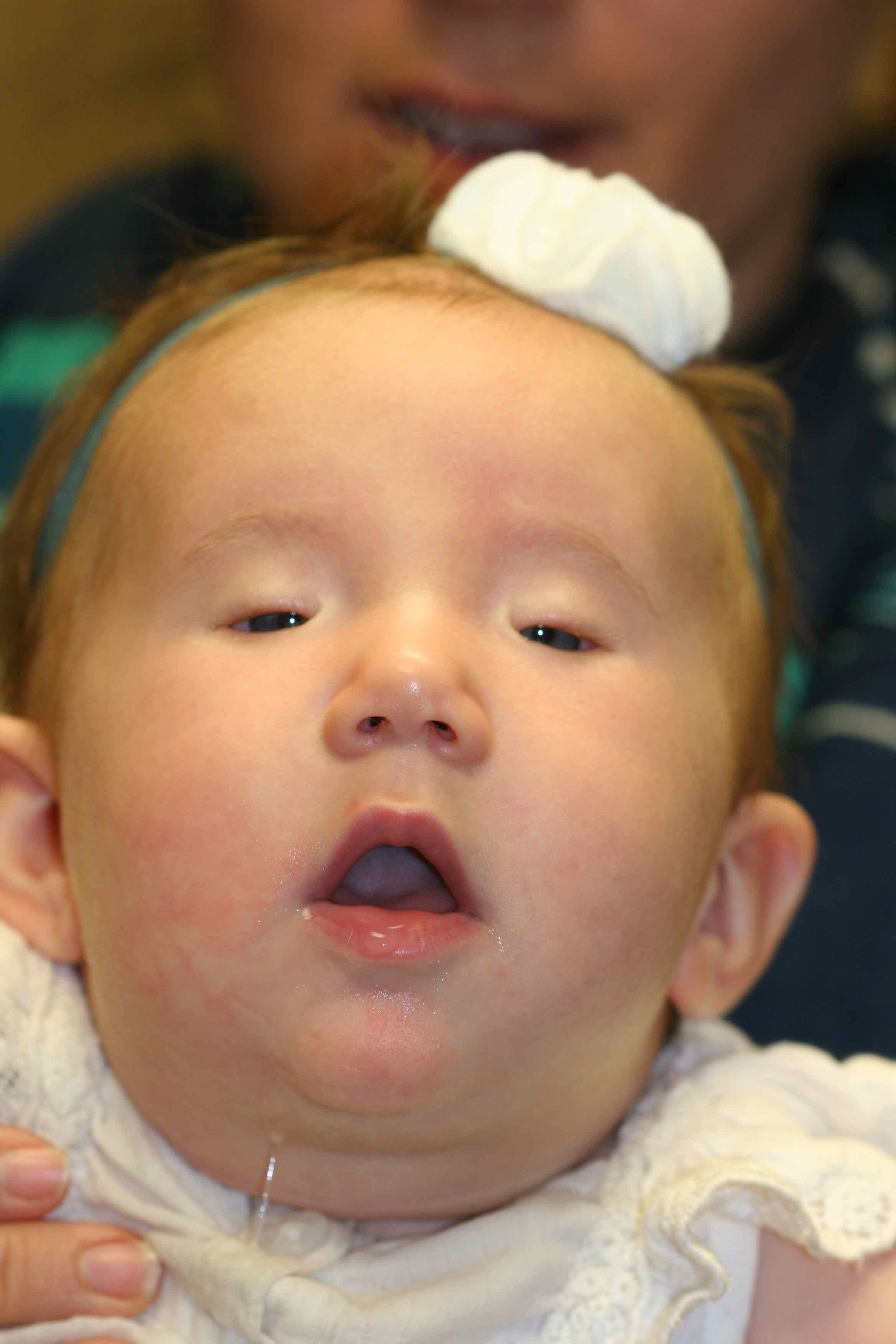
Blepharophimosis Syndrome, Severe Bilateral Ptosis. The image shows an infant with severe bilateral ptosis with the chin-up position. Blepharophimosis syndrome is frequently termed "BPES" for its components of blepharophimosis, ptosis, and epicanthus inverses. Other components include telecanthus and lateral canthal dystopia. There are two types of BPES: type I is found in females with associated premature ovarian insufficiency leading to infertility. Type II has no systemic associations. BPES is almost always inherited in an autosomal dominant manner. Spontaneous genetic mutations can also occur.
Contributed by BCK Patel, MD, FRCS
(Click Image to Enlarge)
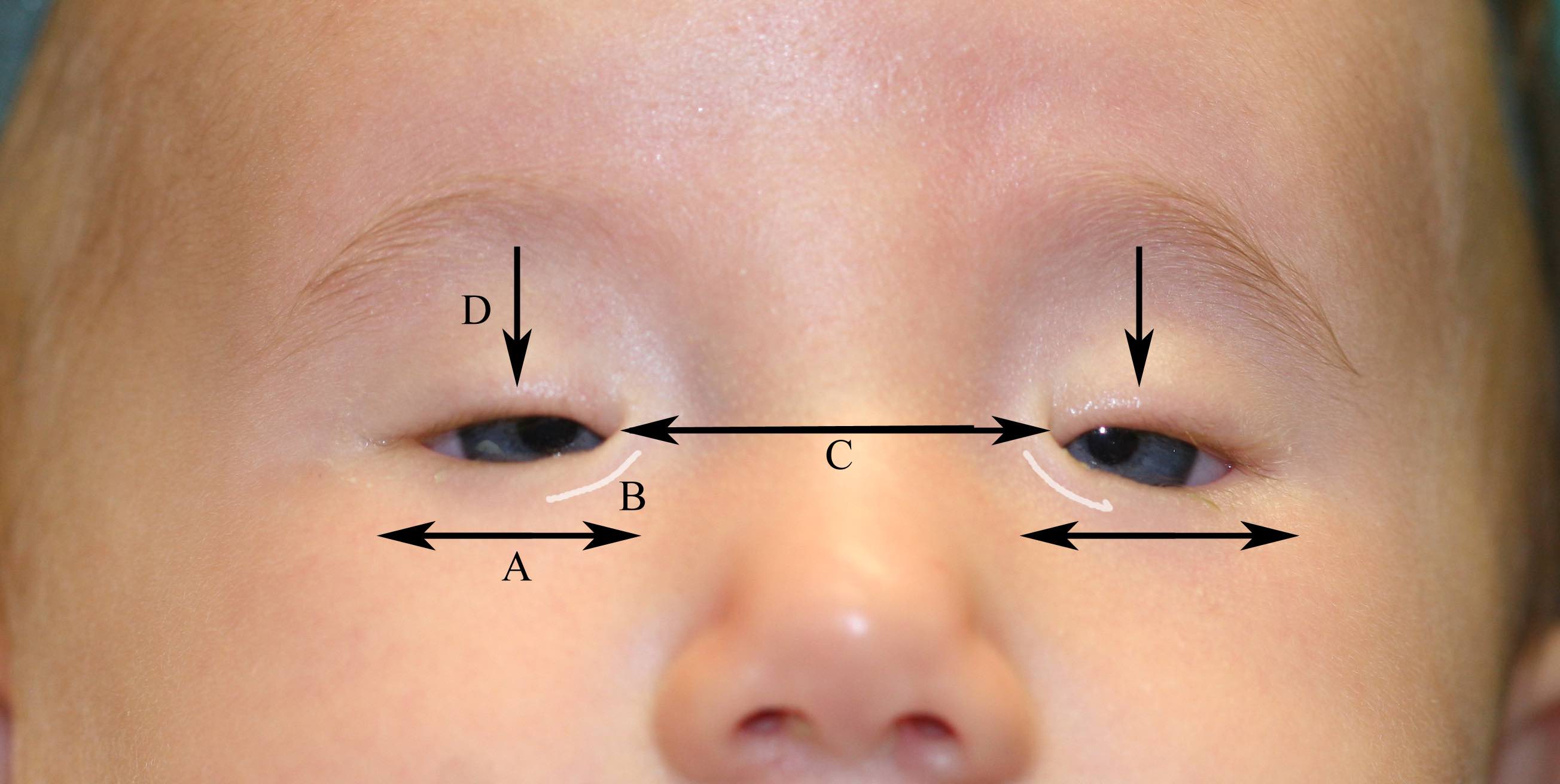
Blepharophimosis Syndrome. The arrows show blepharophimosis (A), epicanthus inversus (B), telecanthus (C), and ptosis (D).
Contributed by BCK Patel, MD, FRCS
(Click Image to Enlarge)
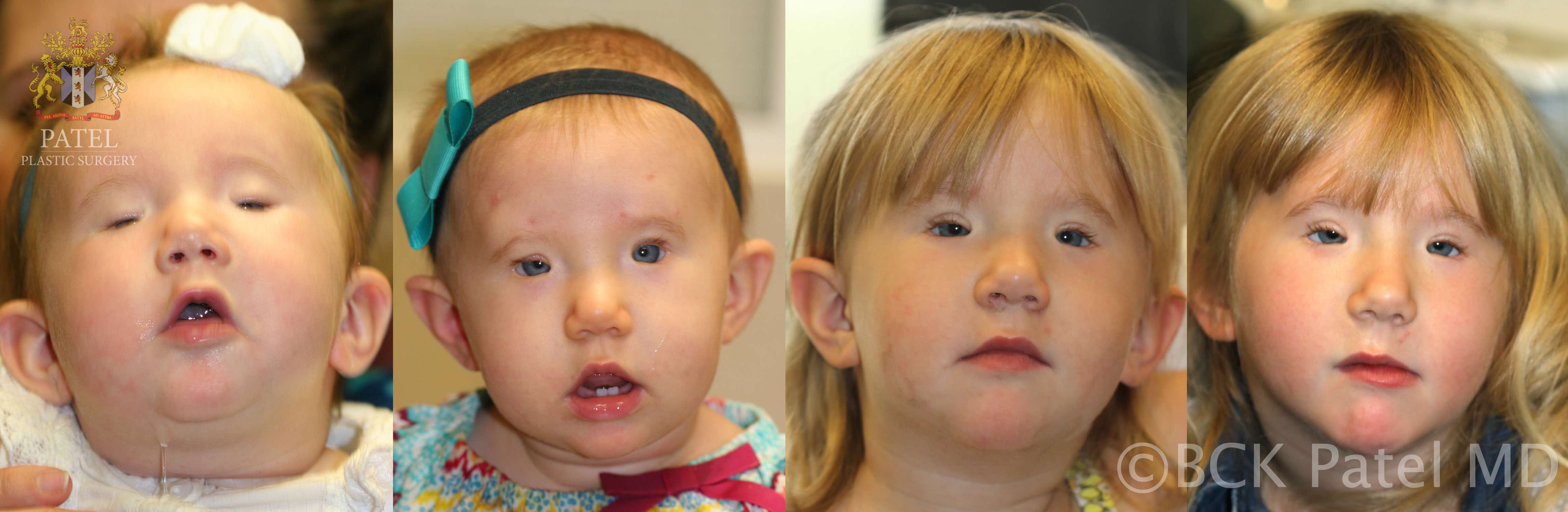
Blepharophimosis Syndrome, Correction of Ptosis. The image shows the correction of the ptosis with Goretex frontalis slings, demonstrating the longevity of the slings.
Contributed by BCK Patel, MD, FRCS
(Click Image to Enlarge)
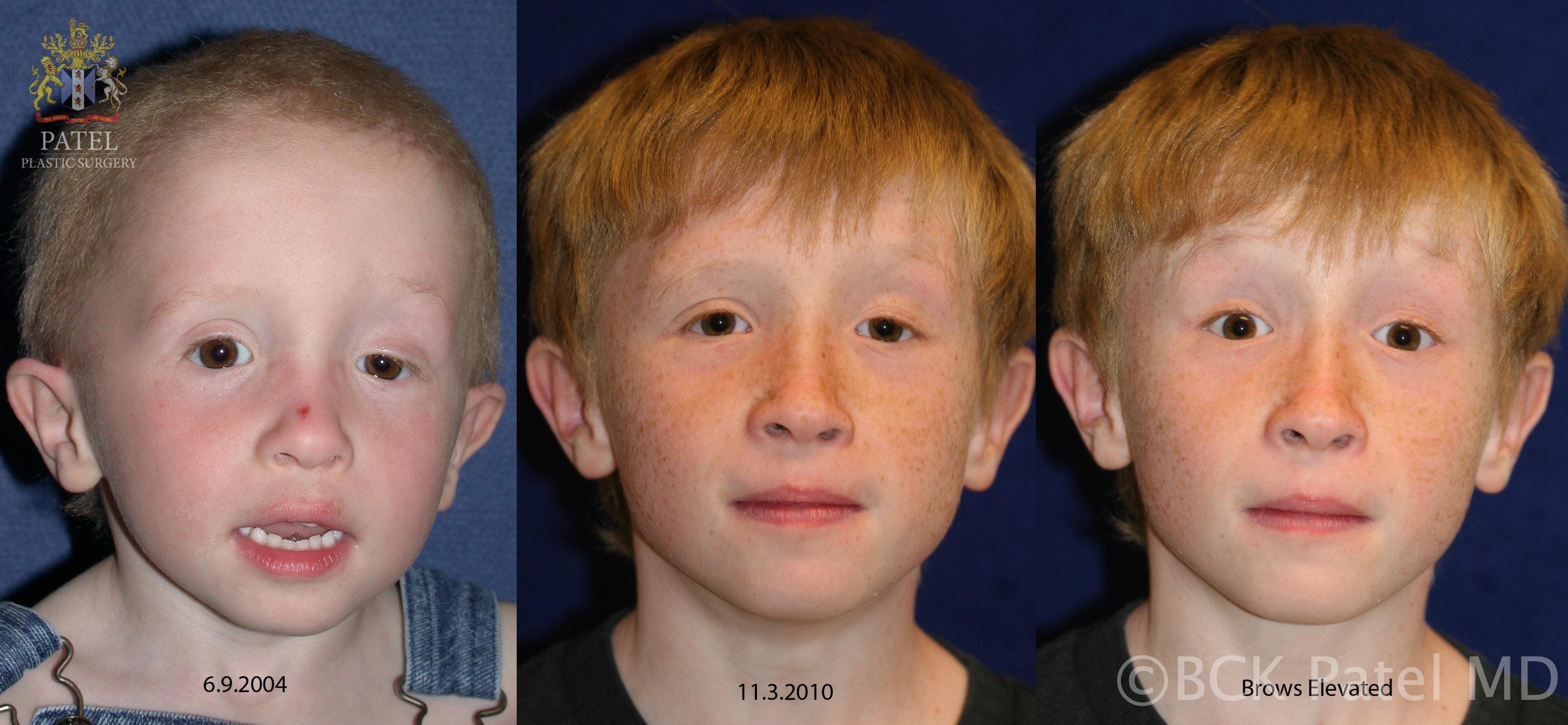
Severe Congenital Ptosis. An image depicting severe left congenital ptosis with poor levator function treated with a left frontalis sling. Photographs show the improved left eyelid height and how using the frontalis muscle allows the left upper eyelid to be lifted more. At rest, in unilateral cases, the ptotic corrected eyelid will invariably be a little lower than the opposite side unless the opposite normal levator muscle is removed and bilateral frontalis slings are performed (the Beard procedure).
Contributed by BCK Patel, MD, FRCS
(Click Image to Enlarge)
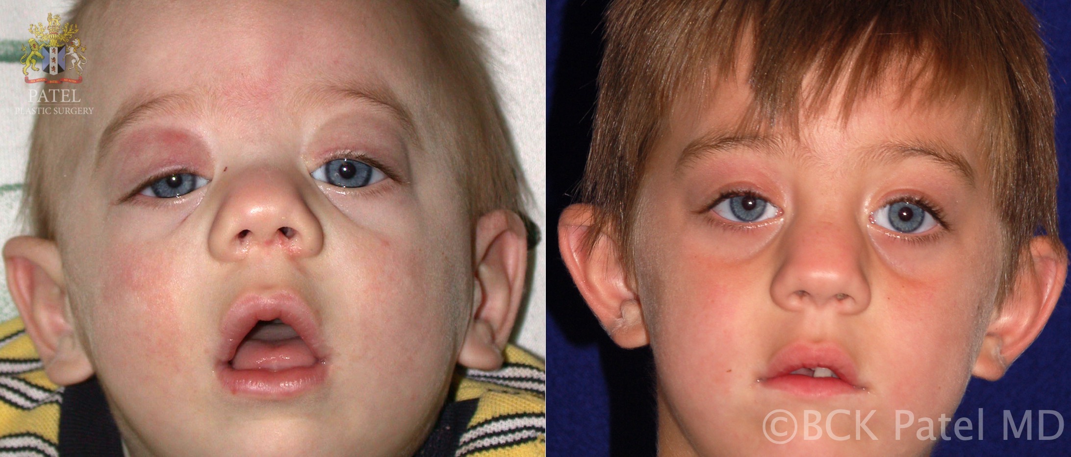
Congenital Ptosis. The image shows severe bilateral congenital ptosis with the chin-up position and poor levator function with overaction of the frontalis muscles. Bilateral frontalis slings correct the eyelid position and the chin-up position.
Contributed by BCK Patel, MD, FRCS
References
Marenco M, Macchi I, Macchi I, Galassi E, Massaro-Giordano M, Lambiase A. Clinical presentation and management of congenital ptosis. Clinical ophthalmology (Auckland, N.Z.). 2017:11():453-463. doi: 10.2147/OPTH.S111118. Epub 2017 Feb 27 [PubMed PMID: 28280295]
Pavone P, Cho SY, Praticò AD, Falsaperla R, Ruggieri M, Jin DK. Ptosis in childhood: A clinical sign of several disorders: Case series reports and literature review. Medicine. 2018 Sep:97(36):e12124. doi: 10.1097/MD.0000000000012124. Epub [PubMed PMID: 30200099]
Level 2 (mid-level) evidenceSooHoo JR, Davies BW, Allard FD, Durairaj VD. Congenital ptosis. Survey of ophthalmology. 2014 Sep-Oct:59(5):483-92. doi: 10.1016/j.survophthal.2014.01.005. Epub 2014 Feb 5 [PubMed PMID: 24657037]
Level 3 (low-level) evidenceSenthilkumar VA, Tripathy K. Marcus Gunn Jaw-Winking Syndrome. StatPearls. 2025 Jan:(): [PubMed PMID: 32644484]
Adam MP, Feldman J, Mirzaa GM, Pagon RA, Wallace SE, Amemiya A, Verdin H, Matton C, De Baere E. Blepharophimosis, Ptosis, and Epicanthus Inversus Syndrome. GeneReviews(®). 1993:(): [PubMed PMID: 20301614]
Lyons CJ, Godoy F, ALQahtani E. Cranial nerve palsies in childhood. Eye (London, England). 2015 Feb:29(2):246-51. doi: 10.1038/eye.2014.292. Epub 2015 Jan 9 [PubMed PMID: 25572578]
Khan Z, Bollu PC. Horner Syndrome. StatPearls. 2025 Jan:(): [PubMed PMID: 29763176]
Griepentrog GJ, Diehl NN, Mohney BG. Incidence and demographics of childhood ptosis. Ophthalmology. 2011 Jun:118(6):1180-3. doi: 10.1016/j.ophtha.2010.10.026. Epub 2011 Apr 15 [PubMed PMID: 21496927]
Level 2 (mid-level) evidenceGazzola R, Piozzi E, Vaienti L, Wilhelm Baruffaldi Preis F. Therapeutic Algorithm for Congenital Ptosis Repair with Levator Resection and Frontalis Suspension: Results and Literature Review. Seminars in ophthalmology. 2018:33(4):454-460. doi: 10.1080/08820538.2017.1297840. Epub 2017 Mar 15 [PubMed PMID: 28296517]
Koka K, Patel BC. Ptosis Correction. StatPearls. 2024 Jan:(): [PubMed PMID: 30969650]
Nemet AY. Is Hering's law as important in congenital blepharoptosis as in acquired ptosis? Aesthetic surgery journal. 2013 Nov 1:33(8):1110-5. doi: 10.1177/1090820X13511630. Epub 2013 Nov 5 [PubMed PMID: 24194587]
Level 2 (mid-level) evidenceFinsterer J. Ptosis: causes, presentation, and management. Aesthetic plastic surgery. 2003 May-Jun:27(3):193-204 [PubMed PMID: 12925861]