 Tools
Tools
 Anatomy, Thorax, Muscles
Introduction
The thoracic wall is composed of 5 muscle groups: the external intercostal muscles, internal intercostal muscles, innermost intercostal muscles, subcostal muscles, and transversus thoracis muscles. These muscles are primarily responsible for changing the volume of the thoracic cavity during respiration. Other muscles that attach to but do not form the thoracic wall include the pectoralis major, pectoralis minor, subclavius, and serratus anterior muscles anteriorly and the levatores costarum, serratus posterior superior, and serratus posterior inferior muscles posteriorly. The muscles of the anterior thorax produce movement of the upper extremity and shoulder, whereas the muscles of the posterior thorax assist with changes in thoracic volume during respiration and reinforce the thoracic wall. The diaphragm is the primary muscle of inspiration and forms the floor of the thorax, separating the thoracic and abdominal contents. Additional accessory muscles attached to the thorax include the scalene and sternocleidomastoid muscles, which may provide limited assistance during respiration.
Structure and Function
Register For Free And Read The Full Article
 Search engine and full access to all medical articles
- 10 free questions in your specialty
- Free CME/CE Activities
- Free daily question in your email
- Save favorite articles to your dashboard
- Emails offering discounts
Learn more about a Subscription to StatPearls Point-of-Care
Structure and Function
The muscles that make up the thoracic wall include the 3 layers of intercostal muscles, the external, internal, and innermost intercostal muscles, as well as the subcostal and transversus thoracis muscles. Eleven pairs of intercostal muscles occupy the intercostal spaces and are arranged from superficial to deep. The most superficial layer is the external intercostal muscle, which originates from the inferior border of the rib above and inserts on the superior border of the rib below in an inferomedial direction. These muscles extend from the rib tubercle posteriorly to the costochondral junction anteriorly, where they continue as thin connective tissue aponeuroses known as the external intercostal membranes.
During inspiration, the external intercostal muscles contract and elevate the lateral portions of the ribs, increasing the transverse diameter of the thorax in a bucket-handle motion. The internal intercostal muscles form the intermediate layer. These muscles originate from the lateral aspect of the costal groove of the rib above and insert on the superior border of the rib below in a direction perpendicular to the external intercostal muscles. This orientation allows the internal intercostal muscles to depress the ribs and reduce thoracic volume during forced expiration. Additionally, these muscles extend from the sternum anteriorly to the angles of the ribs posteriorly, where they continue as the internal intercostal membranes. The deepest layer of the thoracic wall is formed by the innermost intercostal muscles. These muscles originate from the medial aspect of the costal groove of the rib above and insert on the internal aspect of the rib below. The endothoracic fascia lines their internal surface and lies immediately superficial to the parietal pleura. The innermost intercostal muscles are thought to assist the internal intercostal muscles during forced expiration.[1][2]
In addition to the intercostal muscles, the subcostal and transversus thoracis muscles contribute to the thoracic wall. The subcostal muscles occupy the same layer as the innermost intercostal muscles and are most abundant in the lower posterior thoracic wall. They originate from the internal aspect of a lower rib and insert on the internal aspect of the second or third rib below. The transversus thoracis muscles also occupy the same layer as the innermost intercostal muscles. They originate from the posterior surface of the lower sternum, spread across the inner surface of the thoracic cage, and insert on ribs 2 through 6. Both muscle groups assist with rib depression during forced expiration.[2]
Muscles of the posterior thorax, such as the levatores costarum and serratus posterior superior and serratus posterior inferior, may also aid respiration. The levatores costarum muscles originate from the transverse processes of C7 through T11 and insert on the rib below. These muscles provide limited assistance with inspiration by elevating the ribs. The serratus posterior superior muscle attaches to ribs 2 through 5 and elevates them during inspiration, whereas the serratus posterior inferior muscle connects the vertebrae to ribs 8 through 12 and depresses them during forced expiration.[2] Together with the muscles of the thoracic wall, these muscles alter thoracic volume during respiration and reinforce the thoracic wall.
The primary muscle of inspiration is the diaphragm. The diaphragm is attached peripherally to the xiphoid process through its sternal portion, the costal margin of the thoracic wall and lower 6 ribs through its costal portion, and the lumbar vertebrae through its lumbar portion. During inspiration, the muscle contracts and draws its central tendon inferiorly, flattening the diaphragm. Diaphragmatic contraction increases the vertical diameter of the thorax and decreases intrathoracic pressure, drawing air into the lungs. During expiration, the diaphragm relaxes and elevates, allowing air within the lungs to be expelled. Other accessory muscles that aid inspiration include the scalene muscles, which elevate the first and second ribs, and the sternocleidomastoid muscle, which assists with elevation of the sternum. In addition to respiration, the diaphragm increases intra-abdominal pressure during micturition, defecation, and weightlifting.[2][3]
Other muscles of the thorax contribute to upper extremity movement and include the pectoralis major, pectoralis minor, subclavius, and serratus anterior muscles. The pectoralis major originates from the medial half of the clavicle, anterior sternum, first 7 costal cartilages, and aponeurosis of the external oblique muscle and inserts on the lateral lip of the intertubercular sulcus of the humerus. This muscle flexes, adducts, and medially rotates the arm at the glenohumeral joint. Its clavicular head flexes an extended arm, whereas its sternocostal head extends a flexed arm.[4] The pectoralis minor muscle originates from the anterior surfaces of ribs 3 through 5 and the deep fascia overlying the related intercostal spaces and inserts on the coracoid process of the scapula. The muscle primarily depresses the shoulder and protracts the scapula. Pectoralis minor may also assist inspiration by elevating the third, fourth, and fifth ribs.[2] The subclavius muscle originates at the costochondral junction of the first rib and inserts on the subclavian groove of the clavicle. This muscle stabilizes the clavicle.[5]
The serratus anterior muscle originates from the superolateral surfaces of the first through eighth ribs or the first through ninth ribs along the lateral thoracic wall and inserts along the superior angle, medial border, and inferior angle of the scapula. The muscle primarily protracts and rotates the scapula, including during punching movements. The serratus anterior is therefore sometimes called the boxer's muscle. The serratus anterior may also assist inspiration by elevating the ribs when the shoulder girdle is fixed.[6]
Embryology
Skeletal muscle develops through the differentiation of mesoderm. During the fourth through eighth weeks of human development, paraxial mesoderm organizes into paired clusters alongside the neural tube to form somites. These somites then differentiate into 2 cell populations: the dorsolateral dermomyotome and the ventromedial sclerotome. The dermomyotome develops into skeletal muscle, including the thoracic muscles, and the dermis, whereas the sclerotome forms the axial skeleton.[1][4]
Blood Supply and Lymphatics
The intercostal arteries mainly supply the muscles of the thoracic wall and posterior thorax. Each intercostal space is supplied by 3 arteries, including 1 posterior intercostal artery and 2 anterior intercostal arteries, which travel between the internal and innermost intercostal muscles within the costal groove and anastomose laterally. The posterior intercostal arteries of the first 2 intercostal spaces arise from the superior, or supreme, intercostal artery, which originates from the costocervical trunk of the subclavian artery. The remaining posterior intercostal arteries supplying the third through 11th intercostal spaces, along with a pair of subcostal arteries, arise directly from the descending thoracic aorta. Their corresponding veins drain into the azygos or hemiazygos systems. The anterior intercostal arteries of the first 6 intercostal spaces are branches of the internal thoracic artery, which arises from the first portion of the subclavian artery. The remaining anterior intercostal arteries supplying the seventh through ninth intercostal spaces arise from branches of the musculophrenic artery, a terminal branch of the internal thoracic artery. Their corresponding veins drain into the internal thoracic or musculophrenic veins. Lymphatic drainage of the thoracic wall occurs through the parasternal and intercostal lymph nodes. The parasternal lymph nodes of the upper thorax drain into the bronchomediastinal trunk. The intercostal lymph nodes of the upper thorax also drain into the bronchomediastinal trunk, whereas those of the lower thorax drain into the thoracic duct.[1][2]
The pectoralis major and minor muscles are supplied by the pectoral branch of the thoracoacromial trunk, the second branch of the axillary artery. Their venous drainage occurs through the pectoral vein into the subclavian vein.[4] The subclavius receives vascular supply from the clavicular branch of the thoracoacromial trunk. The serratus anterior is supplied by the lateral thoracic artery, the superior thoracic artery, and the thoracodorsal artery.[6] Lymphatic drainage of the superficial regions of the thoracic wall typically involves the axillary lymph nodes or the parasternal nodes. The diaphragm receives arterial blood from multiple vessels, including the musculophrenic branch of the internal thoracic artery, the superior phrenic branches of the aorta, the lower 5 intercostal arteries, the subcostal artery, and the inferior phrenic arteries.[3] Similarly, an extensive lymphatic network drains the diaphragm toward mediastinal lymph nodes and ultimately connects with the bloodstream through the thoracic duct.[7]
Nerves
The muscles that comprise the thoracic wall and posterior thorax are innervated by the intercostal nerves, which mainly arise from the anterior rami of spinal nerves T1 through T11. The anterior ramus of spinal nerve T12 forms the subcostal nerve. Each intercostal nerve supplies a dermatome and a myotome. Afferent fibers transmit sensory information from the overlying skin, whereas efferent fibers provide motor innervation to the respiratory muscles. Notably, only a portion of the anterior ramus of spinal nerve T1 contributes to the lower trunk of the brachial plexus, whereas the remaining intercostal nerves do not form plexuses.[1][2]
Innervation of the muscles of the anterior thorax arises from different branches of the brachial plexus. The pectoralis major is innervated by the lateral pectoral nerve, which supplies the clavicular head, and the medial pectoral nerve, which supplies the sternocostal head. The pectoralis minor receives innervation from the medial pectoral nerve. The lateral pectoral nerve arises from the lateral cord of the brachial plexus, whereas the medial pectoral nerve arises from the medial cord.[4][8] The nerve to the subclavius innervates the subclavius muscle and arises from the superior trunk, primarily from the C5 and C6 nerve roots. When present, the accessory phrenic nerve, which most commonly contains fibers from C5, may also provide motor innervation to the subclavius muscle.[5] Finally, the serratus anterior is innervated by the long thoracic nerve, which originates from the anterior rami of C5 to C7.[6]
The diaphragm receives innervation from the right and left phrenic nerves, which originate from the anterior rami of C3 through C5. The phrenic nerves provide motor innervation for diaphragmatic contraction during inspiration and sensory innervation to the parietal pleura and peritoneum covering the central portion of the diaphragm. The lower 6 intercostal nerves provide sensory innervation to the peripheral portion of the diaphragm.[3][5]
Muscles
The muscles of the thorax discussed in this article include the following:
- Thoracic wall
- Intercostal muscles
- External intercostal muscles
- Internal intercostal muscles
- Innermost intercostal muscles
- Subcostalis
- Transversus thoracis
- Intercostal muscles
- Posterior thorax
- Levatores costarum
- Serratus posterior superior and inferior muscles
- Anterior and superficial thorax
- Pectoralis major and minor muscles
- Subclavius
- Serratus anterior
- Floor
- Diaphragm
Physiologic Variants
Results from multiple studies demonstrated anatomical variations of the thoracic muscles, including supernumerary muscles and congenital anomalies. The sternalis muscle is a rare supernumerary variant of the anterior thoracic wall present in approximately 8% of the population. This vertically oriented muscle lies between the superficial and pectoral fasciae, parallel to the right sternal margin, and measures approximately 7.0 cm in length and 2.9 cm in width.[9] The sternalis muscle may be unilateral in 4.5% of individuals or bilateral in less than 1.7% and may contribute to shoulder movement or serve as an accessory muscle during elevation of the lower chest wall. Its innervation and embryologic origin remain uncertain. Reports indicate that the muscle receives innervation from the external or internal thoracic nerves in 55% of individuals, the intercostal nerves in 43%, or both in 2%. Moreover, some studies suggest that the sternalis muscle derives from the hypaxial myotomes or dermomyotomes, which give rise to the ventral and lateral body wall muscles of the thorax and abdomen. Conversely, results from other studies suggested that the muscle develops from the rectus abdominis sheath or pectoralis major because of abnormal muscle patterning.
Reports have not identified clinical symptoms directly attributable to the sternalis muscle. However, its presence may produce electrocardiographic alterations or lead to misdiagnosis of breast masses, such as breast carcinoma or hematoma, on routine mammography because of its parasternal location and relative unfamiliarity among radiologists. An undetected sternalis muscle may also interfere with or prolong breast and cardiothoracic surgical procedures. When identified preoperatively, surgeons may use the sternalis muscle as a muscular flap during reconstructive procedures involving the anterior chest wall, head and neck, or breast.[10][11]
Anatomical variations of the thoracic muscles may also result from congenital anomalies of varying severity. Poland syndrome is characterized by absence of the sternocostal head of the pectoralis major muscle, with variable hypoplasia or absence of the pectoralis minor muscle and associated digital anomalies. Other abnormalities described in Poland syndrome include absence or hypoplasia of the ipsilateral breast, pectus excavatum, and rib aplasia. These defects are most commonly unilateral and typically occur on the right side, although results from some studies documented rare bilateral manifestations. Numerous variations of the syndrome have been described, ranging from mild hypoplasia of the pectoralis major muscle to severe hypoplasia of the thoracic wall.[12]
Surgical Considerations
Surgical interventions that involve the thorax muscles typically involve chest tube placement and needle therapy for either decompression or anesthesia.[1][2]
Tube thoracostomy: Tube thoracostomy is performed to evacuate pathologic air or fluid from the pleural cavity. A chest tube is usually placed between the anterior and midaxillary lines within the fourth or fifth intercostal space, immediately superior to the rib, to reduce the risk of injury to the blood vessels and nerves along the inferior aspect of each rib. After the incision is made and the tube is inserted, the tube passes through the skin, superficial fascia, serratus anterior muscle, external intercostal muscle, internal intercostal muscle, innermost intercostal muscle, and parietal pleura before entering the pleural cavity.
Needle thoracostomy: Needle thoracostomy is an emergency decompression procedure typically performed fortension pneumothorax. Needle placement may occur at the second intercostal space along the midclavicular line or within the fourth or fifth intercostal space along the anterior axillary line. In either location, the needle should enter immediately superior to the rib to minimize neurovascular injury.
Intercostal nerve block: An intercostal nerve block is usually performed to alleviate pain associated with rib fractures or herpes zoster. The needle is inserted near the inferior border of the rib and passes through the skin, superficial fascia, serratus anterior muscle, external intercostal muscle, and internal intercostal muscle to reach the intercostal nerve. Clinicians should also anesthetize the adjacent intercostal nerves to achieve adequate pain relief because collateral branches and substantial overlap between contiguous dermatomes may result in incomplete blockade.
Clinical Significance
Respiratory muscles are essential for sustaining life and supporting daily activities. The use of accessory muscles to assist respiration may be a normal finding during strenuous activities with high physiologic demands, such as exercise or singing. However, accessory muscle use at rest may indicate a pathologic condition, such as an asthma exacerbation, and warrants further evaluation and treatment. Accessory respiratory muscles include the scalene, sternocleidomastoid, internal intercostal, transversus thoracis, pectoralis major, pectoralis minor, serratus anterior, serratus posterior superior, serratus posterior inferior, latissimus dorsi, trapezius, and abdominal muscles.[2]
Media
(Click Image to Enlarge)
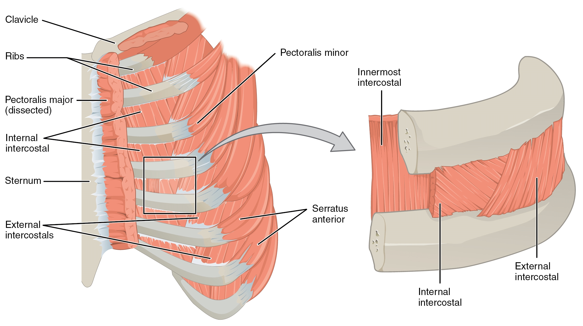
The figure shows a thoracic section and related muscles. Contributed by Bruno Bordoni, PhD
References
Hussain A, Burns B. Anatomy, Thorax, Wall. StatPearls. 2026 Jan:(): [PubMed PMID: 30571035]
Donley ER, Holme MR, Loyd JW. Anatomy, Thorax, Wall Movements. StatPearls. 2026 Jan:(): [PubMed PMID: 30252279]
Bains KNS, Kashyap S, Lappin SL. Anatomy, Thorax: Diaphragm. StatPearls. 2026 Jan:(): [PubMed PMID: 30137842]
Solari F, Burns B. Anatomy, Thorax, Pectoralis Major Major. StatPearls. 2026 Jan:(): [PubMed PMID: 30252247]
Oliver KA, Ashurst JV. Anatomy, Thorax, Phrenic Nerves. StatPearls. 2026 Jan:(): [PubMed PMID: 30020697]
Lung K, St Lucia K, Lui F. Anatomy, Thorax, Serratus Anterior Muscles. StatPearls. 2026 Jan:(): [PubMed PMID: 30285352]
Souilamas R, Hidden G, Riquet M. Mediastinal lymphatic efferents from the diaphragm. Surgical and radiologic anatomy : SRA. 2001 Jun:23(3):159-62 [PubMed PMID: 11490925]
Porzionato A, Macchi V, Stecco C, Loukas M, Tubbs RS, De Caro R. Surgical anatomy of the pectoral nerves and the pectoral musculature. Clinical anatomy (New York, N.Y.). 2012 Jul:25(5):559-75. doi: 10.1002/ca.21301. Epub 2011 Nov 28 [PubMed PMID: 22125052]
Level 1 (high-level) evidenceMier-Garcia JF, López Subias J, Peña Tobar CE, Baena-Caldas GP. [Supernumerary muscular masses of the anterior thoracic wall. Case report and review]. Revista de la Facultad de Ciencias Medicas (Cordoba, Argentina). 2016:73(4):291-296 [PubMed PMID: 28152370]
Level 3 (low-level) evidenceSnosek M, Tubbs RS, Loukas M. Sternalis muscle, what every anatomist and clinician should know. Clinical anatomy (New York, N.Y.). 2014 Sep:27(6):866-84. doi: 10.1002/ca.22361. Epub 2014 Jan 16 [PubMed PMID: 24431029]
Raikos A, Paraskevas GK, Tzika M, Faustmann P, Triaridis S, Kordali P, Kitsoulis P, Brand-Saberi B. Sternalis muscle: an underestimated anterior chest wall anatomical variant. Journal of cardiothoracic surgery. 2011 May 16:6():73. doi: 10.1186/1749-8090-6-73. Epub 2011 May 16 [PubMed PMID: 21575244]
Level 3 (low-level) evidenceTafti D, Cecava ND. Poland Syndrome(Archived). StatPearls. 2026 Jan:(): [PubMed PMID: 30335292]