 Tools
Tools
 Anatomy, Abdomen and Pelvis, Pancreas
Introduction
The pancreas is an elongated accessory digestive gland situated retroperitoneally, crossing the bodies of the L1 and L2 vertebrae on the posterior abdominal wall. The pancreas lies transversely in the upper abdomen between the duodenum on the right and the spleen on the left (see Image. Pancreatic Ducts and Duodenum Anatomy). The gland is divided into the head, neck, body, and tail. The head lies on the inferior vena cava and the renal vein and is surrounded by the C loop of the duodenum. The tail of the pancreas courses toward the splenic hilum. The pancreas produces exocrine secretion (pancreatic juice from acinar cells) that enters the duodenum through the main and accessory pancreatic ducts, and endocrine secretions (glucagon and insulin from the pancreatic islets of Langerhans) that enter the blood.
A detailed understanding of pancreatic structure and disease patterns improves recognition of conditions such as annular pancreas, choledochal cysts, pancreatitis, and pancreatic cancer. Anatomic precision supports safe surgical decision-making, particularly when managing obstruction, perforation, or resection of malignant tissue. Insight into endocrine and exocrine impairment guides metabolic control and symptom-directed treatment.
Structure and Function
Register For Free And Read The Full Article
 Search engine and full access to all medical articles
- 10 free questions in your specialty
- Free CME/CE Activities
- Free daily question in your email
- Save favorite articles to your dashboard
- Emails offering discounts
Learn more about a Subscription to StatPearls Point-of-Care
Structure and Function
Divisions
The pancreas is divided into 4 parts: head, neck, body, and tail. The head of the pancreas is the enlarged portion of the gland surrounded by the C-shaped curve of the duodenum. On its way to the descending part of the duodenum, the bile duct lies in a groove on the posterosuperior surface of the head or is embedded within the glandular substance. The body of the pancreas continues from the neck and passes over the aorta and the L2 vertebra. The anterior surface of the body of the pancreas is covered with peritoneum. The posterior surface of the body is devoid of peritoneum. The posterior surface is in contact with the aorta, the superior mesenteric artery (SMA), the left suprarenal gland, the left kidney, and the renal vessels.
The neck of the pancreas is short. The tail of the pancreas lies anterior to the left kidney and is closely related to the splenic hilum and the left colic flexure. The main pancreatic duct, carrying pancreatic secretions, joins the bile duct to form the hepatopancreatic ampulla, which opens into the descending part of the duodenum. The hepatopancreatic sphincter of Oddi, around the hepatopancreatic ampulla, is a smooth muscle sphincter that regulates the flow of bile and pancreatic juice into the ampulla and inhibits reflux of duodenal contents into the ductal system.
Cell Types
The majority of the pancreas (approximately 80%) consists of exocrine pancreatic tissue. This tissue is composed of pancreatic acini, formed by pyramidal acinar cells with apices directed toward the lumen. These cells contain dense zymogen granules in the apical region, whereas the basal region contains the nucleus and endoplasmic reticulum responsible for digestive enzyme synthesis. These enzymes are stored in secretory vesicles associated with the Golgi complex. The basolateral membrane of the acinar cells contains multiple receptors for neurotransmitters, including secretin, cholecystokinin, and acetylcholine, which regulate exocytosis of digestive enzymes.
The pancreas also contains the islets of Langerhans, which include endocrine cells. Unlike exocrine enzymes released by exocytosis, endocrine secretions enter the bloodstream through a complex capillary network integrated into pancreatic blood flow. Four endocrine cell types are present. A cells secrete glucagon. B cells release insulin. D cells synthesize somatostatin. F cells generate pancreatic polypeptide.
Stellate cells are a direct formation of epithelial structures within the pancreas. In conditions such as chronic pancreatitis, these cells promote inflammation and fibrosis.
Embryology
The pancreas develops from the posterior foregut endoderm. At approximately 4 weeks of gestation, this endoderm gives rise to dorsal and ventral buds, which gradually elongate.[1] Around week 6, the ventral bud rotates around the developing duodenum and ultimately fuses with the dorsal bud at approximately the 17th week of gestation to form the pancreas. The dorsal bud forms the upper portion of the pancreatic head, the body, and the tail, whereas the ventral bud forms the lower portion of the pancreatic head and the uncinate process.
The pancreatic enzymes are drained by 2 pancreatic ducts: the duct of Wirsung (major pancreatic duct) and the duct of Santorini (minor pancreatic duct). The ventral duct forms the proximal portion of the major pancreatic duct, which opens into the duodenum via the ampulla of Vater. The dorsal duct forms part of the major ducts and the minor duct (accessory duct of Santorini). The minor duct usually empties through the ampulla of Vater but may empty independently in approximately 5% of individuals.
Blood Supply and Lymphatics
Blood supply to the pancreas is provided by branches of the splenic artery (a branch of the celiac trunk), the SMA, and the common hepatic artery.[2][3] The gastroduodenal artery, a branch of the common hepatic artery, supplies the head and uncinate process via the pancreaticoduodenal artery. The inferior portion of the head receives additional supply from the inferior pancreaticoduodenal artery, which arises from the SMA. The body and tail receive arterial supply from the splenic artery and its branches.
Venous drainage of the pancreas is region-specific. The head drains into the superior mesenteric vein, whereas the body and neck drain into the splenic vein. The superior mesenteric and splenic veins converge to form the portal vein.
Nerves
The pancreas has a complex network of parasympathetic, sympathetic, and sensory innervations.[4] An intrinsic nerve plexus is also present within the gland. Sympathetic and parasympathetic fibers innervate pancreatic acinar cells. Parasympathetic fibers arise from the posterior vagal trunk and function as secretomotor fibers. Pancreatic secretions are predominantly regulated by cholecystokinin and secretin, hormones produced by epithelial cells of the duodenum and proximal intestinal mucosa, which are stimulated by acidic gastric contents. Sympathetic innervation of the pancreas originates from the splanchnic nerves of T6 to T10 and the celiac plexus.
Physiologic Variants
Partial or dorsal agenesis of the pancreas is often asymptomatic. This condition may be associated with diabetes, polysplenia, malabsorption syndrome, or recurrent pancreatitis. Ectopic pancreatic tissue occurs most commonly in the stomach or small intestine. Such tissue is present in approximately 3% to 5% of the general population and is usually identified incidentally during upper gastrointestinal endoscopy or barium contrast studies. Umbilicated appearance typically characterizes these lesions, which are most often clinically insignificant. Rarely, a pancreatic rest in the small intestine may serve as the lead point for intussusception or cause bowel obstruction. Pancreatic dysgenesis or dysfunction is a component of certain syndromic associations, including Johanson-Blizzard syndrome and Schwachmann-Diamond syndrome.
Pancreas divisum represents the most common developmental anomaly of the pancreas.[5] This anomaly occurs in approximately 10% to 15% of the general population and results from the failure of the pancreatic buds to fuse. Consequently, the tail, body, and part of the head of the pancreas drain through the accessory duct of Santorini rather than the major duct. Pancreas divisum has been historically associated with recurrent pancreatitis, potentially due to stenosis of the sphincter obstructing ventral pancreatic outflow.
Diagnosis is established using endoscopic retrograde cholangiopancreatography or magnetic resonance cholangiopancreatography, with endoscopic ultrasound increasingly employed for detection. In cases of recurrent pancreatitis, pancreas divisum is managed with stent insertion and sphincterotomy under endoscopic retrograde cholangiopancreatography guidance.
Surgical Considerations
Annular pancreas results from incomplete rotation of the ventral pancreatic bud during development. This anomaly may be associated with autosomal recessive mutations. Presentation typically occurs in infancy with complete or partial bowel obstruction. Maternal polyhydramnios is frequently reported in affected pregnancies. Annular pancreas is commonly associated with additional anomalies, including trisomy 21, tracheoesophageal fistula, malrotation, cardiac or renal abnormalities, and components of vertebral, anal, cardiac, tracheoesophageal, renal, and limb anomalies (VACTERL). The condition may also be complicated by pancreatitis. Management typically involves surgical correction via duodenojejunostomy.
Choledochal cysts are congenital dilations of the biliary tract. Clinical presentation commonly includes jaundice and abdominal pain, with fever occurring in some cases. Laboratory evaluation demonstrates elevated direct bilirubin levels with normal transaminases. Diagnosis is usually established through imaging modalities such as ultrasonography, computed tomography, hepatobiliary iminodiacetic acid scan, or magnetic resonance cholangiopancreatography.
Clinical Significance
Perforation
Perforation of the pancreas results in the release of digestive enzymes, including amylase and lipase, into the abdominal cavity, causing pancreatic self-digestion. Surgical removal of the pancreas is possible. However, lifelong management with blood glucose regulation and pancreatic enzyme supplementation is required to maintain digestion.
Cancer
Pancreatic cancers, particularly pancreatic adenocarcinoma, are difficult to treat and are often diagnosed at a stage unsuitable for surgical intervention. Pancreatic cancer is uncommon in younger patients, with a median age of diagnosis of 71 years. Established risk factors include smoking, obesity, diabetes, multiple endocrine neoplasia type 1, and hereditary nonpolyposis colon cancer.
Diabetes Mellitus
Diabetes mellitus type 1 is an autoimmune disorder characterized by immune-mediated destruction of insulin-secreting cells in the pancreas, resulting in decreased insulin production. This form of diabetes typically develops during childhood and requires lifelong insulin therapy for survival. Diabetes mellitus type 2 is the most common form of the disease and is associated with hyperglycemia caused by a combination of insulin resistance and impaired insulin secretion. Management includes modifications in diet and physical activity, as well as pharmacologic therapy with agents such as biguanides, including metformin.
Inflammation
Pancreatic inflammation, or pancreatitis, is associated with recurrent gallstones, alcohol use, measles, mumps, medications, α1-antitrypsin deficiency, and scorpion stings. The condition produces intense central abdominal pain that radiates to the back and may be accompanied by jaundice. Clinical presentation often includes pale stools and dark urine.
Media
(Click Image to Enlarge)
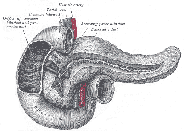
Pancreatic Ducts and Duodenum Anatomy. This illustration shows the pancreas nestled in the C-shaped curve of the duodenum, highlighting the main pancreatic duct, accessory pancreatic duct, and the convergence of these conduits with the common bile duct. The hepatic artery and portal vein are also depicted to demonstrate the close relationship of the pancreatic head and bile duct to the major vascular structures supplying and draining the liver.
Henry Vandyke Carter, Public Domain, via Wikimedia Commons
References
Slack JM. Developmental biology of the pancreas. Development (Cambridge, England). 1995 Jun:121(6):1569-80 [PubMed PMID: 7600975]
Level 3 (low-level) evidenceKulenović A, Sarac-Hadzihalilović A. Blood vessels distribution in body and tail of pancreas- a comparative study of age related variation. Bosnian journal of basic medical sciences. 2010 May:10(2):89-93 [PubMed PMID: 20507286]
Level 2 (mid-level) evidenceLewis MP, Reber HA, Ashley SW. Pancreatic blood flow and its role in the pathophysiology of pancreatitis. The Journal of surgical research. 1998 Feb 15:75(1):81-9 [PubMed PMID: 9614861]
Love JA, Yi E, Smith TG. Autonomic pathways regulating pancreatic exocrine secretion. Autonomic neuroscience : basic & clinical. 2007 Apr 30:133(1):19-34 [PubMed PMID: 17113358]
Level 3 (low-level) evidenceTürkvatan A, Erden A, Türkoğlu MA, Yener Ö. Congenital variants and anomalies of the pancreas and pancreatic duct: imaging by magnetic resonance cholangiopancreaticography and multidetector computed tomography. Korean journal of radiology. 2013 Nov-Dec:14(6):905-13. doi: 10.3348/kjr.2013.14.6.905. Epub 2013 Nov 5 [PubMed PMID: 24265565]