 Tools
Tools
 Intraductal Papillary Mucinous Neoplasm of the Pancreas
Introduction
Intraductal papillary mucinous neoplasms (IPMNs) are the most common pancreatic cystic neoplasms and represent a clinically important spectrum of premalignant lesions that can progress to invasive pancreatic carcinoma.[1][2][3] Analogous to pancreatic intraepithelial neoplasia in conventional pancreatic ductal adenocarcinoma (PDAC), IPMNs are well-established precursor lesions arising from the pancreatic ductal epithelium. Although a definitive etiology has not been identified, risk factors including smoking, obesity, and hereditary syndromes such as Peutz-Jeghers syndrome and familial adenomatous polyposis have been associated with their development and progression.
At the molecular level, IPMNs are driven by somatic mutations, particularly in KRAS and GNAS, which are considered early events in tumorigenesis, as well as additional alterations that contribute to dysplastic progression and malignant transformation.[4][5] IPMNs account for approximately 38% of all resected pancreatic cysts.[6] Intraductal oncocytic papillary neoplasms and intraductal tubulopapillary neoplasms were historically grouped with IPMNs as mucinous pancreatic lesions but are now recognized as distinct entities under the 2019 World Health Organization (WHO) classification.
IPMNs are classified into 3 groups based on ductal involvement: main-duct type (MD-IPMN), branch-duct type (BD-IPMN), and mixed type (MT-IPMN). This distinction has direct clinical relevance, as MD-IPMNs carry a substantially higher risk of malignancy and more frequently harbor high-grade dysplasia or invasive carcinoma than branch duct-type variants.[7] This risk is pronounced in larger lesions or those exhibiting high-risk stigmata, worrisome features, or positive cytology.[8] MT-IPMNs represent a third group with intermediate malignant potential.[9]
Given their heterogeneous malignant potential, management relies on individualized risk stratification integrating clinical, radiographic, and molecular features. Surgical resection is recommended for MD- and MT-IPMNs and for lesions demonstrating high-risk stigmata, whereas BD-IPMNs without high-risk features are managed with active surveillance. Periodic magnetic resonance imaging (MRI) with magnetic resonance cholangiopancreatography (MRCP) serves as the cornerstone of follow-up, with endoscopic ultrasound (EUS) selectively employed when imaging findings are indeterminate or may alter management.[10]
Etiology
Register For Free And Read The Full Article
 Search engine and full access to all medical articles
- 10 free questions in your specialty
- Free CME/CE Activities
- Free daily question in your email
- Save favorite articles to your dashboard
- Emails offering discounts
Learn more about a Subscription to StatPearls Point-of-Care
Etiology
A definitive cause for IPMNs has not been identified. Smoking and obesity are independent modifiable risk factors associated with increased risk of progression to invasive carcinoma.[11] Several hereditary syndromes are associated with an elevated incidence of IPMN, including Peutz-Jeghers syndrome, familial adenomatous polyposis, and McCune-Albright syndrome.[12] At the molecular level, IPMNs arise from polyclonal ductal epithelial proliferation driven by somatic mutations. KRAS and GNAS mutations are the earliest driver events; GNAS mutations are highly specific to IPMNs and are found in approximately 60% of cases. RNF43 mutations represent a later event associated with progression from low- to high-grade dysplasia, while accumulation of alterations in TP53, SMAD4, and p16/CDKN2A drives the final transition to invasive carcinoma.[4][13][14]
Epidemiology
IPMNs are the most common pancreatic cystic neoplasms, with a prevalence of 2.4% to 3.5% in the general population.[15] A United States population-based study identified IPMNs on abdominal computed tomography (CT) in 10.9% of patients aged 50 or older, with BD-IPMNs comprising 90.9% of cases; after a median follow-up of 12 years, 4 cases of incident pancreatic cancer were diagnosed.[8] In an asymptomatic Korean cohort undergoing abdominal CT, pancreatic cysts were detected in 2.2% of patients, with IPMNs constituting 82% of these lesions.[16]
Prevalence increases markedly with age and is higher when MRI/MRCP is the detection modality. A meta-analysis of 17 studies encompassing 48,860 patients reported a pooled prevalence of 9% for incidentally detected pancreatic cystic lesions, the majority being IPMNs. A subsequent meta-analysis estimated age-specific prevalence of 9% in individuals aged 50 to 59, rising to 38% in those 80 years and older; among studies reporting etiology, IPMNs accounted for 46.2% of all pancreatic cystic lesions.[17][18] Large population-based analyses demonstrate no clear sex predominance, though isolated cohorts from the United States, Italy, and Denmark suggest a possible female preponderance in IPMN diagnosis.[8][19][20] No racial or ethnic predispositions have been definitively established.
Pathophysiology
IPMNs are heterogeneous precancerous lesions arising from polyclonal epithelial duct proliferation driven by acquired somatic mutations. An estimated 60% of IPMNs arise from multiple independent clones, each harboring distinct early driver mutations.[13] While KRAS, p16/CDKN2A, SMAD4, and TP53 mutations are common to IPMN and pancreatic ductal adenocarcinoma pathogenesis, activating GNAS mutations are highly specific to IPMNs, present in approximately 60% of cases (range 40%–75%), with prevalence varying markedly by epithelial subtype; approaching 100% in intestinal-type and near-absent in intraductal oncocytic papillary neoplasms (IOPNs), which the 2019 WHO classification now recognizes as a distinct entity.[12][13][14] Alongside KRAS alterations, GNAS mutations are considered the earliest driver events. RNF43 mutations represent an intermediate event associated with progression within the noninvasive spectrum, while mutations in SMAD4, TGFBR2, and TP53 more specifically drive definitive progression to invasive carcinoma.[14][21]
MD-IPMNs exhibit significantly higher rates of high-grade dysplasia and invasive cancer (62%; range 36%–100%) compared to BD-IPMNs (31%; range 15%–48%).[10] Among these, invasive carcinoma is present in 43% (range, 11%–81%) of resected MD-IPMNs and 19% (range, 6%–38%) of resected BD-IPMNs, underscoring the clinical importance of distinguishing the ductal type.[10] MD-IPMNs cause segmental or diffuse main pancreatic duct dilation, impairing exocrine drainage and predisposing to recurrent pancreatitis and obstructive symptoms. BD-IPMNs arise from secondary ducts, grow more indolently, and most commonly present incidentally. Importantly, acute pancreatitis in the context of a known IPMN should raise concern, as ductal obstruction by mucin both produces pancreatitis and may accelerate dysplastic progression through a pro-inflammatory microenvironment, making it a designated worrisome feature in the 2024 Kyoto guidelines.[10]
A recent next-generation sequencing (NGS) analysis of cyst fluid demonstrated that MT-IPMNs frequently harbor high-risk mutations in TP53, SMAD4, CTNNB1, and mTOR at frequencies significantly exceeding those in BD-IPMNs (31% vs 9.3%), corroborating the heightened malignant potential of mixed-duct variants.[22] The "field effect" of the pancreatic ductal epithelium explains the frequent multifocality of IPMNs. Synchronous lesions represent independent clonal events with potentially varying degrees of dysplasia rather than intraluminal spread. Furthermore, the field effect extends beyond multifocal IPMNs: the pancreatic ductal epithelium remains at elevated risk for concomitant pancreatic ductal adenocarcinoma (PDAC) arising de novo at a distinct site from the IPMN, a biologically and clinically distinct entity from IPMN-derived carcinoma, distinguishable by molecular concordance of KRAS and GNAS mutations.[4][13]
Histopathology
Beyond grading morphologic dysplasia, IPMNs are classified by the histologic architecture of their neoplastic papillae into 3 subtypes: gastric, intestinal, and pancreatobiliary. Each subtype exhibits a unique mucin protein expression pattern identifiable via immunohistochemistry.[5][21]
- Gastric-type IPMNs:
- The most common variant (~70%), expressing MUC5AC and/or MUC6. They frequently manifest as BD-IPMNs and are associated with a favorable prognosis, typically harboring only low-grade dysplasia.
- Intestinal-type IPMNs
- Accounting for ~20% of cases, these share similarities with gastrointestinal villous adenomas. They express MUC2 and MUC5AC and often present with high-grade dysplasia.
- Pancreatobiliary-type IPMNs
- The least common variant (~10%), expressing MUC1, MUC5AC, and MUC6. They carry the highest risk of malignant progression and frequently carry high-grade dysplasia.
While all histologic subtypes of IPMNs possess malignant potential, the pancreatobiliary and intestinal variants pose the greatest risk. When an invasive component develops, the entity is termed intraductal papillary mucinous carcinoma (IPMC); these tumors typically present morphologically as either tubular or colloid carcinoma. Tubular carcinomas are more common and typically arise from pancreatobiliary- or gastric-type IPMNs. Colloid carcinomas account for 30% of IPMCs and arise almost exclusively from intestinal-type IPMNs.[23][24]
History and Physical
Most patients are asymptomatic; when symptoms do arise, they are typically secondary to ductal obstruction or mass effect. While the exact prevalence of symptomatic presentation is unknown, surgical series indicate that approximately 29% of patients referred for consultation are symptomatic.[25] Common manifestations include abdominal pain, acute pancreatitis, weight loss, and, less frequently, jaundice or back pain.[26] Patients may also present with new-onset or worsening diabetes, increased abdominal girth, or nonspecific postprandial fullness. New-onset diabetes in a patient without traditional risk factors should prompt evaluation for an underlying pancreatic lesion.
Physical examination is generally unremarkable in asymptomatic individuals. In those who are symptomatic, findings may include jaundice, cachexia, signs of malnutrition and malabsorption, increased abdominal girth, or a palpable mass in the upper abdomen.[6] A fish-mouth or patulous ampulla, caused by mucin extrusion into the duodenum, is a pathognomonic endoscopic finding present in approximately 50% of MD-IPMNs.[27] New-onset jaundice, unexplained weight loss, or progressive abdominal or back pain in the setting of a known pancreatic cyst are red-flag symptoms requiring expedited evaluation for high-risk features.
Evaluation
Evaluation
Given their frequently incidental nature, the diagnostic approach for IPMNs centers on contrast-enhanced MRI with MRCP, which is preferred for its superior cyst characterization and ability to delineate relationships with the main and branch pancreatic ducts. Acceptable alternatives include a multiphase pancreatic protocol CT with intravenous contrast or noncontrast MRI with MRCP.[28] The diagnostic workup should also include a complete blood count, a comprehensive metabolic panel, HbA1c, and serum tumor markers (CA 19-9 and CEA [carcinoembryonic antigen]) to estimate malignancy risk. This risk stratification determines whether surgical resection or active surveillance is indicated, taking into account the patient's life expectancy, surgical fitness, and preferences.[10]
MD-IPMNs
Main pancreatic duct involvement may be segmental or diffuse. Differentiating this from chronic pancreatitis is critical; a history of recurrent pancreatitis, multifocal strictures, and intraductal calcifications favors the latter. Any new focal pancreatic duct stricture or abrupt duct cutoff demands further investigation for an underlying mass, as this finding strongly correlates with pancreatic cancer.[29] Classic MRI features of MD-IPMNs include ductal dilation without strictures, a bulging ampulla, and intraductal nodules.[30][31]
BD-IPMNs
These connect strictly to secondary ducts without involvement of the main duct; a main pancreatic duct diameter less than 5 mm is a reliable surrogate for noninvolvement.[32] They are typically located in the pancreatic head or neck, lack calcifications, and may appear as unilocular or multilocular lesions with septations and lobulated margins.[32][33] A classic "cluster of grapes" appearance is frequently described on MRI.[34]
MT-IPMNs
These exhibit main pancreatic duct dilation of 5 mm or greater, considered worrisome at 5 to 9 mm and high-risk at 10 mm or greater. On MRI with MRCP, this appears as an enlarged main duct communicating with multiple dilated branch ducts.[29][33] The international Kyoto guidelines include serum CA 19-9 as part of the BD-IPMN workup.
While its sensitivity for identifying an invasive component is low (41%–74%), its specificity is high (85%–96%), making it useful.[10][35] However, an elevated CA 19-9 greater than 37 U/mL is not pathognomonic for an invasive component and may arise from biliary obstruction alone. This antigen is also naturally absent in approximately 10% of the population. A serum CEA cutoff of 5 µg/L or greater, combined with a CA 19-9 level of 37 U/mL or greater, was elevated in 80% of patients with IPMNs harboring invasive carcinoma in a single-center study, compared with 18% of noninvasive cases.[36] Tumor markers serve an adjunct role and should not be used in isolation to drive decisions about resection versus surveillance.
Risk Stratification
A set of imaging and clinical characteristics that estimate malignancy risk by predicting the probability of high-grade dysplasia or invasive carcinoma has been developed: the high-risk stigmata and the worrisome features. The high-risk stigmata predict a higher probability of dysplastic progression, with a positive predictive value of 56% to 98%.[37] Worrisome features are also associated with dysplastic progression but carry lower risk than high-risk stigmata.[38]
High-risk stigmata include obstructive jaundice; an enhancing mural nodule measuring 5 mm or greater or containing a solid cyst component; main pancreatic duct dilation of 10 mm or greater; positive cyst fluid cytology for high-grade dysplasia or adenocarcinoma; and suspicious cytology on fine-needle aspiration.[10][37] Worrisome features include acute pancreatitis, elevated serum CA 19-9, new-onset or exacerbated diabetes within the past year, cyst size of 30 mm or greater, an enhancing mural nodule less than 5 mm, thickened or enhancing cyst walls, main pancreatic duct dilation of 5 to 9 mm, an abrupt duct caliber change with distal pancreatic atrophy, lymphadenopathy, and a rapid cyst growth rate of 2.5 mm per year or greater.[10]
Endoscopic Ultrasound
EUS is unnecessary if clear indications for surgery are already established, such as the presence of high-risk stigmata. The American Gastroenterological Association (AGA) 2015 guidelines recommend further evaluation with EUS-guided fine-needle aspiration if a cyst has 2 or more worrisome features, defined as a size of 3 cm or greater, a solid component, or a dilated main pancreatic duct.[39] However, later published data found the AGA guidelines missed 45% of IPMNs with high-grade dysplasia or invasive carcinoma and had low specificity (62%).[40] Accordingly, the Kyoto guidelines recommend EUS for IPMNs with a single worrisome feature only if the findings would alter clinical management.[10]
EUS evaluation has evolved substantially over recent decades. Initially used for biochemical fluid analysis via fine-needle aspiration to differentiate mucinous from nonmucinous lesions, EUS traditionally relied on a cyst fluid CEA cutoff of 192 ng/mL or higher (58% sensitivity, 87% specificity).[32] A higher cutoff of 250 ng/mL improves specificity to 85% at the cost of sensitivity (53%).[41][42]
Cyst fluid CEA cannot differentiate between mucinous cystic lesions or predict the presence of high-grade dysplasia or invasive carcinoma. More recently, cyst fluid glucose of 50 mg/dL or less has demonstrated superior diagnostic utility, identifying mucinous lesions with 93% sensitivity and 89% specificity per the 2024 Kyoto guidelines.[10][43] Contrast-enhanced EUS improves detection of epithelial mural nodules that predict high-grade dysplasia or invasive carcinoma, with sensitivity exceeding 90% in characterizing mural nodules.[44]
Molecular analysis of cyst fluid from EUS-guided fine-needle aspiration has further expanded diagnostic capabilities. Combined KRAS and GNAS mutation analysis identifies mucinous lesions with 79% sensitivity and 98% specificity, but does not predict the presence of high-grade dysplasia or invasive carcinoma.[10][44] Cyst fluid cytology is highly specific for high-grade dysplasia or invasive carcinoma but has a low sensitivity of approximately 50%; negative cytology does not rule out advanced neoplasia.[45][46] NGS of cyst fluid, evaluating TP53, PIK3CA, and PTEN alterations, strongly predicts high-grade dysplasia or invasive carcinoma, with 89% sensitivity and 100% specificity.[47]
Treatment / Management
The varying degrees of progression to malignancy with IPMNs, depending on the type, size, associated risk factors, molecular makeup, and patient factors, preclude a one-size-fits-all approach. Management is individualized based on estimated malignancy risk, life expectancy, surgical fitness, medical comorbidities, and patient preferences. The goal is to selectively resect IPMNs harboring high-grade dysplasia before invasive carcinoma develops, while avoiding the substantial morbidity of partial pancreatectomy for low-risk lesions.[10]
Due to the high risk of dysplastic progression, resection is recommended for MD- and MT-IPMNs in surgically fit individuals. For BD-IPMNs, management is dictated by risk stratification based on the presence of high-risk stigmata and worrisome features. The presence of any high-risk stigmata warrants surgical resection. In the absence of high-risk stigmata, the Kyoto guidelines advise careful consideration of surgery when worrisome features are present alongside recurrent acute pancreatitis, when multiple worrisome features coexist, or after individualized weighing of the patient's age, comorbidities, and life expectancy.[10]
Surveillance
An analysis from the United States estimated that approximately 10% of pancreatectomies were performed for IPMNs, and only 23% of resected surgical specimens contained high-grade dysplasia or invasive carcinoma.[48] Accordingly, considerable effort has been directed toward identifying patients who do not benefit from resection but remain at risk for dysplastic progression.[49] The incidence of progression in BD-IPMNs varies with initial cyst size at diagnosis.[1]
For patients with BD-IPMNs without clear indications for resection, active surveillance is recommended. Intervals are dictated by cyst size per the 2024 Kyoto guidelines: cysts less than 20 mm require MRI with MRCP every 18 months; cysts 20 to 29 mm require imaging at 6 months initially, then annually if stable; and cysts 30 mm or greater require imaging every 6 months. Surveillance should include a physical examination, MRI with MRCP, tumor markers, and assessment for new-onset diabetes; CT and EUS should be considered if any concerning changes are identified. Discontinuation of surveillance may be considered for stable lesions less than 20 mm after 5 years in patients whose life expectancy, comorbidities, or preferences make them unsuitable for surgery. However, this option does not apply to younger patients or those with familial or genetic risk factors.[10]
Differential Diagnosis
The most common pancreatic lesions in the differential diagnosis of IPMNs are serous cystic neoplasms (SCNs), mucinous cystic neoplasms (MCNs), and pseudocysts, followed by solid pseudopapillary tumors (SPTs), cystic pancreatic neuroendocrine tumors (cPNETs), and highly mucinous or cystic degeneration of PDAC. Rarely, true epithelial cysts, lymphoepithelial cysts, and mucinous nonneoplastic cysts can be on the differential diagnosis of undetermined pancreatic cystic lesions. The primary aims of cross-sectional imaging are to distinguish mucinous from nonmucinous cysts, to stratify the risk of malignancy, and to detect features that necessitate further diagnostic characterization or surgical resection.[29]
There are key characteristics in the differential diagnoses of IPMNs (see Image. Differential Diagnosis of Pancreatic Cystic Neoplasms). They are:
- MCNs
- These usually present in the fourth to seventh decades of life, predominantly affect women (9:1), and have a 10% to 34% malignant potential. They are typically unilocular or septate macrocystic lesions with thickened walls, and 25% exhibit pathognomonic eggshell calcifications. They commonly affect the body and the tail of the pancreas and can present with abdominal pain. Cyst fluid demonstrates elevated CEA, low glucose, and low amylase.[46][50]
- SPNs
- They predominantly affect young women in the second to fourth decades of life and possess low malignant potential (10%-15%). Imaging shows mixed solid and cystic masses with hemorrhagic degeneration, often accompanied by calcifications in the head or tail of the pancreas. A CTNNB1 mutation in cyst fluid characterizes them.[51] Their cyst fluid CEA content is variable, while glucose and amylase are normal.[50]
- SCNs
- These are also known as serous cystic adenomas (SCAs), which are benign pancreatic cysts with a microcystic “honeycomb” appearance and a central scar that does not communicate with the main pancreatic duct. They are lined by a layer of glycogen-rich “serous” cells, have a 3-to-1 female predominance, and present in the fifth to sixth decade of life, more commonly in the head and uncinate process. They can present in association with Von-Hippel-Lindau syndrome or sporadically. Their cyst fluid shows low CEA, high glucose, and low amylase, and frequently harbors a Von-Hippel-Lindau mutation on NGS.[46]
- cPNET
- Represents 10% to 36% of all PNETs, possessing a 5% to 10% malignant potential. They classically present as cysts with a hyperenhancing rim on the pancreatic phase, and 10% are functioning. Rarely, they can present with abdominal pain and mass effect symptoms. MEN1 mutations in cyst fluid are highly specific.[32][52]
- PDAC
- Although rare, they can manifest with cystic features in 8% of cases (ie, cystic degeneration of PDAC unrelated to IPMN with invasive carcinoma).[32] Their cystic degeneration may be caused by ductal obstruction associated with retention cysts or pseudocysts, or by large-duct-type ectasia, increased mucinous content, or cystic necrosis.[53]
- Pseudocysts
- Encapsulated fluid collections lacking an epithelial lining, primarily developing secondary to pancreatitis. They feature markedly elevated cyst fluid amylase because they are often associated with the main pancreatic duct. Their epidemiological profile mirrors that of pancreatitis, with a predominantly male population in their third to seventh decade of life. On imaging, they are classically unilocular lesions with thick, enhancing walls that may overlap with those of mucinous cystic neoplasms. Notably, the presence of a (true) pancreatic cyst in a patient with pancreatitis should be identified as a concerning feature because the cyst could be the cause of pancreatitis rather than its consequence.[33]
Surgical Oncology
Partial organ-preserving pancreatectomy, open or minimally invasive, without lymphadenectomy, is recommended when surgical indications exist. Minimally invasive approaches, including laparoscopic and robotic pancreatectomy, are endorsed by the 2024 Kyoto guidelines and offer oncologic outcomes comparable to those of open resection. Radical pancreatectomy with regional lymphadenectomy is strictly reserved for cases with suspected or proven invasive carcinoma. The operative goal is to obtain negative margins; intraoperative frozen sections demonstrating high-grade dysplasia or invasive carcinoma dictate extending the resection, whereas low-grade dysplasia at the margin does not require re-resection.[10]
The surgical strategy is dictated by the lesion's relationship to the superior mesenteric vein-portal vein confluence, which serves as the anatomical landmark determining whether a pancreatoduodenectomy or distal pancreatectomy is indicated. The pancreatic neck is defined as the portion of the pancreas lying directly anterior to this confluence, representing the boundary between the pancreatic head and body. The uncinate process is a continuation of the head, lying posterior to the superior mesenteric vein and superior mesenteric artery.[54]
- Pancreatoduodenectomy (Whipple procedure)
- This is indicated for IPMNs of the head, uncinate process, or neck with bile duct involvement. The pancreas is transected at the neck, just to the left of the superior mesenteric-portal venous confluence.[54]
- Distal pancreatectomy
- Central pancreatectomy
Despite the high frequency of multifocality and the field effect of the pancreatic ductal epithelium, prophylactic total pancreatectomy is not routinely recommended. In cases of multifocal IPMNs, resection should be tailored to target the lesion at highest risk of progression.[10]
Radiation Oncology
Radiotherapy has no established role in the management of noninvasive IPMNs. For invasive IPMC, prospective evidence is lacking; retrospective and Surveillance, Epidemiology, and End Results (SEER) Program data suggest a possible survival benefit for adjuvant chemoradiation in node-positive or T3/T4 disease. Still, no high-level evidence demonstrates superiority over conventional management of pancreatic adenocarcinoma.[58]
Medical Oncology
IPMNs without invasive carcinoma do not benefit from systemic treatment. When an invasive component is present, management follows guidelines for conventional pancreatic ductal adenocarcinoma. Multiagent chemotherapy regimens for fit patients include modified FOLFIRINOX, gemcitabine plus nab-paclitaxel, and NALIRIFOX, the latter of which received United States Food and Drug Administration approval in 2024 as a first-line option for metastatic disease based on the NAPOLI-3 trial.[58][59][60]
However, emerging data specific to invasive intraductal papillary mucinous carcinoma suggest that the benefit of adjuvant chemotherapy may not be uniform. Results from a 2024 international multicenter study found that adjuvant chemotherapy after resection of invasive IPMC was not associated with reduced recurrence or improved survival in the overall cohort.[61] A separate multicenter analysis identified a survival benefit restricted to node-positive patients with elevated CA 19-9, while node-negative patients derived no benefit regardless of tumor marker level.[62] These findings underscore that treatment decisions should be individualized, and that prospective trials specific to this entity remain an important unmet need.
Staging
IPMNs without invasive carcinoma do not require a formal staging workup. If there is a proven invasive pancreatic cancer component, the staging workup follows the recommendations for pancreatic adenocarcinoma.
Prognosis
Noninvasive IPMNs are not associated with high disease-specific mortality. IPMCs have been associated with a relatively favorable prognosis compared with conventional PDAC arising from pancreatic intraepithelial neoplasia.[63] A recent study from Finland evaluated 88 pancreatic resections performed for IPMNs between 2000 and 2008; the results demonstrated 10-year pancreatic cancer mortality rates of 5% in patients with low-grade dysplasia, 9.1% in those with high-grade dysplasia without invasive carcinoma, and 71.8% when invasive carcinoma was found in the surgical specimen.
Complications
In high-volume centers, rates of in-hospital mortality after pancreatic surgical resection range from 1.4% to 2.3%, with severe complications (Clavien-Dindo grade 3 or higher) occurring in 15.8% to 20% of cases.[64][65][66] Given that IPMNs should ideally be resected before invasive carcinoma has developed, understanding complications is critical for patient counseling and surgical decision-making.
Short-Term Complications
Postoperative pancreatic fistula is diagnosed by identification of pancreatic fluid in a drain on or after the third postoperative day, with amylase greater than 3 times the upper limit of normal serum amylase.[67][68] Risk factors include a soft pancreatic texture, a pancreatic duct less than 3 mm, obesity, and nonmalignant pathology.[68] Grading ranges from grade A (biochemical leak without clinical impact) to grade B (clinically relevant, requiring intervention such as drainage or antibiotics) to grade C (associated with organ failure and high mortality, potentially requiring surgical reintervention).[69][70]
Preventive strategies include external pancreatic duct stenting, somatostatin analogs (octreotide, pasireotide), ulinastatin, pancreaticogastrostomy, invagination pancreatojejunostomy technique, and early drain removal on or before postoperative day 4.[71][72] Delayed gastric emptying, occurring in 16% to 40% of pancreatoduodenectomies, presents as vomiting and gastric distention with prolonged hospitalization. Management includes nasogastric decompression, electrolyte repletion, and prokinetic agents; enteral jejunostomy has been associated with reduced rates and improved nutritional status.[73] Other short-term complications include post-pancreatectomy hemorrhage, anastomotic stricture, and surgical site infections.
Long-Term Complications
New-onset diabetes mellitus develops in 14% to 22% of patients after pancreatoduodenectomy and 23% to 38% after distal pancreatectomy, with substantially lower rates (5%) after central pancreatectomy. Management includes insulin and metformin; GLP-1 agonists, sulfonylureas, and SGLT2 inhibitors are generally contraindicated. Screening is recommended within 3 to 6 months of surgery and annually thereafter.[66][74][75]
Exocrine pancreatic insufficiency correlates with the extent of resection; incidence ranges from 25% to 81% after pancreatoduodenectomy to 12% to 28% after distal pancreatectomy, and is lowest after central pancreatectomy (5%–8%). Results from a retrospective study of resected low-grade IPMNs reported a 39.3% incidence of exocrine insufficiency, with 51.3% requiring pancreatic enzyme replacement therapy, the cornerstone of management alongside proton pump inhibitors, nutritional counseling, and vitamin supplementation.[66] Other long-term complications include incisional hernia, weight loss, and nutritional deficiencies.[66][76]
Postoperative and Rehabilitation Care
Postoperative care after pancreatic resection for IPMNs requires a comprehensive, multidisciplinary approach that addresses surgical recovery and functional rehabilitation. The implementation of Enhanced Recovery After Surgery (ERAS) protocols after pancreaticoduodenectomy has significantly reduced hospital lengths of stay and complication rates, resulting in accelerated functional recovery.[77][78] The first recommendations for ERAS after pancreaticoduodenectomy were published in 2012 and updated in 2019; they prioritize early mobilization, early oral intake, early drain removal, multimodal analgesia, antithrombotic prophylaxis, postoperative glycemic control, and regular audit and feedback.[79] Furthermore, an algorithm-based approach for the early recognition of complications, involving daily structured evaluations of vital signs, inflammatory markers, and drain output, has been associated with improved clinical outcomes, as captured by a primary composite outcome that included bleeding requiring invasive intervention, new-onset organ failure, and 90-day mortality, compared with usual care.[80]
Consultations
Multidisciplinary teams and an evidence-based approach for the management of patients undergoing evaluation and surgical treatment of IPMNs are associated with improved outcomes. The participation of multidisciplinary teams, including pancreatic/hepatobiliary surgery and/or surgical oncology, gastroenterology and interventional endoscopy, registered dietitians and nutritionists, endocrinologists, physical therapy and rehabilitation medicine specialists, with inclusion of palliative care specialists, medical oncologists, and radiation oncologists when needed.
Deterrence and Patient Education
Patients diagnosed with an IPMN should understand that while these are premalignant cysts, the majority will not progress to invasive cancer. Reassurance is important, but should be paired with a clear explanation of why ongoing monitoring matters. Adherence to surveillance imaging is the most reliable method for detecting high-risk changes before invasive carcinoma develops, and patients should understand that missing scheduled imaging may delay the identification of lesions that require surgical intervention.
Patients should be counseled on actionable lifestyle modifications, specifically smoking cessation and weight management, as both are associated with an increased risk of malignant progression.[11] Patients must also be instructed to promptly report any new or worsening symptoms, including jaundice, unexplained weight loss, or progressive abdominal or back pain, as these may indicate high-risk features requiring expedited evaluation. For patients who have undergone surgical resection, education should address the risk of new lesions developing in the remaining pancreas and the importance of continued long-term surveillance.[10]
Pearls and Other Issues
Clinical pearls pertaining to IPMNs include:
- IPMN surveillance must be individualized based on cyst type, size, and risk features. Initial MRI with MRCP is mandatory at the time of diagnosis.[10]
- For MD- and MT-IPMNs, resection is recommended for surgically fit individuals, with decisions individualized based on comorbidities, life expectancy, and patient preference.[10]
- For BD-IPMNs, risk stratification based on high-risk stigmata and worrisome features dictates management. Surveillance intervals for stable lesions without resection indications: cysts less than 20 mm every 18 months; cysts 20 to 29 mm at 6 months initially, then annually if stable; cysts 30 mm or greater every 6 months.[10]
- EUS is indicated for BD-IPMNs with new or indeterminate worrisome features when findings would alter clinical management.[10]
- Surveillance discontinuation may be considered after 5 years of stability in patients with limited life expectancy or those unfit for surgery, but not in younger patients or those with familial or genetic risk factors.[10]
- After surgical resection, long-term surveillance of the pancreatic remnant is required regardless of the pathologic grade of the resected specimen, given the field effect and the risk of metachronous or concomitant lesions.[10]
Enhancing Healthcare Team Outcomes
Patients with IPMNs are at risk for progression to invasive pancreatic cancer, making early identification, accurate risk stratification, and appropriate management essential to reducing morbidity and mortality. Optimal care requires close collaboration among hepatobiliary surgeons, gastroenterologists, radiologists, pathologists, medical oncologists, primary care clinicians, advanced practice providers, nurses, pharmacists, dietitians, and genetic counselors. Clinicians must recognize high-risk stigmata and worrisome features, understand the complementary roles of magnetic resonance imaging with MRCP, EUS, cyst fluid analysis, and histopathology, and apply evidence-based guidelines to determine whether surveillance or surgical resection is appropriate. Advanced practice clinicians coordinate surveillance, provide patient counseling, and reinforce adherence to long-term monitoring, while nurses facilitate education, procedural preparation, symptom assessment, postoperative care, and continuity across care settings. Patient education regarding surveillance adherence and modifiable risk factors, including smoking cessation, is essential to reduce malignant progression.[11][10]
A strategic, multidisciplinary approach is critical to optimizing surveillance intervals, selecting appropriate surgical candidates, and balancing the morbidity of pancreatic resection against the risk of malignancy. Ethical decision-making requires informed consent, shared decision-making, and respect for patient autonomy. Surgeons perform complex pancreatic resections, pharmacists optimize perioperative medication management, pancreatic enzyme replacement therapy, and glycemic control when indicated, and dietitians provide nutritional support before and after surgery. Multidisciplinary pancreatic conferences and tumor boards promote effective communication, coordinated treatment planning, and seamless transitions from incidental diagnosis through long-term surveillance or postoperative recovery, improving patient safety, reducing unnecessary pancreatic resections, and enhancing oncologic and functional outcomes.[10]
Media
(Click Image to Enlarge)
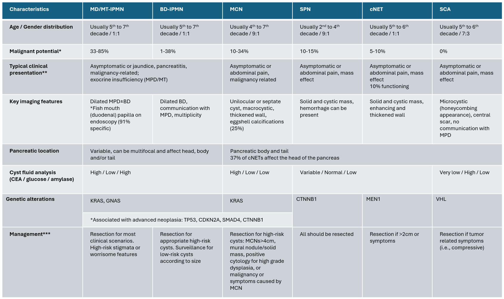
Differential Diagnosis of Pancreatic Cystic Neoplasms.
Contributed by V Dominguez, MD
References
Oyama H, Tada M, Takagi K, Tateishi K, Hamada T, Nakai Y, Hakuta R, Ijichi H, Ishigaki K, Kanai S, Kogure H, Mizuno S, Saito K, Saito T, Sato T, Suzuki T, Takahara N, Morishita Y, Arita J, Hasegawa K, Tanaka M, Fukayama M, Koike K. Long-term Risk of Malignancy in Branch-Duct Intraductal Papillary Mucinous Neoplasms. Gastroenterology. 2020 Jan:158(1):226-237.e5. doi: 10.1053/j.gastro.2019.08.032. Epub 2019 Aug 29 [PubMed PMID: 31473224]
Assawasirisin C, Fagenholz P, Qadan M, Hernandez-Barco Y, Aimprasittichai S, Kambadakone A, Mino-Kenudson M, Ike A, Chen SY, Sheng C, Brugge W, Warshaw AL, Lillemoe KD, Fernández-Del Castillo C. Unraveling the Long-term Natural History of Branch Duct Intraductal Papillary Mucinous Neoplasm: Beyond 10 years. Annals of surgery. 2025 Jan 1:281(1):154-160. doi: 10.1097/SLA.0000000000006535. Epub 2024 Sep 10 [PubMed PMID: 39253809]
Hamada T, Oyama H, Nakai Y, TOP-CREATE Study Group. Pancreatic Cysts. The New England journal of medicine. 2024 Nov 28:391(21):2063-2064. doi: 10.1056/NEJMc2412611. Epub [PubMed PMID: 39602646]
Omori Y, Ono Y, Tanino M, Karasaki H, Yamaguchi H, Furukawa T, Enomoto K, Ueda J, Sumi A, Katayama J, Muraki M, Taniue K, Takahashi K, Ambo Y, Shinohara T, Nishihara H, Sasajima J, Maguchi H, Mizukami Y, Okumura T, Tanaka S. Pathways of Progression From Intraductal Papillary Mucinous Neoplasm to Pancreatic Ductal Adenocarcinoma Based on Molecular Features. Gastroenterology. 2019 Feb:156(3):647-661.e2. doi: 10.1053/j.gastro.2018.10.029. Epub 2018 Oct 17 [PubMed PMID: 30342036]
Ozcan K, Klimstra DS. A Review of Mucinous Cystic and Intraductal Neoplasms of the Pancreatobiliary Tract. Archives of pathology & laboratory medicine. 2022 Mar 1:146(3):298-311. doi: 10.5858/arpa.2021-0399-RA. Epub [PubMed PMID: 35192699]
Valsangkar NP, Morales-Oyarvide V, Thayer SP, Ferrone CR, Wargo JA, Warshaw AL, Fernández-del Castillo C. 851 resected cystic tumors of the pancreas: a 33-year experience at the Massachusetts General Hospital. Surgery. 2012 Sep:152(3 Suppl 1):S4-12. doi: 10.1016/j.surg.2012.05.033. Epub 2012 Jul 6 [PubMed PMID: 22770958]
Choi SH, Park SH, Kim KW, Lee JY, Lee SS. Progression of Unresected Intraductal Papillary Mucinous Neoplasms of the Pancreas to Cancer: A Systematic Review and Meta-analysis. Clinical gastroenterology and hepatology : the official clinical practice journal of the American Gastroenterological Association. 2017 Oct:15(10):1509-1520.e4. doi: 10.1016/j.cgh.2017.03.020. Epub 2017 Mar 22 [PubMed PMID: 28342950]
Level 1 (high-level) evidencede la Fuente J, Chatterjee A, Lui J, Nehra AK, Bell MG, Lennon RJ, Kassmeyer BA, Graham RP, Nagayama H, Schulte PJ, Doering KA, Delgado AM, Vege SS, Chari ST, Takahashi N, Majumder S. Long-Term Outcomes and Risk of Pancreatic Cancer in Intraductal Papillary Mucinous Neoplasms. JAMA network open. 2023 Oct 2:6(10):e2337799. doi: 10.1001/jamanetworkopen.2023.37799. Epub 2023 Oct 2 [PubMed PMID: 37847503]
Fritz S, Klauss M, Bergmann F, Strobel O, Schneider L, Werner J, Hackert T, Büchler MW. Pancreatic main-duct involvement in branch-duct IPMNs: an underestimated risk. Annals of surgery. 2014 Nov:260(5):848-55; discussion 855-6. doi: 10.1097/SLA.0000000000000980. Epub [PubMed PMID: 25379856]
Ohtsuka T, Fernandez-Del Castillo C, Furukawa T, Hijioka S, Jang JY, Lennon AM, Miyasaka Y, Ohno E, Salvia R, Wolfgang CL, Wood LD. International evidence-based Kyoto guidelines for the management of intraductal papillary mucinous neoplasm of the pancreas. Pancreatology : official journal of the International Association of Pancreatology (IAP) ... [et al.]. 2024 Mar:24(2):255-270. doi: 10.1016/j.pan.2023.12.009. Epub 2023 Dec 28 [PubMed PMID: 38182527]
Moris M, Raimondo M, Woodward TA, Skinner V, Arcidiacono PG, Petrone MC, De Angelis C, Manfrè S, Fusaroli P, Wallace MB. Risk factors for malignant progression of intraductal papillary mucinous neoplasms. Digestive and liver disease : official journal of the Italian Society of Gastroenterology and the Italian Association for the Study of the Liver. 2015 Jun:47(6):495-501. doi: 10.1016/j.dld.2015.03.007. Epub 2015 Mar 17 [PubMed PMID: 25869552]
Nagtegaal ID, Odze RD, Klimstra D, Paradis V, Rugge M, Schirmacher P, Washington KM, Carneiro F, Cree IA, WHO Classification of Tumours Editorial Board. The 2019 WHO classification of tumours of the digestive system. Histopathology. 2020 Jan:76(2):182-188. doi: 10.1111/his.13975. Epub 2019 Nov 13 [PubMed PMID: 31433515]
Level 3 (low-level) evidenceFischer CG, Beleva Guthrie V, Braxton AM, Zheng L, Wang P, Song Q, Griffin JF, Chianchiano PE, Hosoda W, Niknafs N, Springer S, Dal Molin M, Masica D, Scharpf RB, Thompson ED, He J, Wolfgang CL, Hruban RH, Roberts NJ, Lennon AM, Jiao Y, Karchin R, Wood LD. Intraductal Papillary Mucinous Neoplasms Arise From Multiple Independent Clones, Each With Distinct Mutations. Gastroenterology. 2019 Oct:157(4):1123-1137.e22. doi: 10.1053/j.gastro.2019.06.001. Epub 2019 Jun 5 [PubMed PMID: 31175866]
Noë M, Niknafs N, Fischer CG, Hackeng WM, Beleva Guthrie V, Hosoda W, Debeljak M, Papp E, Adleff V, White JR, Luchini C, Pea A, Scarpa A, Butturini G, Zamboni G, Castelli P, Hong SM, Yachida S, Hiraoka N, Gill AJ, Samra JS, Offerhaus GJA, Hoorens A, Verheij J, Jansen C, Adsay NV, Jiang W, Winter J, Albores-Saavedra J, Terris B, Thompson ED, Roberts NJ, Hruban RH, Karchin R, Scharpf RB, Brosens LAA, Velculescu VE, Wood LD. Genomic characterization of malignant progression in neoplastic pancreatic cysts. Nature communications. 2020 Aug 14:11(1):4085. doi: 10.1038/s41467-020-17917-8. Epub 2020 Aug 14 [PubMed PMID: 32796935]
Elta GH, Enestvedt BK, Sauer BG, Lennon AM. ACG Clinical Guideline: Diagnosis and Management of Pancreatic Cysts. The American journal of gastroenterology. 2018 Apr:113(4):464-479. doi: 10.1038/ajg.2018.14. Epub 2018 Feb 27 [PubMed PMID: 29485131]
Chang YR, Park JK, Jang JY, Kwon W, Yoon JH, Kim SW. Incidental pancreatic cystic neoplasms in an asymptomatic healthy population of 21,745 individuals: Large-scale, single-center cohort study. Medicine. 2016 Dec:95(51):e5535. doi: 10.1097/MD.0000000000005535. Epub [PubMed PMID: 28002329]
Zerboni G, Signoretti M, Crippa S, Falconi M, Arcidiacono PG, Capurso G. Systematic review and meta-analysis: Prevalence of incidentally detected pancreatic cystic lesions in asymptomatic individuals. Pancreatology : official journal of the International Association of Pancreatology (IAP) ... [et al.]. 2019 Jan:19(1):2-9. doi: 10.1016/j.pan.2018.11.014. Epub 2018 Nov 28 [PubMed PMID: 30503370]
Level 1 (high-level) evidenceVilela A, Quingalahua E, Vargas A, Hawa F, Shannon C, Carpenter ES, Shi J, Krishna SG, Lee UJ, Chalhoub JM, Machicado JD. Global Prevalence of Pancreatic Cystic Lesions in the General Population on Magnetic Resonance Imaging: A Systematic Review and Meta-analysis. Clinical gastroenterology and hepatology : the official clinical practice journal of the American Gastroenterological Association. 2024 Sep:22(9):1798-1809.e6. doi: 10.1016/j.cgh.2024.02.018. Epub 2024 Feb 28 [PubMed PMID: 38423346]
Level 1 (high-level) evidenceRicci C, Migliori M, Imbrogno A, Mazzotta E, Felicani C, Serra C, Bergonzoni B, Calculli L, Casadei R. Prevalence of Asymptomatic Intraductal Papillary Mucinous Neoplasms in Healthy and Ill Populations Detected by Ultrasonography: A Single-Center Study of 6353 Outpatients. Pancreas. 2019 Jan:48(1):113-120. doi: 10.1097/MPA.0000000000001205. Epub [PubMed PMID: 30451793]
Abdolalizadeh B, Scheller NM, Lunetcas M, Rosenkrantz O, Jawad S, Kristensen TS, Axelsen T, Hansen CP, Ewertsen C. Pancreatic IPMN in clinical practice: descriptive analysis of 1082 patients referred to multidisciplinary evaluation. Acta radiologica (Stockholm, Sweden : 1987). 2026 Jan:67(1):67-74. doi: 10.1177/02841851251389575. Epub 2025 Nov 4 [PubMed PMID: 41186496]
Wood LD, Adsay NV, Basturk O, Brosens LAA, Fukushima N, Hong SM, Kim SJ, Lee JW, Luchini C, Noë M, Pitman MB, Scarpa A, Singhi AD, Tanaka M, Furukawa T. Systematic review of challenging issues in pathology of intraductal papillary mucinous neoplasms. Pancreatology : official journal of the International Association of Pancreatology (IAP) ... [et al.]. 2023 Nov:23(7):878-891. doi: 10.1016/j.pan.2023.08.002. Epub 2023 Aug 11 [PubMed PMID: 37604731]
Level 1 (high-level) evidenceKannan A, Murimwa GZ, Mansour JC, Whitham ZA, Jones AR, Sheth MK, Abbas A, Deverakonda A, Sawas T, Ellis DJ, Vanderveldt HS, Coleman V, Foley S, Tavakkoli A, Goldschmeidt M, Rajamohan N, Khatri G, Brand RE, Nikiforova MN, Paniccia A, Zureikat AH, Lee KKW, Fleming JB, Zeh HJ, Kubiliun NM, Singhi AD, Polanco PM. Molecular Analysis of Mixed-Type and Branch-Duct Intraductal Papillary Mucinous Neoplasms. JAMA surgery. 2026 Feb 1:161(2):171-178. doi: 10.1001/jamasurg.2025.5692. Epub [PubMed PMID: 41432930]
Mino-Kenudson M, Fernández-del Castillo C, Baba Y, Valsangkar NP, Liss AS, Hsu M, Correa-Gallego C, Ingkakul T, Perez Johnston R, Turner BG, Androutsopoulos V, Deshpande V, McGrath D, Sahani DV, Brugge WR, Ogino S, Pitman MB, Warshaw AL, Thayer SP. Prognosis of invasive intraductal papillary mucinous neoplasm depends on histological and precursor epithelial subtypes. Gut. 2011 Dec:60(12):1712-20. doi: 10.1136/gut.2010.232272. Epub 2011 Apr 20 [PubMed PMID: 21508421]
Hirono S, Shimizu Y, Ohtsuka T, Kin T, Hara K, Kanno A, Koshita S, Hanada K, Kitano M, Inoue H, Itoi T, Ueki T, Shimokawa T, Hijioka S, Yanagisawa A, Nakamura M, Okazaki K, Yamaue H. Recurrence patterns after surgical resection of intraductal papillary mucinous neoplasm (IPMN) of the pancreas; a multicenter, retrospective study of 1074 IPMN patients by the Japan Pancreas Society. Journal of gastroenterology. 2020 Jan:55(1):86-99. doi: 10.1007/s00535-019-01617-2. Epub 2019 Aug 28 [PubMed PMID: 31463655]
Level 2 (mid-level) evidenceFerrone CR, Correa-Gallego C, Warshaw AL, Brugge WR, Forcione DG, Thayer SP, Fernández-del Castillo C. Current trends in pancreatic cystic neoplasms. Archives of surgery (Chicago, Ill. : 1960). 2009 May:144(5):448-54. doi: 10.1001/archsurg.2009.36. Epub [PubMed PMID: 19451487]
Level 2 (mid-level) evidenceFernández-del Castillo C, Targarona J, Thayer SP, Rattner DW, Brugge WR, Warshaw AL. Incidental pancreatic cysts: clinicopathologic characteristics and comparison with symptomatic patients. Archives of surgery (Chicago, Ill. : 1960). 2003 Apr:138(4):427-3; discussion 433-4 [PubMed PMID: 12686529]
Level 2 (mid-level) evidenceAsokkumar R, Chin YK. Fish-Mouth Papilla. Clinical gastroenterology and hepatology : the official clinical practice journal of the American Gastroenterological Association. 2018 Sep:16(9):A34. doi: 10.1016/j.cgh.2017.11.029. Epub 2018 Apr 18 [PubMed PMID: 29678437]
Expert Panel on Gastrointestinal Imaging, Fábrega-Foster K, Kamel IR, Horowitz JM, Arif-Tiwari H, Bashir MR, Chernyak V, Goldstein A, Grajo JR, Hindman NM, Kamaya A, McNamara MM, Porter KK, Scheiman JM, Solnes LB, Srivastava PK, Zaheer A, Carucci LR. ACR Appropriateness Criteria® Pancreatic Cyst. Journal of the American College of Radiology : JACR. 2020 May:17(5S):S198-S206. doi: 10.1016/j.jacr.2020.01.021. Epub [PubMed PMID: 32370963]
Taya M, Hecht EM, Huang C, Lo GC. Pancreatic Cystic Lesions: Imaging Techniques and Diagnostic Features. Gastrointestinal endoscopy clinics of North America. 2023 Jul:33(3):497-518. doi: 10.1016/j.giec.2023.03.007. Epub 2023 Apr 26 [PubMed PMID: 37245932]
Kim JH, Hong SS, Kim YJ, Kim JK, Eun HW. Intraductal papillary mucinous neoplasm of the pancreas: differentiate from chronic pancreatits by MR imaging. European journal of radiology. 2012 Apr:81(4):671-6. doi: 10.1016/j.ejrad.2011.01.066. Epub 2011 Feb 15 [PubMed PMID: 21324627]
Kim SH, Lee JM, Lee ES, Baek JH, Kim JH, Han JK, Choi BI. Intraductal papillary mucinous neoplasms of the pancreas: evaluation of malignant potential and surgical resectability by using MR imaging with MR cholangiography. Radiology. 2015 Mar:274(3):723-33. doi: 10.1148/radiol.14132960. Epub 2014 Oct 8 [PubMed PMID: 25302831]
Huang C, Chopra S, Bolan CW, Chandarana H, Harfouch N, Hecht EM, Lo GC, Megibow AJ. Pancreatic Cystic Lesions: Next Generation of Radiologic Assessment. Gastrointestinal endoscopy clinics of North America. 2023 Jul:33(3):533-546. doi: 10.1016/j.giec.2023.03.004. Epub 2023 Apr 8 [PubMed PMID: 37245934]
Megibow AJ. Pancreatic Cysts: Radiology. Gastrointestinal endoscopy clinics of North America. 2023 Jul:33(3):519-531. doi: 10.1016/j.giec.2023.03.008. Epub 2023 Apr 24 [PubMed PMID: 37245933]
Kovač JD, Janković A, Mašulović D. The "bunch of grapes" pattern of branch-duct IPMN. Abdominal radiology (New York). 2020 Jan:45(1):249-250. doi: 10.1007/s00261-019-02266-y. Epub [PubMed PMID: 31605187]
Shimizu Y, Hijioka S, Hirono S, Kin T, Ohtsuka T, Kanno A, Koshita S, Hanada K, Kitano M, Inoue H, Itoi T, Ueki T, Matsuo K, Yanagisawa A, Yamaue H, Sugiyama M, Okazaki K. New Model for Predicting Malignancy in Patients With Intraductal Papillary Mucinous Neoplasm. Annals of surgery. 2020 Jul:272(1):155-162. doi: 10.1097/SLA.0000000000003108. Epub [PubMed PMID: 30499803]
Fritz S, Hackert T, Hinz U, Hartwig W, Büchler MW, Werner J. Role of serum carbohydrate antigen 19-9 and carcinoembryonic antigen in distinguishing between benign and invasive intraductal papillary mucinous neoplasm of the pancreas. The British journal of surgery. 2011 Jan:98(1):104-10. doi: 10.1002/bjs.7280. Epub 2010 Oct 14 [PubMed PMID: 20949535]
Duvvuri A, Bandla H, Thoguluva VC, Dasari C, Desai M, Nutalapati V, Moole V, Anvesh N, Harsh P, Gress F, Sharma P, Kohli DR. Comparing accuracy of high-risk features for detecting advanced neoplasia in pancreatic cystic lesions: a systematic review and meta-analysis. Annals of gastroenterology. 2021 Sep-Oct:34(5):743-750. doi: 10.20524/aog.2021.0630. Epub 2021 May 27 [PubMed PMID: 34475747]
Level 1 (high-level) evidenceIzumo W, Higuchi R, Furukawa T, Yazawa T, Uemura S, Shiihara M, Yamamoto M. Importance of each high-risk stigmata and worrisome features as a predictor of high-grade dysplasia in intraductal papillary mucinous neoplasms of the pancreas. Pancreatology : official journal of the International Association of Pancreatology (IAP) ... [et al.]. 2020 Jul:20(5):895-901. doi: 10.1016/j.pan.2020.06.011. Epub 2020 Jun 20 [PubMed PMID: 32624417]
Vege SS, Ziring B, Jain R, Moayyedi P, Clinical Guidelines Committee, American Gastroenterology Association. American gastroenterological association institute guideline on the diagnosis and management of asymptomatic neoplastic pancreatic cysts. Gastroenterology. 2015 Apr:148(4):819-22; quize12-3. doi: 10.1053/j.gastro.2015.01.015. Epub [PubMed PMID: 25805375]
Singhi AD, Zeh HJ, Brand RE, Nikiforova MN, Chennat JS, Fasanella KE, Khalid A, Papachristou GI, Slivka A, Hogg M, Lee KK, Tsung A, Zureikat AH, McGrath K. American Gastroenterological Association guidelines are inaccurate in detecting pancreatic cysts with advanced neoplasia: a clinicopathologic study of 225 patients with supporting molecular data. Gastrointestinal endoscopy. 2016 Jun:83(6):1107-1117.e2. doi: 10.1016/j.gie.2015.12.009. Epub 2015 Dec 18 [PubMed PMID: 26709110]
Kwan MC, Pitman MB, Fernandez-Del Castillo C, Zhang ML. Revisiting the performance of cyst fluid carcinoembryonic antigen as a diagnostic marker for pancreatic mucinous cysts: a comprehensive 20-year institutional review. Gut. 2024 Mar 7:73(4):629-638. doi: 10.1136/gutjnl-2023-331138. Epub 2024 Mar 7 [PubMed PMID: 38195219]
Pflüger MJ, Jamouss KT, Afghani E, Lim SJ, Rodriguez Franco S, Mayo H, Spann M, Wang H, Singhi A, Lennon AM, Wood LD. Predictive ability of pancreatic cyst fluid biomarkers: A systematic review and meta-analysis. Pancreatology : official journal of the International Association of Pancreatology (IAP) ... [et al.]. 2023 Nov:23(7):868-877. doi: 10.1016/j.pan.2023.05.005. Epub 2023 May 15 [PubMed PMID: 37230894]
Level 1 (high-level) evidenceMohan BP, Madhu D, Khan SR, Kassab LL, Ponnada S, Chandan S, Facciorusso A, Crino SF, Barresi L, McDonough S, Adler DG. Intracystic Glucose Levels in Differentiating Mucinous From Nonmucinous Pancreatic Cysts: A Systematic Review and Meta-analysis. Journal of clinical gastroenterology. 2022 Feb 1:56(2):e131-e136. doi: 10.1097/MCG.0000000000001507. Epub [PubMed PMID: 33731599]
Level 1 (high-level) evidenceLisotti A, Napoleon B, Facciorusso A, Cominardi A, Crinò SF, Brighi N, Gincul R, Kitano M, Yamashita Y, Marchegiani G, Fusaroli P. Contrast-enhanced EUS for the characterization of mural nodules within pancreatic cystic neoplasms: systematic review and meta-analysis. Gastrointestinal endoscopy. 2021 Nov:94(5):881-889.e5. doi: 10.1016/j.gie.2021.06.028. Epub 2021 Jul 1 [PubMed PMID: 34217751]
Level 1 (high-level) evidenceTacelli M, Celsa C, Magro B, Barchiesi M, Barresi L, Capurso G, Arcidiacono PG, Cammà C, Crinò SF. Diagnostic performance of endoscopic ultrasound through-the-needle microforceps biopsy of pancreatic cystic lesions: Systematic review with meta-analysis. Digestive endoscopy : official journal of the Japan Gastroenterological Endoscopy Society. 2020 Nov:32(7):1018-1030. doi: 10.1111/den.13626. Epub 2020 Feb 26 [PubMed PMID: 31912580]
Level 1 (high-level) evidenceGonda TA, Cahen DL, Farrell JJ. Pancreatic Cysts. The New England journal of medicine. 2024 Sep 5:391(9):832-843. doi: 10.1056/NEJMra2309041. Epub [PubMed PMID: 39231345]
Singhi AD, McGrath K, Brand RE, Khalid A, Zeh HJ, Chennat JS, Fasanella KE, Papachristou GI, Slivka A, Bartlett DL, Dasyam AK, Hogg M, Lee KK, Marsh JW, Monaco SE, Ohori NP, Pingpank JF, Tsung A, Zureikat AH, Wald AI, Nikiforova MN. Preoperative next-generation sequencing of pancreatic cyst fluid is highly accurate in cyst classification and detection of advanced neoplasia. Gut. 2018 Dec:67(12):2131-2141. doi: 10.1136/gutjnl-2016-313586. Epub 2017 Sep 28 [PubMed PMID: 28970292]
Khoury RE, Kabir C, Maker VK, Banulescu M, Wasserman M, Maker AV. What is the Incidence of Malignancy in Resected Intraductal Papillary Mucinous Neoplasms? An Analysis of Over 100 US Institutions in a Single Year. Annals of surgical oncology. 2018 Jun:25(6):1746-1751. doi: 10.1245/s10434-018-6425-6. Epub 2018 Mar 20 [PubMed PMID: 29560572]
Pollini T, Wong P, Maker AV. Current Management of Intraductal Papillary Mucinous Neoplasms. Hematology/oncology clinics of North America. 2026 Feb:40(1):65-77. doi: 10.1016/j.hoc.2025.07.006. Epub [PubMed PMID: 41224443]
Pollini T, Todeschini L, Maker AV. Pancreas Cyst Diagnosis and Advances in Cyst Fluid Analysis. The Surgical clinics of North America. 2024 Oct:104(5):965-974. doi: 10.1016/j.suc.2024.03.003. Epub 2024 Apr 23 [PubMed PMID: 39237171]
Level 3 (low-level) evidenceWu J, Jiao Y, Dal Molin M, Maitra A, de Wilde RF, Wood LD, Eshleman JR, Goggins MG, Wolfgang CL, Canto MI, Schulick RD, Edil BH, Choti MA, Adsay V, Klimstra DS, Offerhaus GJ, Klein AP, Kopelovich L, Carter H, Karchin R, Allen PJ, Schmidt CM, Naito Y, Diaz LA Jr, Kinzler KW, Papadopoulos N, Hruban RH, Vogelstein B. Whole-exome sequencing of neoplastic cysts of the pancreas reveals recurrent mutations in components of ubiquitin-dependent pathways. Proceedings of the National Academy of Sciences of the United States of America. 2011 Dec 27:108(52):21188-93. doi: 10.1073/pnas.1118046108. Epub 2011 Dec 8 [PubMed PMID: 22158988]
Nakashima Y, Ohtsuka T, Nakamura S, Mori Y, Nakata K, Miyasaka Y, Ishigami K, Matsuda R, Oda Y, Nakamura M. Clinicopathological characteristics of non-functioning cystic pancreatic neuroendocrine tumors. Pancreatology : official journal of the International Association of Pancreatology (IAP) ... [et al.]. 2019 Jan:19(1):50-56. doi: 10.1016/j.pan.2018.11.010. Epub 2018 Nov 22 [PubMed PMID: 30497875]
D'Onofrio M, De Robertis R, Capelli P, Tinazzi Martini P, Crosara S, Gobbo S, Butturini G, Salvia R, Barbi E, Girelli R, Bassi C, Pederzoli P. Uncommon presentations of common pancreatic neoplasms: a pictorial essay. Abdominal imaging. 2015 Aug:40(6):1629-44. doi: 10.1007/s00261-015-0388-x. Epub [PubMed PMID: 25772002]
Kaderi ASA, Munagala PA, Mavroeidis VK, MacCormick A, Aroori S. The Concept of Anatomical Analogy Between the Liver and the Pancreas. Clinical anatomy (New York, N.Y.). 2025 Dec 15:():. doi: 10.1002/ca.70058. Epub 2025 Dec 15 [PubMed PMID: 41395890]
Rieser C, Hays S, Hogg ME. Techniques for Robotic Pancreatectomy. Surgical oncology clinics of North America. 2026 Jan:35(1):89-102. doi: 10.1016/j.soc.2025.03.001. Epub 2025 Apr 12 [PubMed PMID: 41233075]
Goudard Y, Gaujoux S, Dokmak S, Cros J, Couvelard A, Palazzo M, Ronot M, Vullierme MP, Ruszniewski P, Belghiti J, Sauvanet A. Reappraisal of central pancreatectomy a 12-year single-center experience. JAMA surgery. 2014 Apr:149(4):356-63. doi: 10.1001/jamasurg.2013.4146. Epub [PubMed PMID: 24740703]
Sauvanet A, Gaujoux S, Blanc B, Couvelard A, Dokmak S, Vullierme MP, Ruszniewski P, Belghiti J, Lévy P. Parenchyma-sparing pancreatectomy for presumed noninvasive intraductal papillary mucinous neoplasms of the pancreas. Annals of surgery. 2014 Aug:260(2):364-71. doi: 10.1097/SLA.0000000000000601. Epub [PubMed PMID: 24646561]
Stoop TF, Javed AA, Oba A, Koerkamp BG, Seufferlein T, Wilmink JW, Besselink MG. Pancreatic cancer. Lancet (London, England). 2025 Apr 5:405(10485):1182-1202. doi: 10.1016/S0140-6736(25)00261-2. Epub [PubMed PMID: 40187844]
Thalji SZ, Clarke CN. Perioperative Chemotherapy and Total Neoadjuvant Chemotherapy for Pancreatic Adenocarcinoma. Surgical oncology clinics of North America. 2026 Jan:35(1):71-87. doi: 10.1016/j.soc.2025.05.003. Epub 2025 Jun 13 [PubMed PMID: 41233074]
Yang G, Li M, Yang Q, Zhai X, Halima J, Hu Q, Lei C, Dang R. Bta-miR-127 inhibits secretion, proliferation and promotes apoptosis by targeting ITGA6 in bovine Sertoli cell. International journal of biological macromolecules. 2023 Dec 31:253(Pt 3):126838. doi: 10.1016/j.ijbiomac.2023.126838. Epub 2023 Sep 13 [PubMed PMID: 37714242]
Lucocq J, Hawkyard J, Haugk B, Mownah O, Menon K, Furukawa T, Inoue Y, Hirose Y, Sasahira N, Feretis M, Balakrishnan A, Ceresa C, Davidson B, Pande R, Dasari B, Tanno L, Karavias D, Helliwell J, Young A, Nunes Q, Urbonas T, Silva M, Gordon-Weeks A, Barrie J, Gomez D, Van Laarhoven S, Robertson F, Nawara H, Doyle J, Bhogal R, Harrison E, Roalso M, Ciprani D, Aroori S, Ratnayake B, Koea J, Capurso G, Bellotti R, Stättner S, Alsaoudi T, Bhardwaj N, Rajesh S, Jeffery F, Connor S, Cameron A, Jamieson N, Sheen A, Mittal A, Samra J, Gill A, Roberts K, Søreide K, Pandanaboyana S. Adjuvant chemotherapy for adenocarcinoma arising from intraductal papillary mucinous neoplasia: multicentre ADENO-IPMN study. The British journal of surgery. 2024 Apr 3:111(4):. pii: znae100. doi: 10.1093/bjs/znae100. Epub [PubMed PMID: 38659247]
Habib JR, Kinny-Köster B, Javed AA, Zelga P, Saadat LV, Kim RC, Gorris M, Allegrini V, Watanabe S, Sharib J, Arcerito M, Kaiser J, Lafaro KJ, Tu M, Bhandre M, Shi C, Kim MP, Correa C, Daamen LA, Oberstein PE, Schmidt CM, Hanna NN, Allen P, Loos M, Shrikhande SV, Molenaar IQ, Frigerio I, Katz MHG, Soares KC, Miao Y, Del Chiaro M, He J, Hackert T, Salvia R, Büchler MW, Castillo CF, Besselink MG, Marchegiani G, Wolfgang CL, Verona IPMN Consortium. Impact of Adjuvant Chemotherapy on Resected Intraductal Papillary Mucinous Neoplasm-Derived Pancreatic Cancer: Results From an International Multicenter Study. Journal of clinical oncology : official journal of the American Society of Clinical Oncology. 2024 Dec 20:42(36):4317-4326. doi: 10.1200/JCO.23.02313. Epub 2024 Sep 10 [PubMed PMID: 39255450]
Level 2 (mid-level) evidenceZiogas IA, Rodriguez Franco S, Schmoke N, Meguid C, Murphy C, Al-Musawi M, Alexopoulos SP, Schulick RD, Del Chiaro M. Comparison of Invasive Pancreatic Ductal Adenocarcinoma versus Intraductal Papillary Mucinous Neoplasm: A National Cancer Database Analysis. Cancers. 2023 Feb 13:15(4):. doi: 10.3390/cancers15041185. Epub 2023 Feb 13 [PubMed PMID: 36831527]
Cameron JL, He J. Two thousand consecutive pancreaticoduodenectomies. Journal of the American College of Surgeons. 2015 Apr:220(4):530-6. doi: 10.1016/j.jamcollsurg.2014.12.031. Epub 2015 Jan 6 [PubMed PMID: 25724606]
Bassi C, Marchegiani G, Giuliani T, Di Gioia A, Andrianello S, Zingaretti CC, Brentegani G, De Pastena M, Fontana M, Pea A, Paiella S, Malleo G, Tuveri M, Landoni L, Esposito A, Casetti L, Butturini G, Falconi M, Salvia R. Pancreatoduodenectomy at the Verona Pancreas Institute: the Evolution of Indications, Surgical Techniques, and Outcomes: A Retrospective Analysis of 3000 Consecutive Cases. Annals of surgery. 2022 Dec 1:276(6):1029-1038. doi: 10.1097/SLA.0000000000004753. Epub 2021 Jan 15 [PubMed PMID: 33630454]
Level 2 (mid-level) evidenceAleotti F, Crippa S, Belfiori G, Tamburrino D, Partelli S, Longo E, Palumbo D, Pecorelli N, Lena MS, Capurso G, Arcidiacono PG, Falconi M. Pancreatic resections for benign intraductal papillary mucinous neoplasms: Collateral damages from friendly fire. Surgery. 2022 Oct:172(4):1202-1209. doi: 10.1016/j.surg.2022.04.036. Epub 2022 Jun 3 [PubMed PMID: 35667898]
Bassi C, Dervenis C, Butturini G, Fingerhut A, Yeo C, Izbicki J, Neoptolemos J, Sarr M, Traverso W, Buchler M, International Study Group on Pancreatic Fistula Definition. Postoperative pancreatic fistula: an international study group (ISGPF) definition. Surgery. 2005 Jul:138(1):8-13 [PubMed PMID: 16003309]
Bassi C, Marchegiani G, Dervenis C, Sarr M, Abu Hilal M, Adham M, Allen P, Andersson R, Asbun HJ, Besselink MG, Conlon K, Del Chiaro M, Falconi M, Fernandez-Cruz L, Fernandez-Del Castillo C, Fingerhut A, Friess H, Gouma DJ, Hackert T, Izbicki J, Lillemoe KD, Neoptolemos JP, Olah A, Schulick R, Shrikhande SV, Takada T, Takaori K, Traverso W, Vollmer CM, Wolfgang CL, Yeo CJ, Salvia R, Buchler M, International Study Group on Pancreatic Surgery (ISGPS). The 2016 update of the International Study Group (ISGPS) definition and grading of postoperative pancreatic fistula: 11 Years After. Surgery. 2017 Mar:161(3):584-591. doi: 10.1016/j.surg.2016.11.014. Epub 2016 Dec 28 [PubMed PMID: 28040257]
Gianotti L, Besselink MG, Sandini M, Hackert T, Conlon K, Gerritsen A, Griffin O, Fingerhut A, Probst P, Abu Hilal M, Marchegiani G, Nappo G, Zerbi A, Amodio A, Perinel J, Adham M, Raimondo M, Asbun HJ, Sato A, Takaori K, Shrikhande SV, Del Chiaro M, Bockhorn M, Izbicki JR, Dervenis C, Charnley RM, Martignoni ME, Friess H, de Pretis N, Radenkovic D, Montorsi M, Sarr MG, Vollmer CM, Frulloni L, Büchler MW, Bassi C. Nutritional support and therapy in pancreatic surgery: A position paper of the International Study Group on Pancreatic Surgery (ISGPS). Surgery. 2018 Nov:164(5):1035-1048. doi: 10.1016/j.surg.2018.05.040. Epub 2018 Jul 17 [PubMed PMID: 30029989]
Nebbia M, Capretti G, Nappo G, Zerbi A. Updates in the management of postoperative pancreatic fistula. International journal of surgery (London, England). 2024 Oct 1:110(10):6135-6144. doi: 10.1097/JS9.0000000000001395. Epub 2024 Oct 1 [PubMed PMID: 38518082]
Magnin J, Jolissaint JS, Fuchs HE, Seier K, Gonen M, Barekzai A, Balachandran VP, D'Angelica MI, Drebin JA, Kingham TP, Soares K, Wei AC, Jarnagin WR. Safety of Selective Operative Drainage After Pancreatic Resection: A Retrospective Analysis of 2607 Patients. Annals of surgery. 2026 Mar 1:283(3):479-487. doi: 10.1097/SLA.0000000000006425. Epub 2024 Jun 28 [PubMed PMID: 38939927]
Level 2 (mid-level) evidencePARANOIA Study Group:, Writing group:, Halle-Smith JM, Pande R, Hall L, Hodson J, Roberts KJ, Steering committee:, Arshad A, Connor S, Conlon KCP, Dickson EJ, Giovinazzo F, Harrison E, de Liguori Carino N, Hore T, Knight SR, Loveday B, Magill L, Mirza D, Mitta A, Pandanaboyana S, Perry RJ, Pinkney T, Samra J, Siriwardena AK, Satoi S, Skipworth J, Stättner S, Sutcliffe RP, Tingstedt B, Roberts KJ. Perioperative interventions to reduce pancreatic fistula following pancreatoduodenectomy: meta-analysis. The British journal of surgery. 2022 Aug 16:109(9):812-821. doi: 10.1093/bjs/znac074. Epub [PubMed PMID: 35727956]
Level 1 (high-level) evidenceSnyder RA, Ewing JA, Parikh AA. Delayed gastric emptying after pancreaticoduodenectomy: A study of the national surgical quality improvement program. Pancreatology : official journal of the International Association of Pancreatology (IAP) ... [et al.]. 2020 Mar:20(2):205-210. doi: 10.1016/j.pan.2019.12.003. Epub 2019 Dec 16 [PubMed PMID: 31875832]
Level 2 (mid-level) evidenceMaeda H, Hanazaki K. Pancreatogenic diabetes after pancreatic resection. Pancreatology : official journal of the International Association of Pancreatology (IAP) ... [et al.]. 2011:11(2):268-76. doi: 10.1159/000328785. Epub 2011 Jul 5 [PubMed PMID: 21734430]
American Diabetes Association Professional Practice Committee for Diabetes*. 2. Diagnosis and Classification of Diabetes: Standards of Care in Diabetes-2026. Diabetes care. 2026 Jan 1:49(Supplement_1):S27-S49. doi: 10.2337/dc26-S002. Epub [PubMed PMID: 41358893]
Maina RM, Wancata L. Palliative Care and Patient Quality of Life in Pancreatic Adenocarcinoma. Surgical oncology clinics of North America. 2026 Jan:35(1):181-190. doi: 10.1016/j.soc.2025.05.006. Epub 2025 Jun 23 [PubMed PMID: 41233071]
Level 2 (mid-level) evidenceKuemmerli C, Tschuor C, Kasai M, Alseidi AA, Balzano G, Bouwense S, Braga M, Coolsen M, Daniel SK, Dervenis C, Falconi M, Hwang DW, Kagedan DJ, Kim SC, Lavu H, Liang T, Nussbaum D, Partelli S, Passeri MJ, Pecorelli N, Pillai SA, Pillarisetty VG, Pucci MJ, Su W, Sutcliffe RP, Tingstedt B, van der Kolk M, Vrochides D, Wei A, Williamsson C, Yeo CJ, Zani S, Zouros E, Abu Hilal M. Impact of enhanced recovery protocols after pancreatoduodenectomy: meta-analysis. The British journal of surgery. 2022 Feb 24:109(3):256-266. doi: 10.1093/bjs/znab436. Epub [PubMed PMID: 35037019]
Level 1 (high-level) evidenceIm K, O'Connor VV. Enhanced Recovery After Surgery (ERAS) After Pancreatectomy: Interventions and Outcomes at an ERAS Qualified Pancreatectomy Center. The American surgeon. 2025 Oct:91(10):1786-1791. doi: 10.1177/00031348251359117. Epub 2025 Jul 9 [PubMed PMID: 40631431]
Melloul E, Lassen K, Roulin D, Grass F, Perinel J, Adham M, Wellge EB, Kunzler F, Besselink MG, Asbun H, Scott MJ, Dejong CHC, Vrochides D, Aloia T, Izbicki JR, Demartines N. Guidelines for Perioperative Care for Pancreatoduodenectomy: Enhanced Recovery After Surgery (ERAS) Recommendations 2019. World journal of surgery. 2020 Jul:44(7):2056-2084. doi: 10.1007/s00268-020-05462-w. Epub [PubMed PMID: 32161987]
Smits FJ, Henry AC, Besselink MG, Busch OR, van Eijck CH, Arntz M, Bollen TL, van Delden OM, van den Heuvel D, van der Leij C, van Lienden KP, Moelker A, Bonsing BA, Borel Rinkes IH, Bosscha K, van Dam RM, Derksen WJM, den Dulk M, Festen S, Groot Koerkamp B, de Haas RJ, Hagendoorn J, van der Harst E, de Hingh IH, Kazemier G, van der Kolk M, Liem M, Lips DJ, Luyer MD, de Meijer VE, Mieog JS, Nieuwenhuijs VB, Patijn GA, Te Riele WW, Roos D, Schreinemakers JM, Stommel MWJ, Wit F, Zonderhuis BA, Daamen LA, van Werkhoven CH, Molenaar IQ, van Santvoort HC, Dutch Pancreatic Cancer Group. Algorithm-based care versus usual care for the early recognition and management of complications after pancreatic resection in the Netherlands: an open-label, nationwide, stepped-wedge cluster-randomised trial. Lancet (London, England). 2022 May 14:399(10338):1867-1875. doi: 10.1016/S0140-6736(22)00182-9. Epub 2022 Apr 28 [PubMed PMID: 35490691]
Level 1 (high-level) evidence