 Tools
Tools
 Inner Ear Anatomy and Cochlear Physiology
Introduction
The inner ear is a highly specialized sensory organ that perceives sound and maintains balance and spatial orientation. Encased within the dense petrous portion of the temporal bone, the inner ear is one of the most anatomically complex and physiologically sophisticated structures in the human body. Despite occupying only a few cubic centimeters of space, the inner ear contains intricate sensory epithelia that convert mechanical stimuli into precisely coded neural signals, enabling hearing, gaze stabilization, postural control, and spatial navigation. The remarkable sensitivity of these structures permits detection of acoustic vibrations on the nanometer scale while simultaneously monitoring angular and linear accelerations generated during everyday movement.[1]
Unlike most sensory systems, the inner ear simultaneously performs mechanical signal detection, frequency analysis, neural encoding, and vestibular integration within a compact osseous structure. This combination of sensory specialization and anatomic complexity explains both the precision of normal auditory and vestibular function and the profound consequences of disease affecting even small portions of the labyrinth.
Although traditionally associated with otolaryngology, a thorough understanding of inner ear anatomy and physiology is important for physicians across numerous medical specialties. Primary care physicians, emergency medicine clinicians, neurologists, neurosurgeons, radiologists, geriatricians, audiologists, physical therapists, and rehabilitation specialists frequently encounter patients with hearing loss, tinnitus, vertigo, imbalance, falls, and facial nerve (cranial nerve VII) disorders. Dizziness alone accounts for millions of healthcare visits annually and ranks among the most common neurologic and otologic complaints in clinical practice. Accurate diagnosis often requires an understanding of the intricate relationships among the peripheral vestibular apparatus, vestibular nerve (cranial nerve VIII), brainstem pathways, cerebellum, and higher cortical centers. Consequently, familiarity with inner ear anatomy extends beyond otology and provides a foundation for evaluating numerous neurologic and systemic disorders.
From an anatomic perspective, the inner ear consists of a bony labyrinth and a membranous labyrinth.[2] The bony labyrinth comprises the cochlea, vestibule, and semicircular canals, which contain perilymph and are embedded within the otic capsule. Suspended within this framework, the membranous labyrinth contains endolymph and houses the sensory organs responsible for auditory and vestibular transduction. The cochlea contains the organ of Corti, the primary receptor organ for hearing. In contrast, the vestibular system consists of the utricle, saccule, and 3 semicircular ducts that collectively detect linear acceleration, gravitational forces, and angular head movement. These structures communicate with the central nervous system (CNS) through the vestibulocochlear nerve (cranial nerve VIII), which conveys highly organized sensory information to specialized brainstem and cortical pathways.
The neural pathways arising from the inner ear are among the most extensively organized sensory systems in the body. A distinguishing feature of the auditory system is the early bilateral distribution of neural projections within the brainstem. Consequently, unilateral lesions above the cochlear nuclei rarely produce complete unilateral deafness. In contrast, lesions involving the cochlea, cochlear nerve (cranial nerve VIII), or cochlear nucleus may result in profound ipsilateral hearing loss.
Auditory information travels from cochlear hair cells through the spiral ganglion and the cochlear nerve to the cochlear nuclei of the brainstem before ascending through the superior olivary complex, lateral lemniscus, inferior colliculus, and medial geniculate body to the auditory cortex. Vestibular information follows an equally complex course through the vestibular nerve to the vestibular nuclei and cerebellum, where integration with visual and proprioceptive inputs maintains balance, coordinates eye movements, and preserves postural stability. The vestibulo-ocular reflex, one of the most clinically important reflex pathways in medicine, stabilizes visual fixation during head movement and provides the physiologic basis for many bedside vestibular examinations.
Understanding these pathways is particularly important for distinguishing peripheral vestibular disorders from CNS disease. Peripheral causes of vertigo, such as benign paroxysmal positional vertigo, vestibular neuritis, labyrinthitis, and Ménière disease, arise from pathology involving the labyrinth or vestibular nerve. In contrast, central causes originate within the brainstem, cerebellum, or higher vestibular processing centers and may reflect serious conditions, including ischemic stroke, demyelinating disease, neoplasms, and neurodegenerative disorders. Since symptoms frequently overlap, detailed knowledge of vestibular anatomy, neural connections, and physiologic function is essential for accurate localization and timely diagnosis. Distinguishing benign peripheral vestibulopathy from a potentially life-threatening central lesion requires a thorough understanding of inner ear anatomy.[3]
The inner ear occupies a central position within the temporal bone and maintains critical relationships with numerous neurovascular structures. The facial nerve traverses the temporal bone in close proximity to the labyrinth, particularly along the labyrinthine segment and geniculate ganglion. The internal auditory canal transmits the facial and vestibulocochlear nerves, while the cochlear aqueduct, vestibular aqueduct, jugular bulb, carotid canal, and petrous apex constitute additional clinically important landmarks. These relationships are particularly relevant during otologic and lateral skull base surgery, where detailed knowledge of labyrinthine anatomy is essential to minimize morbidity and preserve auditory and vestibular function.
Embryologically, the inner ear is derived from the otic placode, a thickening of surface ectoderm that appears during the 3rd week of gestation.[4][5] Progressive invagination forms the otic vesicle, which subsequently differentiates into the cochlear and vestibular components of the membranous labyrinth. Developmental abnormalities during this process may result in a spectrum of congenital malformations, ranging from isolated enlargement of the vestibular aqueduct to complete labyrinthine aplasia. Modern radiologic classifications of congenital inner ear anomalies are largely based on patterns of embryologic arrest, making an understanding of embryology essential for interpreting temporal bone imaging and for surgical planning. Knowledge of these developmental pathways provides important insight into congenital hearing loss, vestibular dysfunction, and modern cochlear implantation strategies.
The physiologic uniqueness of the inner ear largely stems from its highly regulated fluid compartments.[6][7] Endolymph, characterized by a high potassium and low sodium concentration, differs markedly from perilymph and resembles intracellular fluid. Maintenance of this ionic gradient by the stria vascularis generates the endocochlear potential, a critical electrochemical driving force for hair-cell transduction. The stria vascularis is among the most metabolically active tissues in the human body and generates an endocochlear potential of approximately +80 mV, one of the largest bioelectric gradients in mammalian physiology. Disruption of fluid homeostasis or sensory hair-cell function underlies many common otologic disorders, including Ménière disease, sudden sensorineural hearing loss (SSNHL), ototoxic injury, autoimmune inner ear disease, and age-related hearing loss.
Advances in microscopy, molecular biology, electrophysiology, and radiographic imaging have transformed understanding of inner ear structure and function. Contemporary investigations have revealed complex interactions among hair cells, supporting cells, afferent and efferent neural pathways, ion transport systems, and central vestibular processing networks. These discoveries continue to influence clinical practice through improvements in cochlear implantation, vestibular rehabilitation, hearing preservation surgery, gene therapy, regenerative medicine, and lateral skull base reconstruction.[8][9][10][11]
For the otolaryngologist, mastery of inner ear anatomy extends beyond simple structural recognition. A detailed knowledge of labyrinthine architecture forms the foundation for interpreting audiometric and vestibular testing, diagnosing disorders of hearing and balance, interpreting temporal bone imaging, and performing advanced otologic and neurotologic procedures. For physicians in other disciplines, a broad grasp of inner ear anatomy and physiology provides an essential framework for evaluating dizziness, hearing loss, tinnitus, gait instability, falls, cranial neuropathies, and disorders affecting the brainstem and cerebellum.
Clinical Pearls
- The inner ear occupies a unique position at the intersection of otology, neurology, and skull base surgery. Understanding the anatomy of this sensory organ requires more than recognition of individual structures. Appreciation of the integration between peripheral sensory organs and central auditory and vestibular pathways is also necessary. This relationship explains why disorders originating within the cochlea or vestibular labyrinth may mimic CNS disease and vice versa.
- Knowledge of inner ear anatomy is crucial to localization. Peripheral vestibular disorders originate within the labyrinth or vestibular nerve, whereas central vestibular disorders arise from the brainstem, cerebellum, or higher cortical pathways. Distinguishing between these entities is essential because benign vestibulopathy and posterior circulation stroke may initially present with remarkably similar symptoms.
- Hearing and balance are often discussed together, yet the cochlear and vestibular systems function as distinct sensory organs with separate receptor populations, neural pathways, and vascular supplies. Consequently, a patient may lose hearing while retaining normal vestibular function, or vice versa, depending on lesion location.[12]
Structure and Function
Register For Free And Read The Full Article
 Search engine and full access to all medical articles
- 10 free questions in your specialty
- Free CME/CE Activities
- Free daily question in your email
- Save favorite articles to your dashboard
- Emails offering discounts
Learn more about a Subscription to StatPearls Point-of-Care
Structure and Function
The organ of Corti is the specialized sensory epithelium of the cochlea responsible for auditory transduction. Located within the scala media and resting on the basilar membrane, the organ of Corti converts mechanical vibrations generated by sound into neural signals, ultimately interpreted by the auditory cortex (see Image. Cochlear Cross-Section and Organ of Corti). The organ of Corti serves as the functional receptor organ of hearing and is among the most highly specialized sensory organs in the human body.[13][14][15][16][17][18] Modern advances in cochlear implantation, electrophysiologic testing, hearing preservation surgery, and regenerative therapies all rely on a thorough understanding of cochlear anatomy and physiology.[19]
Cochlear Location and Organization
The organ of Corti is situated within the scala media of the cochlea and extends throughout the entire cochlear spiral. The organ of Corti rests on the basilar membrane, which separates the scala media from the perilymph-filled scala tympani. Superiorly, the organ of Corti is associated with the tectorial membrane, an acellular gelatinous structure that plays a critical role in auditory transduction. The scala media contains endolymph, a unique extracellular fluid characterized by a high potassium concentration and a positive electrical potential relative to surrounding tissues. The stria vascularis maintains this specialized ionic environment and is essential for normal hair-cell function. The electrochemical gradient established between endolymph and the intracellular environment of hair cells provides the driving force for mechanoelectrical transduction.
Cellular Architecture
The organ of Corti consists of highly specialized sensory hair cells and multiple supporting cell populations that provide structural and functional support.[20][21][22] Patterning of the developing mammalian cochlea into distinct sensory and nonsensory regions, and specification of multiple cell fates within those regions, are critical for proper auditory function.[23] The sensory epithelium contains 1 row of inner hair cells and 3 rows of outer hair cells extending along the length of the cochlea. Inner and outer hair cells differ significantly in structure and function despite similar embryologic origins. Supporting cells include pillar cells, Deiters cells, Hensen cells, Claudius cells, and other specialized cellular elements.[24] These cells help maintain cochlear architecture, ionic homeostasis, mechanical stability, and tissue repair. The pillar cells form the Corti tunnel, a characteristic feature of the mature cochlea that separates the inner and outer hair cell populations.
Sound Transmission to the Cochlea
Auditory transduction begins when sound waves enter the external auditory canal and vibrate the tympanic membrane. These vibrations are transmitted through the ossicular chain of the middle ear, consisting of the malleus, incus, and stapes. Movement of the stapes footplate at the oval window generates pressure waves within the perilymph of the scala vestibuli. These pressure waves travel through the cochlea and ultimately dissipate through the scala tympani and the round window. The pressure wave creates a traveling wave along the basilar membrane. The location of maximal basilar membrane displacement depends on sound frequency and forms the basis of cochlear frequency discrimination.
Mechanism of Auditory Transduction
The primary function of the organ of Corti is the conversion of mechanical energy into electrical activity within the auditory nervous system. A shearing force develops between the tectorial membrane and hair-cell stereocilia as the basilar membrane vibrates. Deflection of stereocilia toward the tallest stereocilium increases tension on specialized tip-link proteins that connect adjacent stereocilia. This tension opens mechanically gated ion channels located near the stereociliary tips. The high potassium concentration and positive electrical potential of endolymph promote rapid potassium influx into hair cells, producing depolarization. Subsequent opening of voltage-gated calcium channels at the basal aspect of the cell triggers neurotransmitter release, primarily glutamate, onto afferent fibers of the spiral ganglion.[25] The resulting action potentials travel along the cochlear division of the vestibulocochlear nerve to the cochlear nuclei in the brainstem and then along higher auditory pathways to the auditory cortex, where sound is ultimately perceived.
Inner Hair Cells: Primary Sensory Receptors
Inner hair cells serve as the principal sensory receptors of hearing. Although inner hair cells account for only a minority of cochlear hair cells, they provide approximately 90% to 95% of the afferent neural input to the auditory nerve.[26] Unlike outer hair cells, inner hair-cell stereocilia are not firmly embedded within the tectorial membrane. Instead, stimulation occurs primarily through fluid motion and local mechanical forces generated by basilar membrane displacement. Activation of inner hair cells directly triggers neurotransmitter release and transmission of auditory information to the CNS. Inner hair cells encode acoustic information. Consequently, injury to inner hair cells often results in significant hearing impairment and reduced speech discrimination.
Spiral ganglion neurons
The cell bodies of primary auditory afferent neurons reside within the spiral ganglion, which is located within the Rosenthal canal of the modiolus. Spiral ganglion neurons form the critical link between sensory hair cells of the organ of Corti and the central auditory pathways.
Two major populations of spiral ganglion neurons are recognized. Type I spiral ganglion neurons account for approximately 90% to 95% of cochlear afferent fibers and primarily innervate inner hair cells. Each inner hair cell receives multiple afferent contacts, allowing highly precise encoding of sound intensity, frequency, and temporal information. In contrast, type II spiral ganglion neurons constitute a small minority of cochlear afferents and innervate multiple outer hair cells. Although the precise physiologic role of type II spiral ganglion neurons is incompletely understood, these neurons may participate in signaling cochlear injury and excessive acoustic stimulation.
Central processes of spiral ganglion neurons converge to form the cochlear division of the vestibulocochlear nerve, which transmits auditory information to the cochlear nuclei of the brainstem. Degeneration of spiral ganglion neurons or disruption of synaptic connections between inner hair cells and auditory nerve fibers may contribute to hearing impairment even when sensory hair cells remain relatively preserved.
Recognition of cochlear synaptopathy, commonly referred to as "hidden hearing loss," arose from this concept.[27][28] Hidden hearing loss describes impaired auditory neural processing despite normal pure-tone audiometric thresholds. Patients frequently report difficulty understanding speech in noisy environments despite a normal audiogram. Noise exposure, aging, ototoxic medications, and peripheral neuropathies have all been implicated in the development of cochlear synaptopathy. These observations highlight the importance of neural integrity within the auditory system and demonstrate that normal hearing thresholds do not necessarily indicate normal auditory function.
Outer hair cells and the cochlear amplifier
Outer hair cells perform a fundamentally different role. Rather than serving primarily as sensory receptors, outer hair cells function as active mechanical amplifiers that enhance cochlear sensitivity and frequency selectivity.[29] Outer hair cells contain the motor protein prestin within their lateral cell membranes. Prestin undergoes rapid conformational changes in response to alterations in membrane potential, allowing outer hair cells to change length at extremely high speeds. Depolarization causes cell shortening, whereas hyperpolarization causes elongation. Prestin must be gated by changes in membrane potential on a cycle-by-cycle basis.[30]
Electromotile responses feed mechanical energy back into the basilar membrane and cochlear partition, amplifying local vibrations at the site of stimulation. As a result, auditory sensitivity increases, frequency tuning becomes sharper, and speech discrimination improves, particularly in complex acoustic environments. The cochlear amplifier generated by outer hair cells accounts for much of the extraordinary sensitivity, frequency selectivity, and dynamic range characteristic of normal hearing.
Tonotopic Organization
A defining feature of cochlear structure is tonotopic organization. The mechanical properties of the basilar membrane vary progressively from base to apex.[31][32] The basal turn of the cochlea is relatively narrow and stiff and, therefore, responds preferentially to high-frequency sounds. In contrast, the apical turn is wider and more compliant, making it more responsive to low-frequency stimulation. As a result, individual frequencies produce maximal activation at specific locations along the cochlear spiral. This frequency map extends throughout the ascending auditory pathways, including the cochlear nuclei, the superior olivary complex, the inferior colliculus, the medial geniculate body, and the auditory cortex. Tonotopic organization is a fundamental principle of auditory processing and neural coding.
The cochlea performs frequency analysis before auditory information reaches the brain. Through the tonotopic organization of the basilar membrane, complex sounds are mechanically separated into individual frequency components, allowing different populations of hair cells to respond preferentially to specific frequencies.
Efferent Modulation of Cochlear Function
Auditory processing is often viewed as an ascending sensory system, though significant descending control also contributes to cochlear function. Efferent fibers originating primarily from the superior olivary complex project to the cochlea through the olivocochlear system, forming a simple reflex pathway.[33][34]
Descending pathways predominantly influence outer hair-cell activity and allow the CNS to regulate cochlear amplification. Efferent modulation contributes to selective auditory attention, protection against acoustic overstimulation, improved signal detection in background noise, and optimization of auditory processing under varying environmental conditions.
Extensive efferent control distinguishes the auditory system from many other sensory systems and highlights the dynamic interaction between peripheral sensory organs and central neural pathways. Dysfunction of the olivocochlear system has been implicated in impaired speech perception in noise, altered auditory attention, tinnitus, and reduced protection from acoustic injury.
Prestin and Outer Hair Cell Electromotility
A defining characteristic of outer hair cells is the ability to undergo rapid, reversible changes in length in response to alterations in membrane potential. Electromotility is mediated by prestin, a specialized motor protein densely expressed within the lateral membrane of outer hair cells.[35]
Unlike conventional molecular motors that rely on adenosine triphosphate–dependent mechanical activity, prestin functions as a voltage-sensitive membrane protein. Depolarization causes outer hair-cell shortening, whereas hyperpolarization causes elongation. These conformational changes occur at extraordinarily high speeds, allowing outer hair cells to respond on a cycle-by-cycle basis, even at frequencies relevant to human hearing.
The mechanical energy generated by prestin-mediated electromotility is fed back into the cochlear partition, amplifying local basilar membrane motion and enhancing stimulation of adjacent inner hair cells. This active process, commonly referred to as the "cochlear amplifier," increases auditory sensitivity by approximately 40 to 60 dB and sharpens frequency selectivity.[36] Consequently, prestin is considered a key molecular determinant of normal hearing and a critical component of cochlear signal processing.
The cochlea is not a passive sensory organ. Outer hair cells actively amplify sound through prestin-mediated electromotility, a process in which specialized motor proteins change cell length, rapidly enhancing basilar membrane motion. As a result, the cochlea functions as both a sensory receptor and a biological amplifier, dramatically improving auditory sensitivity and frequency discrimination.
Clinical Significance
- The cochlea is an active signal processor, not merely a microphone. Through tonotopic organization, cochlear amplification, and efferent modulation, the inner ear performs sophisticated sound analysis before a single action potential reaches the auditory cortex.[37]
- The structure and function of the organ of Corti are directly relevant to numerous clinical disorders encountered across multiple medical specialties. Noise-induced hearing loss, presbycusis, genetic hearing disorders, sensorineural hearing loss (SNHL), autoimmune inner ear disease, and ototoxic injury frequently involve damage to cochlear hair cells or supporting structures.
- A normal audiogram does not always indicate normal hearing. Damage to synapses between inner hair cells and spiral ganglion neurons may impair speech perception in noisy environments despite preservation of pure-tone thresholds, a phenomenon known as hidden hearing loss or cochlear synaptopathy.
- Outer hair-cell dysfunction often manifests as reduced hearing sensitivity and the absence of otoacoustic emissions. In contrast, injury to inner hair cells or auditory neurons may result in disproportionately poor speech discrimination despite relatively preserved cochlear amplification. These distinctions are important in contemporary audiologic assessment and guide diagnostic and therapeutic decision-making.
- Damage to only a few thousand sensory hair cells can produce profound hearing impairment. This remarkable vulnerability reflects the extraordinary specialization of the organ of Corti and the limited regenerative capacity of the mature mammalian cochlea.
Embryology
The inner ear is derived entirely from surface ectoderm and is the first sensory organ to begin development in the human embryo. Formation of the auditory and vestibular systems begins during the 3rd week of gestation and continues throughout fetal development as the membranous and bony labyrinths undergo progressive differentiation. Precise coordination of molecular signaling pathways, cellular migration, and morphogenesis establishes the highly specialized structures responsible for hearing and balance. Disruptions at various stages of development may result in congenital hearing loss, vestibular dysfunction, or temporal bone malformations.[38]
Formation of the Otic Placode and Otocyst
Inner ear development begins with the formation of the otic placodes, paired ectodermal thickenings located lateral to the developing rhombencephalon. Otic placodes become identifiable during the 3rd week of gestation under the influence of signaling molecules originating from the hindbrain, paraxial mesoderm, and surrounding neural crest-derived mesenchyme. Regulatory molecules, including fibroblast growth factors, Wnt signaling proteins, and sonic hedgehog, play critical roles in otic induction and patterning.[39] The hindbrain and cranial paraxial mesoderm have been implicated in inner ear induction and patterning, though their precise roles remain unclear.[40]
The otic placodes invaginate to form otic pits during the 4th week, subsequently detaching from the surface ectoderm to form otic vesicles, also known as otocysts. The otocyst is the primordial structure from which the entire membranous labyrinth develops. Neuroblasts simultaneously delaminate from the otocyst and contribute to the formation of the vestibulocochlear (statoacoustic) ganglion, which later differentiates into distinct vestibular and cochlear divisions.
Development of the Membranous Labyrinth
The otocyst undergoes regional specialization into dorsal and ventral components during development. The dorsal, or utricular, portion gives rise to the vestibular apparatus, including the utricle, semicircular ducts, endolymphatic duct, and endolymphatic sac. The ventral, or saccular, portion forms the saccule and cochlear duct.[41][42] Formation of the semicircular canals begins during the 6th week of gestation through selective outpouching and resorption of portions of the utricular component. By approximately the 8th week, the semicircular canals have assumed their characteristic orthogonal orientation, establishing the structural basis for detecting angular acceleration. The endolymphatic duct and sac develop from a dorsomedial extension of the otocyst and play important roles in endolymph homeostasis. Abnormal development of these structures has been implicated in enlarged vestibular aqueduct syndrome and certain forms of congenital SNHL.
Development of the Cochlea
The cochlea develops from an elongating ventral diverticulum of the saccular portion of the otocyst. The cochlear bud undergoes rapid growth and spirals around a central mesenchymal core that ultimately forms the modiolus, beginning during the 5th week. By the 8th week of gestation, the cochlear duct has completed approximately 2-1/2 turns, closely resembling the mature cochlear configuration.[43] Surrounding mesenchyme condenses to form the otic capsule, which initially develops as cartilaginous tissue. Vacuolization within this cartilage creates the perilymphatic spaces that later become the scala vestibuli and scala tympani. The cochlear duct persists as the scala media and remains separated from the perilymphatic spaces by the Reissner membrane superiorly and the basilar membrane inferiorly. Supporting structures, such as the spiral ligament and osseous spiral lamina, develop concurrently, establishing the architectural framework necessary for mature cochlear function.
Development of the Organ of Corti
The organ of Corti arises from a specialized region of the cochlear duct known as the prosensory domain. Under the influence of tightly regulated molecular signaling pathways, precursor cells differentiate into sensory hair cells and supporting cells. Key regulatory genes involved in this process include SOX2, ATOH1, PAX2, GATA3, EYA1, SIX1, and components of the Notch signaling pathway.[44][45]
Hair-cell differentiation follows a highly ordered pattern that proceeds from the basal turn toward the apex of the cochlea. A single row of inner hair cells and 3 rows of outer hair cells ultimately develop along the length of the basilar membrane. Supporting cells, including pillar cells and Deiters cells, differentiate simultaneously and contribute to the formation of the mature organ of Corti.
The spiral ganglion forms early in development and sends peripheral neurites toward the developing sensory epithelium before the organ of Corti matures. Central projections simultaneously establish connections with the developing cochlear nuclei within the brainstem, creating the foundation of the auditory pathway.
Formation of the Bony Labyrinth
The cartilaginous otic capsule surrounding the membranous labyrinth undergoes progressive ossification between approximately 16 and 24 weeks of gestation, forming the dense otic capsule of the temporal bone. This process creates the mature bony labyrinth, which houses and protects the delicate sensory structures of the inner ear.[46][47] Unlike most skeletal structures, the otic capsule demonstrates minimal postnatal remodeling. This unique characteristic contributes to exceptional preservation of temporal bone anatomy throughout life and explains the dense osseous appearance observed on radiographic imaging.
Clinical Correlations
- Knowledge of inner ear embryology is essential for understanding congenital hearing loss and temporal bone malformations. Developmental arrest at different stages produces a spectrum of abnormalities ranging from complete labyrinthine aplasia (Michel deformity) to cochlear hypoplasia, common cavity deformities, incomplete partition anomalies, enlarged vestibular aqueduct syndrome, and isolated semicircular canal malformations.
- Many genes involved in hair-cell differentiation during embryogenesis, including ATOH1 and SOX2, are important therapeutic targets in contemporary efforts to regenerate hair cells and restore hearing.
- Modern radiologic classification systems used in cochlear implantation and pediatric otology are based largely on embryologic principles. Consequently, understanding the developmental origins of the cochlea, vestibular system, and auditory pathways provides important insight into the diagnosis, prognosis, and surgical management of congenital auditory and vestibular disorders.
- The inner ear, the sensory organ responsible for hearing and balance, contains highly specialized sensory and nonsensory epithelia arranged within a remarkably complex 3-dimensional structure. Development of this architecture requires precise coordination of morphogenesis and cell-fate specification to ensure proper formation of sensory hair cells, supporting cells, and neural pathways necessary for normal auditory and vestibular function.[48]
- Formation of the otic placode and otocyst is governed by a complex network of molecular signaling pathways, including fibroblast growth factor, Wnt, sonic hedgehog, Notch, and multiple transcription factors. These developmental programs are repeatedly utilized throughout inner ear morphogenesis and are important targets for contemporary regenerative and gene-based therapies.[49]
- Many congenital inner ear malformations reflect developmental arrest at specific stages of otic development. Consequently, modern radiologic classifications of inner ear anomalies are fundamentally embryologic classifications.
Blood Supply and Lymphatics
The inner ear possesses a highly specialized vascular system that supports the significant metabolic demands of the cochlea and vestibular apparatus. The sensory hair cells, the stria vascularis, and associated neural structures require a continuous blood supply to maintain ionic gradients and normal sensory transduction. Unlike many head and neck tissues, the inner ear has limited collateral circulation, making it particularly vulnerable to ischemic injury. Even brief interruptions in blood flow may result in irreversible auditory or vestibular dysfunction.
A functional intrastrial fluid-blood barrier is critical for maintaining solute and ion homeostasis within the inner ear and preventing the influx of toxic substances into the stria vascularis. Disruption of the intrastrial fluid-blood barrier has been implicated in various hearing disorders, including autoimmune inner ear disease, Ménière disease, drug-induced hearing loss, noise-induced hearing loss, sudden deafness, and genetically linked hearing dysfunction.[50][51]
Arterial Supply
The labyrinthine artery, also known as the internal auditory artery, provides the primary arterial supply to the inner ear. In most individuals, the labyrinthine artery arises from the anterior inferior cerebellar artery (AICA), although in a minority of cases it arises directly from the basilar artery. The vessel enters the internal auditory canal alongside the facial and vestibulocochlear nerves before dividing into branches supplying the cochlear and vestibular portions of the membranous labyrinth.
Within the internal auditory canal, the labyrinthine artery typically divides into the common cochlear and anterior vestibular arteries. The anterior vestibular artery supplies the utricle, the superior portion of the saccule, and the ampullae of the superior and lateral semicircular canals. The common cochlear artery subsequently divides into the main cochlear and vestibulocochlear arteries, providing vascular supply to the cochlea, posterior semicircular canal, and portions of the saccule.
The cochlea receives its blood supply through a network of spiral vessels that course along the modiolus and osseous spiral lamina. Spiral vessels provide perfusion to the organ of Corti, spiral ganglion, lateral wall structures, and the stria vascularis. The stria vascularis is among the most metabolically active tissues in the body and plays a critical role in generating and maintaining the endocochlear potential required for auditory transduction.
Venous Drainage
Venous drainage generally parallels arterial anatomy. The spiral modiolar and cochlear aqueduct veins collect blood from the cochlea, whereas vestibular structures drain through the veins of the vestibular aqueduct and associated labyrinthine venous channels. These veins ultimately drain into the inferior petrosal, superior petrosal, and sigmoid sinuses or adjacent dural venous systems. Although venous anatomy is more variable than arterial anatomy, understanding these pathways is important during skull base surgery and analysis of temporal bone imaging.
Absence of Lymphatic Drainage
Traditionally, the inner ear was considered devoid of a true lymphatic system. Contemporary research has demonstrated the presence of lymphatic-like drainage pathways and immune-regulatory mechanisms associated with the endolymphatic sac. However, conventional lymphatic vessels comparable to those found elsewhere in the head and neck are absent from the cochlea and vestibular labyrinth.[52][53][54]
The endolymphatic sac is believed to play a crucial role in fluid balance, immune surveillance, antigen processing, and endolymph volume regulation. The endolymphatic sac is part of the membranous labyrinth and is located within the petrous bone and a dural duplication near the cerebellum. Dysfunction of these regulatory mechanisms has been linked to disorders such as Ménière disease and autoimmune inner ear disease, both associated with abnormal endolymphatic fluid dynamics. Endolymphatic hydrops is a pathologic anatomic finding characterized by distension of structures surrounding the endolymphatic space due to increased endolymphatic volume. This condition is associated with a cascade of subtle biochemical and morphologic changes.[55]
Clinical Significance
- The labyrinthine artery functions essentially as an end artery with minimal collateral circulation. Consequently, vascular compromise may result in SNHL, acute vestibular syndromes, or combined audiovestibular deficits. Ischemia involving the labyrinthine artery or its branches may occasionally be the earliest manifestation of vertebrobasilar insufficiency or AICA infarction.
- The close relationship between the labyrinthine artery and the facial and vestibulocochlear nerves within the internal auditory canal is also of considerable surgical importance. Preservation of vascular integrity is a critical objective during vestibular schwannoma surgery, cochlear implantation, and lateral skull base procedures. Injury to the labyrinthine blood supply may result in permanent hearing loss despite preservation of neural structures.[56][57]
- The cochlea has one of the highest metabolic demands of any sensory organ and depends on a terminal arterial supply. This dependence explains why SNHL may occasionally be a vascular event and why recovery of hearing after prolonged ischemia is often limited.
- Research on the human inner ear is challenging due to the surrounding dense bone. However, techniques such as micro-computed tomography (micro-CT), synchrotron radiation phase-contrast imaging (SR-PCI), and 3-dimensional reconstruction have been employed to study major vascular tributaries in undecalcified human temporal bone specimens. These methods have revealed an extensive arterial supply along the plexus arteriosus.[58]
- Although inner ear arterial anatomy receives considerable clinical attention because of its role in sudden audiovestibular ischemia, venous anatomy is often more relevant during temporal bone and lateral skull base surgery. Significant anatomic variability occurs in the jugular bulb, sigmoid sinus, and petrosal venous system, and abnormalities involving these structures may contribute to pulsatile tinnitus, vestibular symptoms, and surgical complications. Consequently, careful evaluation of temporal bone venous anatomy is an essential component of preoperative imaging assessment.
- Unlike most tissues in the head and neck, the cochlea and vestibular labyrinth lack a conventional lymphatic network. Instead, fluid homeostasis and immune regulation are thought to occur largely through the endolymphatic sac. This unique arrangement may help explain why disorders such as Ménière disease and autoimmune inner ear disease differ from inflammatory conditions encountered elsewhere in the body.
- Historically, the inner ear was considered an immunologically privileged organ. However, recent research has shown that the endolymphatic sac participates in immune surveillance and antigen processing. Macrophages expressing ionized calcium-binding adaptor molecule 1 (IBA1) are abundant in the human inner ear and have also been identified in the endolymphatic sac. These findings challenge traditional concepts and provide new insights into mechanisms underlying inflammatory audiovestibular disorders, such as autoimmune inner ear disease and Ménière disease.[59]
Nerves
The sensory functions of the inner ear are transmitted to the CNS through the vestibulocochlear nerve. This nerve consists of distinct cochlear and vestibular divisions that convey auditory and balance information from specialized receptor organs within the membranous labyrinth to the brainstem. Together with the facial nerve, the vestibulocochlear nerve traverses the internal auditory canal and forms one of the most clinically important neurovascular complexes of the temporal bone.
A detailed understanding of inner ear innervation is essential for interpreting audiologic and vestibular testing, evaluating cerebellopontine angle lesions, understanding temporal bone imaging, and performing otologic and lateral skull base surgery. Current audiologic evaluation techniques that integrate mechanism-based considerations and a team-based care approach can contribute significantly to the differential diagnosis of hearing loss.[60]
Cochlear Nerve
The cochlear nerve carries afferent auditory information from the organ of Corti to the central auditory system.[61] The cell bodies of these primary sensory neurons reside within the spiral ganglion located in the Rosenthal canal of the modiolus. Peripheral processes of spiral ganglion neurons innervate cochlear hair cells, whereas central processes converge to form the cochlear nerve. Approximately 90% to 95% of afferent fibers are type I spiral ganglion neurons that form synaptic connections with inner hair cells and transmit the majority of acoustic information to the brain. Type II spiral ganglion neurons account for only a small percentage of cochlear afferents and primarily innervate outer hair cells. The cochlear nerve exits the modiolus through the osseous spiral lamina and joins the vestibular nerve within the internal auditory canal before entering the brainstem at the cerebellopontine angle.
Vestibular Nerve
The vestibular nerve conveys information regarding head position, linear acceleration, and angular acceleration.[62] Sensory receptors are located within the utricle, saccule, and semicircular canals. The cell bodies of vestibular afferent neurons are located within the Scarpa (vestibular) ganglion. Peripheral fibers innervate vestibular hair cells, whereas central fibers project to the vestibular nuclei and cerebellum. The superior vestibular nerve typically supplies the utricle, superior semicircular canal, and lateral semicircular canal. The inferior vestibular nerve innervates the saccule and posterior semicircular canal. Knowledge of this division is important for vestibular testing and localization of lesions affecting specific vestibular structures.
Efferent Innervation
Although most discussions focus on sensory input from the inner ear, significant efferent pathways also contribute to inner ear function. The olivocochlear system originates primarily within the superior olivary complex of the brainstem and projects back to the cochlea.[63]
Medial olivocochlear fibers mainly innervate outer hair cells, influencing cochlear amplification, whereas lateral olivocochlear fibers affect afferent signals near inner hair cells. These pathways play vital roles in protection against acoustic damage. Together, the lateral and medial efferent systems enable sound localization and perception within a 3-dimensional auditory environment. The ear's efferent system serves several key functions: shielding from noise, mediating selective attention, and enhancing signal clarity by improving the signal-to-noise ratio. The efferent system also aids adaptation and frequency tuning by altering the micromechanical properties of outer hair cells. Multiple neurotransmitters participate in the complex mechanisms that precisely regulate this system.[64] Efferent vestibular fibers also project to vestibular sensory organs, although their precise physiologic role is less well understood.[65]
Central Auditory Pathways
Vestibular afferents project primarily to the brainstem vestibular nuclei and the cerebellum. These pathways integrate vestibular information with visual and proprioceptive inputs to maintain balance, posture, and gaze stability. The brain combines vestibular and nonvestibular cues, including visual and proprioceptive information, to estimate self-motion.[66] Outputs from the vestibular nuclei contribute to the vestibulo-ocular reflex, vestibulospinal tracts, cerebellar pathways, and cortical vestibular networks. Dysfunction within these pathways may produce vertigo, oscillopsia, imbalance, abnormal eye movements, and postural instability.
Relationship to the Facial Nerve
The facial nerve and vestibulocochlear nerve share a close anatomic relationship throughout the internal auditory canal. Within the canal, the facial nerve occupies the anterosuperior quadrant, the cochlear nerve occupies the anteroinferior quadrant, and the superior and inferior vestibular nerves occupy the posterior quadrants.[67] This arrangement has important implications for vestibular schwannomas, temporal bone fractures, inflammatory disorders, and lateral skull base surgery. Lesions within the internal auditory canal may affect hearing, balance, facial movement, or a combination of these functions, depending on location and extent.
Clinical Significance
- Disorders affecting the vestibulocochlear nerve may produce hearing loss, tinnitus, vertigo, disequilibrium, or combined audiovestibular dysfunction. Common pathologies include vestibular schwannoma, labyrinthitis, vestibular neuritis, temporal bone trauma, demyelinating disease, and ischemic injury.
- The auditory and vestibular systems become bilateral shortly after entering the brainstem. Consequently, complete unilateral hearing loss usually localizes to the cochlea, cochlear nerve, or cochlear nucleus. In contrast, lesions higher within the central auditory pathways more commonly produce deficits in sound localization, speech discrimination, and auditory processing rather than deafness.
Muscles
Although the inner ear contains no intrinsic skeletal muscles, 2 small middle-ear muscles influence auditory function: the stapedius and tensor tympani. These muscles modify sound transmission through the ossicular chain and help protect the cochlea from excessive acoustic stimulation. The mechanical effects of these muscles on oval window energy transfer have important implications for cochlear physiology, hearing disorders, and facial nerve pathology.
Stapedius
The stapedius is the smallest skeletal muscle in the human body and originates from the pyramidal eminence of the posterior middle ear cavity. The tendon inserts onto the neck of the stapes. Innervated by the nerve to the stapedius, a branch of the facial nerve, contraction of the stapedius pulls the stapes posteriorly. This action reduces the amplitude of stapedial movement at the oval window. The stapedius reduces transmission of low-frequency acoustic energy to the inner ear and is an important component of the acoustic reflex. The stapedius muscle is particularly relevant in disorders affecting the facial nerve. Lesions proximal to the origin of the nerve to the stapedius may result in muscle paralysis and loss of the acoustic reflex. Patients may subsequently experience hyperacusis, characterized by increased sensitivity to sound due to unopposed transmission of acoustic energy through the ossicular chain.[68][69]
Tensor Tympani
The tensor tympani originates from the cartilaginous portion of the auditory tube, the greater wing of the sphenoid, and the adjacent temporal bone. The muscle's tendon wraps around the cochleariform process before inserting onto the handle of the malleus. The tensor tympani is innervated by the mandibular division of the trigeminal nerve (cranial nerve V3). Contraction increases tension on the tympanic membrane and reduces ossicular mobility. Although traditionally considered a protective muscle, the physiologic role of the tensor tympani in humans appears less significant than that of the stapedius. Mastication, swallowing, yawning, and certain tactile or startle stimuli activate the tensor tympani. Abnormal involuntary contraction has been implicated in rare cases of objective tinnitus and middle ear myoclonus.[70]
Acoustic Reflex
The acoustic reflex is an involuntary protective response triggered by exposure to intense sound. Afferent signals originate from the cochlea and travel through the vestibulocochlear nerve to the cochlear nuclei and superior olivary complex. Efferent fibers then activate the facial nerve, resulting in bilateral stapedius muscle contraction. The reflex attenuates the transmission of low-frequency acoustic energy and may help protect the cochlea from prolonged exposure to loud sounds. Clinically, acoustic reflex testing provides valuable information regarding the integrity of the auditory pathway, facial nerve, and brainstem auditory circuits.[71]
Clinical Significance
- The stapedius and the tensor tympani serve as important links between middle ear mechanics and inner ear function. Disorders affecting these muscles or their neural pathways may alter sound perception, acoustic reflex testing, and auditory protection mechanisms.
- Facial nerve disruption proximal to the nerve to the stapedius commonly produces hyperacusis due to stapedius paralysis. Conversely, abnormalities involving the tensor tympani or the stapedius may contribute to objective tinnitus, middle ear myoclonus, and abnormal auditory sensations.
- Hyperacusis in a patient with facial nerve paralysis localizes the lesion proximal to the origin of the nerve to the stapedius. Thus, abnormal sound sensitivity may provide an important clue to the level of facial nerve involvement within the temporal bone.
Physiologic Variants
Although the gross anatomy of the inner ear is remarkably consistent among individuals, substantial physiologic variation exists in auditory and vestibular function. Differences in cochlear mechanics, vestibular responsiveness, neural processing, vascular anatomy, and age-related adaptation contribute to a broad spectrum of normal function. Recognition of these physiologic variants is important when interpreting audiometric testing, vestibular studies, electrophysiologic measurements, and clinical symptoms.
Variations in Auditory Function
Normal hearing thresholds vary among individuals despite structurally normal cochlear anatomy. Differences in basilar membrane mechanics, outer hair cell function, neural synchrony, and central auditory processing contribute to variation in hearing sensitivity and speech perception. Age-related changes occur throughout adulthood and are most pronounced at higher frequencies. Mild elevation of high-frequency thresholds may occur in otherwise healthy individuals without indicating pathologic hearing loss. Similarly, speech recognition performance may vary despite comparable pure-tone thresholds, reflecting differences in neural processing and cognitive integration. Otoacoustic emissions also demonstrate physiologic variability among healthy individuals. Variations in outer hair cell function may influence emission amplitude without necessarily indicating cochlear pathology.
Variations in Vestibular Function
Vestibular responsiveness demonstrates considerable physiologic variation. Differences in semicircular canal sensitivity, otolith function, and vestibulo-ocular reflex gain may occur among asymptomatic individuals. Age-related decline in vestibular hair cells, vestibular neurons, and central processing pathways contributes to gradual reductions in vestibular function. These changes may manifest as mild imbalance, slower postural responses, or reduced vestibulo-ocular reflex performance in older adults without indicating overt vestibular disease. Therefore, variability in vestibular testing results must be interpreted within the context of patient age, symptoms, and overall clinical presentation.
Vestibular Dominance
Evidence suggests that healthy individuals may demonstrate vestibular dominance analogous to cerebral hemispheric dominance.[72] Although both vestibular systems contribute to balance and spatial orientation, one side may contribute more strongly to vestibular perception and postural control. This phenomenon may partially explain interindividual variability in vestibular testing and recovery following unilateral vestibular injury.
Central Compensation
A unique physiologic characteristic of the vestibular system is its capacity for central compensation.[73] Adaptive changes within the brainstem, cerebellum, and higher vestibular pathways may substantially reduce symptoms over time following unilateral vestibular injury. As a result, patients with similar peripheral vestibular deficits may demonstrate markedly different levels of dizziness, imbalance, and functional impairment. This variability reflects differences in neural plasticity rather than differences in peripheral injury severity. Central compensation forms the physiologic basis for vestibular rehabilitation therapy and explains why symptoms often improve despite persistent peripheral vestibular deficits.
Vascular Variability
The vascular supply of the inner ear demonstrates normal anatomic variation. The labyrinthine artery most commonly arises from the AICA but may originate directly from the basilar artery. Variations in arterial branching patterns and venous drainage pathways are frequently encountered and generally do not affect function under normal circumstances. These variants become clinically relevant during temporal bone surgery, skull base procedures, and interpretation of vascular imaging studies.
Clinical Significance
- Audiometric, vestibular, and electrophysiologic testing must always be interpreted within the context of normal biologic variability. Failure to recognize physiologic variants may lead to overdiagnosis, unnecessary testing, or inappropriate treatment.
- For clinicians evaluating hearing loss, tinnitus, vertigo, or imbalance, appreciating normal physiologic variation is often as important as knowledge of pathologic processes.
- The severity of vestibular symptoms often correlates more closely with the effectiveness of central compensation than with the magnitude of the peripheral lesion itself. Consequently, patients with similar vestibular injuries may present with dramatically different levels of dizziness, imbalance, and disability.
- Normal aging affects every level of the auditory and vestibular systems, including hair cells, neural pathways, and central processing centers. Therefore, mild declines in hearing sensitivity, vestibulo-ocular reflex function, and postural stability may represent physiologic aging rather than disease, emphasizing the importance of age-matched interpretation of audiologic and vestibular testing.[74][75][76][77]
Surgical Considerations
The intricate anatomy of the inner ear presents unique challenges during otologic and lateral skull base surgery. Advances in microsurgical technique, image-guided navigation, and implant technology have significantly improved outcomes while reducing complication rates.
Cochlear Implantation
Cochlear implantation has become the standard surgical treatment for appropriately selected patients with severe-to-profound SNHL who derive limited benefit from conventional amplification. Successful implantation requires atraumatic insertion of the electrode array into the scala tympani while preserving residual cochlear structures whenever possible. Contemporary surgical techniques emphasize hearing preservation through soft-surgery principles, including meticulous cochleostomy or round-window insertion, minimizing intracochlear trauma, avoiding excessive force during electrode advancement, and preserving cochlear fluids. These approaches have improved residual hearing preservation rates and expanded indications for hybrid electric-acoustic stimulation devices.[78]
Hearing Preservation Surgery
Preservation of residual hearing has become a major objective in contemporary otologic surgery. Advances in electrode design, soft-surgery techniques, round-window insertion strategies, and reduction of intracochlear trauma have improved rates of hearing preservation following cochlear implantation. Similar principles apply to other otologic and lateral skull base procedures in which maintenance of cochlear function is desirable. Successful hearing preservation depends on minimizing mechanical disruption of intracochlear structures, maintaining cochlear fluid homeostasis, preserving vascular supply, and avoiding inflammatory injury.
Facial Nerve Considerations
The facial nerve is among the most important structures at risk during temporal bone surgery. The nerve's close relationship to the middle ear, mastoid cavity, cochlea, and internal auditory canal necessitates careful preoperative planning and intraoperative identification. Anatomic variations of the facial nerve, including dehiscence of the fallopian canal, aberrant courses, and displacement by congenital anomalies or disease processes, may increase surgical complexity. Facial nerve monitoring is routinely employed during cochlear implantation, vestibular schwannoma surgery, and other lateral skull base procedures to reduce the risk of iatrogenic injury.
Vascular Considerations
The petrous internal carotid artery and jugular bulb lie in proximity to the cochlea and middle ear cavity and are critical surgical landmarks. Vascular variants, such as high-riding jugular bulbs, dehiscent jugular bulbs, and aberrant carotid arteries, may significantly alter the surgical anatomy of the temporal bone.
Preoperative high-resolution computed tomography and, when indicated, magnetic resonance imaging are essential for identifying anatomic variants and planning safe surgical approaches. Failure to recognize these variants may result in catastrophic hemorrhage or compromise of adjacent neurovascular structures.
The labyrinthine artery also warrants special consideration, as it functions largely as a terminal vessel with limited collateral circulation. Vascular injury or interruption of the cochlear blood supply may result in irreversible hearing loss despite otherwise successful surgery.
Vestibular Preservation
Maintenance of vestibular function has become an increasingly important goal in modern ear surgery. Preserving vestibular function is especially crucial for older adults and patients with preexisting contralateral vestibular issues, as postoperative balance problems can significantly affect quality of life and independence. Surgical interventions involving the vestibular labyrinth, endolymphatic system, or vestibular nerve can lead to temporary or permanent vestibular dysfunction.
Whenever possible, preservation of vestibular structures can promote faster recovery after surgery and reduce the incidence of dizziness and balance issues. Preoperative vestibular testing can help identify patients at higher risk of postoperative vestibular complications, supporting more informed surgical decision-making.
Lateral Skull Base Surgery
Lesions involving the internal auditory canal, cerebellopontine angle, petrous apex, or labyrinth often require advanced skull base approaches. Common indications include vestibular schwannomas, meningiomas, epidermoid tumors, temporal bone malignancies, and selected vascular lesions. Selection of a translabyrinthine, retrosigmoid, or middle cranial fossa approach depends on tumor characteristics, hearing status, facial nerve function, and surgeon experience.[79]
Complications
Complications of inner ear surgery may be categorized as minor or major. Minor complications include transient vertigo, tinnitus, wound infection, taste disturbance, and temporary facial weakness. Major complications include cerebrospinal fluid leak, meningitis, permanent facial nerve injury, profound hearing loss, significant vestibular dysfunction, vascular injury, and device failure in cochlear implant recipients. Although complication rates have declined substantially because of advances in surgical technique and implant design, careful patient selection, detailed preoperative imaging, and meticulous surgical execution remain critical determinants of successful outcomes.
Clinical Significance
- Surgical decision-making increasingly emphasizes preserving function in addition to eradicating disease. Whether performing cochlear implantation, hearing preservation surgery, vestibular procedures, or complex lateral skull base operations, the surgeon must balance operative objectives against the potential impact on hearing, balance, facial nerve function, and quality of life.
- The goal of modern otologic surgery is no longer simply treating disease. The highest level of surgical success is achieved when pathology is addressed while preserving the delicate auditory, vestibular, neural, and vascular systems that enable normal hearing and balance. In many cases, preserving function is as important as managing disease, and it has become a defining principle of contemporary otology and neurotology.[80][81][82]
Clinical Significance
The clinical consequences of inner ear disease are often disproportionate to lesion size, with the highly specialized cochlea and vestibular labyrinth possessing limited regenerative capacity. Damage involving only a few millimeters of cochlear, vestibular, neural, or vascular tissue may result in profound hearing loss, disabling vertigo, or permanent imbalance.
Sensorineural Hearing Loss
SNHL is among the most common disorders affecting the inner ear. The condition may arise from damage to the cochlear hair cells, spiral ganglion neurons, cochlear nerve, or central auditory pathways. Age-related hearing loss (presbycusis), excessive noise exposure, genetic disorders, autoimmune disease, infectious processes, and ototoxic medications represent common etiologies.
Noise-induced hearing loss primarily affects outer hair cells within the basal turn of the cochlea, where high-frequency sounds are encoded. Loss of outer hair cell function disrupts cochlear amplification, reducing auditory sensitivity, impairing frequency discrimination, and diminishing speech understanding, particularly in noisy environments. Progressive injury may subsequently involve inner hair cells and spiral ganglion neurons. More recently, cochlear synaptopathy and hidden hearing loss have emerged as important concepts, demonstrating that disruption of synaptic connections between inner hair cells and spiral ganglion neurons may impair auditory performance despite relatively preserved audiometric thresholds.[83]
Ototoxic medications, including aminoglycosides, platinum-based chemotherapeutic agents, loop diuretics, and certain antimalarial agents, may selectively damage cochlear and vestibular hair cells. Minimal regenerative capacity of mature mammalian hair cells often results in permanent hearing loss or vestibular dysfunction following significant injury.
SSNHL is an otologic emergency characterized by rapid loss of cochlear function occurring over hours to days. Although the precise etiology often remains uncertain, proposed mechanisms include viral injury, vascular compromise, autoimmune processes, and inflammatory dysfunction of the cochlea. Dependence of the cochlea on a terminal arterial blood supply and highly specialized sensory epithelium likely contributes to its vulnerability. Prompt recognition and treatment of SSNHL are essential because diagnostic delays may reduce the likelihood of hearing recovery.
Vestibular Disorders
Disorders affecting the vestibular labyrinth commonly present with vertigo, imbalance, oscillopsia, and gait instability. Benign paroxysmal positional vertigo, vestibular neuritis, labyrinthitis, Ménière disease, vestibular migraine, and vestibular schwannoma are important causes of vestibular dysfunction.
Vascular Disorders
The inner ear is particularly vulnerable to ischemic injury because its blood supply is derived primarily from the labyrinthine artery, a terminal vessel with limited collateral circulation. Interruption of cochlear or vestibular blood flow may result in SSNHL, acute vestibular syndromes, or combined audiovestibular dysfunction. In some patients, sudden hearing loss or acute vertigo may represent an early manifestation of vertebrobasilar ischemia or AICA compromise. Consequently, vascular etiologies should be considered in the differential diagnosis of acute audiovestibular symptoms, particularly in patients with cerebrovascular risk factors.
Vestibular Schwannoma and Retrocochlear Disorders
Vestibular schwannomas arise from Schwann cells of the vestibular division of the vestibulocochlear nerve and are the most common tumors of the cerebellopontine angle. Progressive unilateral hearing loss, tinnitus, imbalance, and asymmetric audiometric findings are common presenting features. The close anatomic relationship between the vestibulocochlear and facial nerves within the internal auditory canal accounts for the characteristic symptom complex associated with these lesions and underscores the importance of detailed knowledge of temporal bone anatomy in the evaluation of retrocochlear pathology. Other retrocochlear pathologies, including meningiomas, metastatic lesions, demyelinating disease, and brainstem disorders, may produce similar audiovestibular symptoms.
Cochlear Implantation and Hearing Restoration
Advances in cochlear implantation have transformed the management of severe-to-profound SNHL. Successful implantation relies on preserving cochlear architecture and directly stimulating surviving spiral ganglion neurons. Understanding cochlear anatomy, tonotopic organization, and neural pathways is essential to optimize electrode placement, preserve residual hearing, and maximize postoperative auditory outcomes. Preservation of functional spiral ganglion neurons is critical for successful cochlear implantation because implant electrodes bypass damaged hair cells and directly stimulate the auditory nerve. Ongoing research into hair-cell regeneration, gene therapy, and neural prosthetics continues to expand therapeutic options for patients with inner ear disease.
Other Issues
Aging and Degeneration
Age-related changes affect virtually every component of the inner ear. Progressive loss of sensory hair cells, degeneration of spiral ganglion neurons, decline in strial vascular function, and alterations in central auditory processing contribute to presbycusis, the most common cause of hearing loss worldwide. These changes may occur through sensory, neural, metabolic (strial), or mixed mechanisms, reflecting the multifactorial nature of age-related auditory decline.[84][85]
Similarly, age-related degeneration of vestibular hair cells, vestibular neurons, and central vestibular pathways contributes to impaired balance, increased fall risk, and reduced vestibulo-ocular reflex performance. Gradual progression of these changes often produces subtle symptoms that may overlap with neurologic, ophthalmologic, and musculoskeletal disorders encountered in older adults.
Genetic Disorders
Numerous genetic abnormalities influence inner ear development and function. Mutations affecting hair-cell differentiation, stereociliary architecture, ion transport, synaptic transmission, and neural signaling may result in syndromic or nonsyndromic hearing loss. Advances in molecular genetics have transformed understanding of hereditary auditory disorders and have expanded the role of genetic testing in clinical practice. Genetic characterization increasingly influences prognosis, cochlear implant candidacy, counseling, and the development of targeted therapeutic strategies. In some cases, genetic testing may also help predict cochlear implant outcomes and identify patients who may benefit from emerging molecular therapies.
Immunology and Inflammation
Historically, the inner ear was considered an immunologically privileged organ. Contemporary research has demonstrated that the endolymphatic sac participates in immune surveillance, antigen processing, and regulation of inflammatory responses, suggesting a more active role in immune homeostasis than previously recognized. This evolving understanding has important implications for inflammatory disorders affecting auditory and vestibular function, such as autoimmune inner ear disease and Ménière disease. Ongoing investigations continue to explore mechanisms by which immune-mediated injury contributes to progressive SNHL and vestibular dysfunction.
Hair Cell Regeneration and Gene Therapy
Unlike nonmammalian vertebrates, humans possess limited capacity for spontaneous regeneration of cochlear and vestibular hair cells. Consequently, injury to sensory hair cells frequently results in permanent hearing loss or vestibular dysfunction. Recent advances in stem-cell biology, molecular genetics, and developmental signaling pathways have generated considerable interest in regenerative strategies. Experimental approaches involving ATOH1-based regenerative strategies, supporting-cell transdifferentiation, gene replacement therapies, gene-editing technologies, regenerative medicine techniques, and neural repair strategies may ultimately provide novel treatments that restore sensory and neural function rather than compensate for functional loss.[86][87]
Future Directions
The future of inner ear research increasingly focuses on biological restoration rather than compensation for irreversible damage. Advances in cochlear implant technology, vestibular implants, gene therapy, molecular diagnostics, regenerative medicine, and neural interface technologies continue to reshape the treatment landscape for auditory and vestibular disorders. As understanding of inner ear biology expands, future therapies may move beyond amplification and prosthetic devices toward restoration of native cochlear and vestibular function. The convergence of molecular medicine, neural engineering, and regenerative biology has the potential to fundamentally alter the management of hearing and balance disorders in the coming decades. Future advances may permit individualized treatment strategies based on patient genetic profiles, molecular pathology, and the specific site of auditory or vestibular dysfunction.
Clinical Significance
- The inner ear is part of a complex sensory system. Disorders affecting hearing and balance impose substantial personal, societal, and economic burdens worldwide. Continued advances in molecular biology, neuroscience, genetics, and biomedical engineering are transforming understanding of auditory and vestibular disease.
- Clinicians caring for patients with hearing loss, tinnitus, vertigo, and imbalance must remain familiar not only with traditional anatomy and physiology but also with rapidly evolving advances in genetics, immunology, neural engineering, and regenerative medicine that are likely to influence future diagnostic and therapeutic strategies.
- For much of modern otology, treatment has focused on compensating for lost function through hearing aids, vestibular rehabilitation, and cochlear implantation. The next frontier in neurotology is biologic restoration: repairing or regenerating the sensory, neural, and molecular components of the inner ear. Future hearing and balance care may depend not on bypassing damaged structures but on restoring native function.
Media
(Click Image to Enlarge)
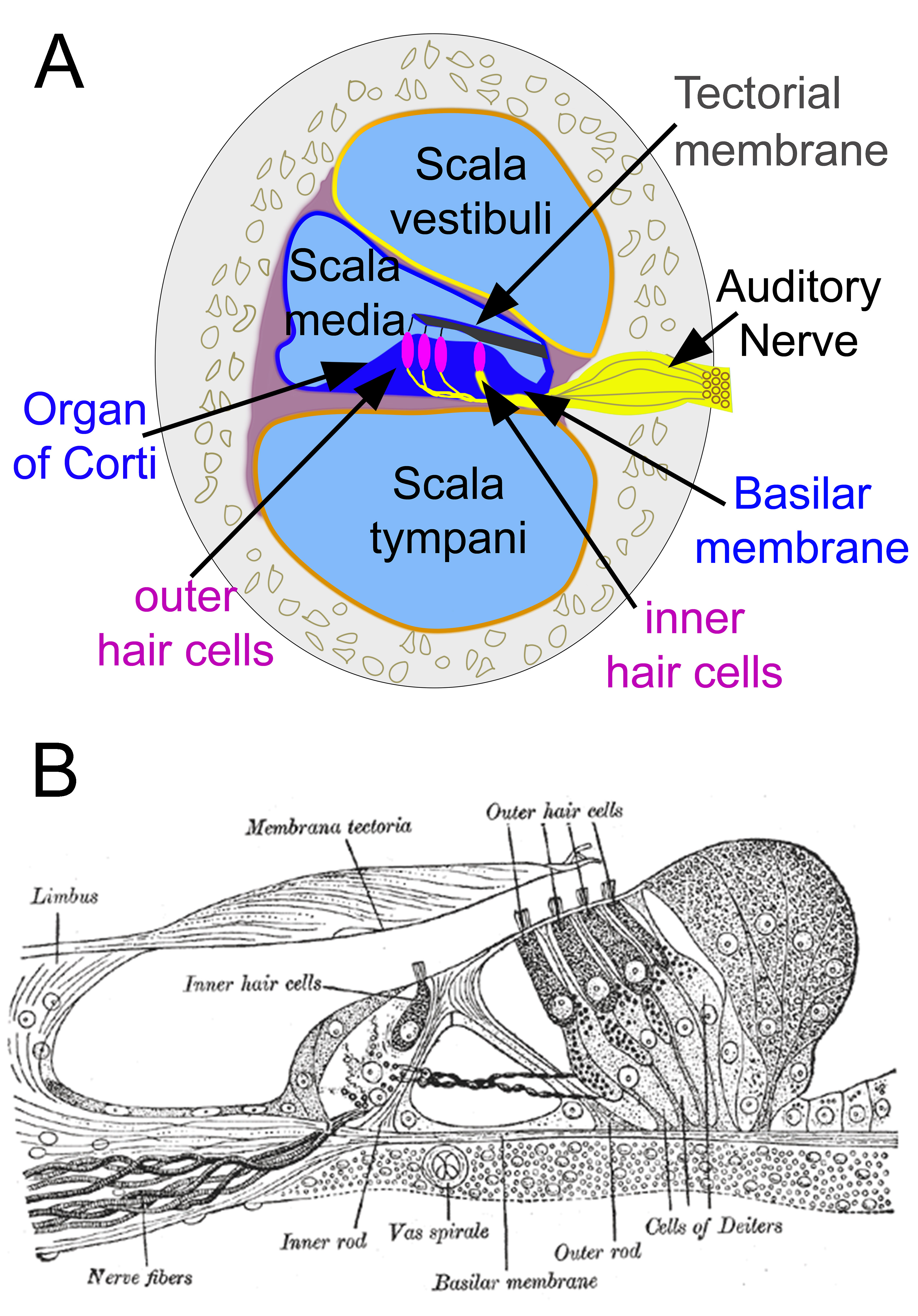
Cochlear Cross-Section and Organ of Corti. Panel A illustrates a cross-sectional view of the cochlea, showing the scala vestibuli, scala media, and scala tympani. The organ of Corti lies within the scala media, rests on the basilar membrane, and is situated beneath the tectorial membrane. Panel B demonstrates the cellular architecture of the organ of Corti, including inner and outer hair cells, supporting cells, the Corti tunnel, and associated nerve fibers.
(A) Contributed by D Peterson, MD
(B) Henry Vandyke Carter, Public Domain, via Wikimedia Commons
References
Sundar PS, Chowdhury C, Kamarthi S. Evaluation of Human Ear Anatomy and Functionality by Axiomatic Design. Biomimetics (Basel, Switzerland). 2021 May 19:6(2):. doi: 10.3390/biomimetics6020031. Epub 2021 May 19 [PubMed PMID: 34069537]
Bruss DM, Shohet JA. Neuroanatomy, Ear. StatPearls. 2026 Jan:(): [PubMed PMID: 31869122]
Thompson TL, Amedee R. Vertigo: a review of common peripheral and central vestibular disorders. Ochsner journal. 2009 Spring:9(1):20-6 [PubMed PMID: 21603405]
Freyer L, Aggarwal V, Morrow BE. Dual embryonic origin of the mammalian otic vesicle forming the inner ear. Development (Cambridge, England). 2011 Dec:138(24):5403-14. doi: 10.1242/dev.069849. Epub [PubMed PMID: 22110056]
Ali MM, Jayabalan S, Machnicki M, Sohal GS. Ventrally emigrating neural tube cells migrate into the developing vestibulocochlear nerve and otic vesicle. International journal of developmental neuroscience : the official journal of the International Society for Developmental Neuroscience. 2003 Jun:21(4):199-208 [PubMed PMID: 12781787]
Nin F, Yoshida T, Sawamura S, Ogata G, Ota T, Higuchi T, Murakami S, Doi K, Kurachi Y, Hibino H. The unique electrical properties in an extracellular fluid of the mammalian cochlea; their functional roles, homeostatic processes, and pathological significance. Pflugers Archiv : European journal of physiology. 2016 Oct:468(10):1637-49. doi: 10.1007/s00424-016-1871-0. Epub 2016 Aug 27 [PubMed PMID: 27568193]
Zhang Q, Ota T, Yoshida T, Ino D, Sato MP, Doi K, Horii A, Nin F, Hibino H. Electrochemical properties of the non-excitable tissue stria vascularis of the mammalian cochlea are sensitive to sounds. The Journal of physiology. 2021 Oct:599(19):4497-4516. doi: 10.1113/JP281981. Epub 2021 Sep 16 [PubMed PMID: 34426971]
Langlie J, Finberg A, Bencie NB, Mittal J, Omidian H, Omidi Y, Mittal R, Eshraghi AA. Recent advancements in cell-based models for auditory disorders. BioImpacts : BI. 2022:12(2):155-169. doi: 10.34172/bi.2022.23900. Epub 2022 Feb 6 [PubMed PMID: 35411298]
Shah JJ, Jimenez-Jaramillo CA, Lybrand ZR, Yuan TT, Erbele ID. Modern In Vitro Techniques for Modeling Hearing Loss. Bioengineering (Basel, Switzerland). 2024 Apr 26:11(5):. doi: 10.3390/bioengineering11050425. Epub 2024 Apr 26 [PubMed PMID: 38790292]
Park S, Kim YJ, Sharma H, Kim D, Gwon Y, Kim W, Park S, Ha CW, Choung YH, Kim J. Graphene Hybrid Inner Ear Organoid with Enhanced Maturity. Nano letters. 2023 Jun 28:23(12):5573-5580. doi: 10.1021/acs.nanolett.3c00988. Epub 2023 Jun 13 [PubMed PMID: 37311113]
Level 3 (low-level) evidenceQi J, Zhang L, Wang X, Chen X, Li Y, Wang T, Wu P, Chai R. Modeling, applications and challenges of inner ear organoid. Smart medicine. 2024 Feb:3(1):e20230028. doi: 10.1002/SMMD.20230028. Epub 2024 Jan 16 [PubMed PMID: 39188517]
Level 2 (mid-level) evidenceCastellucci A, Botti C, Delmonte S, Bettini M, Lusetti F, Brizzi P, Ruberto R, Gamberini L, Martellucci S, Malara P, Armato E, Renna L, Ghidini A, Bianchin G. Vestibular assessment in sudden sensorineural hearing loss: Role in the prediction of hearing outcome and in the early detection of vascular and hydropic pathomechanisms. Frontiers in neurology. 2023:14():1127008. doi: 10.3389/fneur.2023.1127008. Epub 2023 Feb 15 [PubMed PMID: 36873440]
Patuzzi R, Robertson D. Tuning in the mammalian cochlea. Physiological reviews. 1988 Oct:68(4):1009-82 [PubMed PMID: 3054945]
Level 3 (low-level) evidencePeterson DC, Reddy V, Mayes DA, Hamel RN. Neuroanatomy, Auditory Pathway. StatPearls. 2026 Jan:(): [PubMed PMID: 30335344]
Lim DJ. Functional structure of the organ of Corti: a review. Hearing research. 1986:22():117-46 [PubMed PMID: 3525482]
Level 3 (low-level) evidenceWright A. Scanning electron microscopy of the human organ of Corti. Journal of the Royal Society of Medicine. 1983 Apr:76(4):269-78 [PubMed PMID: 6341584]
Ashmore JF. A fast motile response in guinea-pig outer hair cells: the cellular basis of the cochlear amplifier. The Journal of physiology. 1987 Jul:388():323-47 [PubMed PMID: 3656195]
Level 3 (low-level) evidenceAshmore J. Cochlear outer hair cell motility. Physiological reviews. 2008 Jan:88(1):173-210. doi: 10.1152/physrev.00044.2006. Epub [PubMed PMID: 18195086]
Level 3 (low-level) evidenceTarabichi O, Jensen M, Hansen MR. Advances in hearing preservation in cochlear implant surgery. Current opinion in otolaryngology & head and neck surgery. 2021 Oct 1:29(5):385-390. doi: 10.1097/MOO.0000000000000742. Epub [PubMed PMID: 34354014]
Level 3 (low-level) evidenceSpoendlin H. Anatomy of cochlear innervation. American journal of otolaryngology. 1985 Nov-Dec:6(6):453-67 [PubMed PMID: 3909832]
Carricondo F, Romero-Gómez B. The Cochlear Spiral Ganglion Neurons: The Auditory Portion of the VIII Nerve. Anatomical record (Hoboken, N.J. : 2007). 2019 Mar:302(3):463-471. doi: 10.1002/ar.23815. Epub 2018 May 4 [PubMed PMID: 29659185]
Driver EC, Kelley MW. Specification of cell fate in the mammalian cochlea. Birth defects research. Part C, Embryo today : reviews. 2009 Sep:87(3):212-21. doi: 10.1002/bdrc.20154. Epub [PubMed PMID: 19750520]
Maunsell HR, Ellis K, Kelley MW, Driver EC. Lrrn1 Regulates Medial Boundary Formation in the Developing Mouse Organ of Corti. The Journal of neuroscience : the official journal of the Society for Neuroscience. 2023 Jul 19:43(29):5305-5318. doi: 10.1523/JNEUROSCI.2141-22.2023. Epub 2023 Jun 27 [PubMed PMID: 37369584]
Liu H, Chen L, Giffen KP, Stringham ST, Li Y, Judge PD, Beisel KW, He DZZ. Cell-Specific Transcriptome Analysis Shows That Adult Pillar and Deiters' Cells Express Genes Encoding Machinery for Specializations of Cochlear Hair Cells. Frontiers in molecular neuroscience. 2018:11():356. doi: 10.3389/fnmol.2018.00356. Epub 2018 Oct 1 [PubMed PMID: 30327589]
Zdebik AA, Wangemann P, Jentsch TJ. Potassium ion movement in the inner ear: insights from genetic disease and mouse models. Physiology (Bethesda, Md.). 2009 Oct:24():307-16. doi: 10.1152/physiol.00018.2009. Epub [PubMed PMID: 19815857]
McPherson DR. Sensory Hair Cells: An Introduction to Structure and Physiology. Integrative and comparative biology. 2018 Aug 1:58(2):282-300. doi: 10.1093/icb/icy064. Epub [PubMed PMID: 29917041]
Level 2 (mid-level) evidenceC Kohrman D, Wan G, Cassinotti L, Corfas G. Hidden Hearing Loss: A Disorder with Multiple Etiologies and Mechanisms. Cold Spring Harbor perspectives in medicine. 2020 Jan 2:10(1):. doi: 10.1101/cshperspect.a035493. Epub 2020 Jan 2 [PubMed PMID: 30617057]
Level 3 (low-level) evidenceBudak M, Grosh K, Sasmal A, Corfas G, Zochowski M, Booth V. Contrasting mechanisms for hidden hearing loss: Synaptopathy vs myelin defects. PLoS computational biology. 2021 Jan:17(1):e1008499. doi: 10.1371/journal.pcbi.1008499. Epub 2021 Jan 22 [PubMed PMID: 33481777]
Dallos P. Cochlear amplification, outer hair cells and prestin. Current opinion in neurobiology. 2008 Aug:18(4):370-6. doi: 10.1016/j.conb.2008.08.016. Epub 2008 Oct 4 [PubMed PMID: 18809494]
Level 3 (low-level) evidenceJohnson SL, Beurg M, Marcotti W, Fettiplace R. Prestin-driven cochlear amplification is not limited by the outer hair cell membrane time constant. Neuron. 2011 Jun 23:70(6):1143-54. doi: 10.1016/j.neuron.2011.04.024. Epub [PubMed PMID: 21689600]
Ruben RJ. The Developing Concept of Tonotopic Organization of the Inner Ear. Journal of the Association for Research in Otolaryngology : JARO. 2020 Feb:21(1):1-20. doi: 10.1007/s10162-019-00741-3. Epub 2020 Feb 4 [PubMed PMID: 32020418]
Tani T, Koike-Tani M, Tran MT, Shribak M, Levic S. Postnatal structural development of mammalian Basilar Membrane provides anatomical basis for the maturation of tonotopic maps and frequency tuning. Scientific reports. 2021 Apr 7:11(1):7581. doi: 10.1038/s41598-021-87150-w. Epub 2021 Apr 7 [PubMed PMID: 33828185]
Romero GE, Trussell LO. Central circuitry and function of the cochlear efferent systems. Hearing research. 2022 Nov:425():108516. doi: 10.1016/j.heares.2022.108516. Epub 2022 May 11 [PubMed PMID: 35606211]
Torres Cadenas L, Cheng H, Weisz CJC. Synaptic plasticity of inhibitory synapses onto medial olivocochlear efferent neurons. The Journal of physiology. 2022 Jun:600(11):2747-2763. doi: 10.1113/JP282815. Epub 2022 May 13 [PubMed PMID: 35443073]
Bavi N, Clark MD, Contreras GF, Shen R, Reddy BG, Milewski W, Perozo E. The conformational cycle of prestin underlies outer-hair cell electromotility. Nature. 2021 Dec:600(7889):553-558. doi: 10.1038/s41586-021-04152-4. Epub 2021 Oct 25 [PubMed PMID: 34695838]
Oghalai JS. The cochlear amplifier: augmentation of the traveling wave within the inner ear. Current opinion in otolaryngology & head and neck surgery. 2004 Oct:12(5):431-8 [PubMed PMID: 15377957]
Level 3 (low-level) evidenceElliott SJ, Shera CA. The cochlea as a smart structure. Smart materials & structures. 2012 Jun:21(6):64001 [PubMed PMID: 23148128]
Wu DK, Kelley MW. Molecular mechanisms of inner ear development. Cold Spring Harbor perspectives in biology. 2012 Aug 1:4(8):a008409. doi: 10.1101/cshperspect.a008409. Epub 2012 Aug 1 [PubMed PMID: 22855724]
Level 3 (low-level) evidenceChoo D. The role of the hindbrain in patterning of the otocyst. Developmental biology. 2007 Aug 15:308(2):257-65 [PubMed PMID: 17601528]
Kil SH, Streit A, Brown ST, Agrawal N, Collazo A, Zile MH, Groves AK. Distinct roles for hindbrain and paraxial mesoderm in the induction and patterning of the inner ear revealed by a study of vitamin-A-deficient quail. Developmental biology. 2005 Sep 1:285(1):252-71 [PubMed PMID: 16039643]
Arnold WH, Lang T. Development of the membranous labyrinth of human embryos and fetuses using computer aided 3D-reconstruction. Annals of anatomy = Anatomischer Anzeiger : official organ of the Anatomische Gesellschaft. 2001 Jan:183(1):61-6 [PubMed PMID: 11206984]
Toyoda S, Shiraki N, Yamada S, Uwabe C, Imai H, Matsuda T, Yoneyama A, Takeda T, Takakuwa T. Morphogenesis of the inner ear at different stages of normal human development. Anatomical record (Hoboken, N.J. : 2007). 2015 Dec:298(12):2081-90. doi: 10.1002/ar.23268. Epub 2015 Oct 7 [PubMed PMID: 26369281]
Driver EC, Kelley MW. Development of the cochlea. Development (Cambridge, England). 2020 Jun 22:147(12):. doi: 10.1242/dev.162263. Epub 2020 Jun 22 [PubMed PMID: 32571852]
Fritzsch B, Jahan I, Pan N, Kersigo J, Duncan J, Kopecky B. Dissecting the molecular basis of organ of Corti development: Where are we now? Hearing research. 2011 Jun:276(1-2):16-26. doi: 10.1016/j.heares.2011.01.007. Epub 2011 Jan 21 [PubMed PMID: 21256948]
Level 3 (low-level) evidenceJahan I, Pan N, Kersigo J, Fritzsch B. Beyond generalized hair cells: molecular cues for hair cell types. Hearing research. 2013 Mar:297():30-41. doi: 10.1016/j.heares.2012.11.008. Epub 2012 Nov 27 [PubMed PMID: 23201032]
Gunz P, Ramsier M, Kuhrig M, Hublin JJ, Spoor F. The mammalian bony labyrinth reconsidered, introducing a comprehensive geometric morphometric approach. Journal of anatomy. 2012 Jun:220(6):529-43. doi: 10.1111/j.1469-7580.2012.01493.x. Epub 2012 Mar 8 [PubMed PMID: 22404255]
Costeur L, Mennecart B, Müller B, Schulz G. Prenatal growth stages show the development of the ruminant bony labyrinth and petrosal bone. Journal of anatomy. 2017 Feb:230(2):347-353. doi: 10.1111/joa.12549. Epub 2016 Oct 11 [PubMed PMID: 27726136]
Schneider-Maunoury S, Pujades C. Hindbrain signals in otic regionalization: walk on the wild side. The International journal of developmental biology. 2007:51(6-7):495-506 [PubMed PMID: 17891712]
Whitfield TT. Development of the inner ear. Current opinion in genetics & development. 2015 Jun:32():112-8. doi: 10.1016/j.gde.2015.02.006. Epub 2015 Mar 19 [PubMed PMID: 25796080]
Level 3 (low-level) evidenceShi X. Pathophysiology of the cochlear intrastrial fluid-blood barrier (review). Hearing research. 2016 Aug:338():52-63. doi: 10.1016/j.heares.2016.01.010. Epub 2016 Jan 20 [PubMed PMID: 26802581]
Neng L, Zhang F, Kachelmeier A, Shi X. Endothelial cell, pericyte, and perivascular resident macrophage-type melanocyte interactions regulate cochlear intrastrial fluid-blood barrier permeability. Journal of the Association for Research in Otolaryngology : JARO. 2013 Apr:14(2):175-85. doi: 10.1007/s10162-012-0365-9. Epub 2012 Dec 18 [PubMed PMID: 23247886]
Yimtae K, Song H, Billings P, Harris JP, Keithley EM. Connection between the inner ear and the lymphatic system. The Laryngoscope. 2001 Sep:111(9):1631-5 [PubMed PMID: 11568618]
Level 3 (low-level) evidenceSusai S, Motwani R, Chandrupatla M. Tracking Lymphatic Drainage Pathways Through Inner Ear Channels: A Systematic Review. Cureus. 2024 Aug:16(8):e66670. doi: 10.7759/cureus.66670. Epub 2024 Aug 12 [PubMed PMID: 39262517]
Level 1 (high-level) evidenceZhang D, Li X, Lv Y, Song Y, Kong L, Li B, Zheng J, Pérez-Fernández N, Fan Z, Wang H. Lymphatic Vessels in the Inner Ear of Patients With Meniere Disease: A Novel Pathological Finding. OTO open. 2024 Jul-Sep:8(3):e171. doi: 10.1002/oto2.171. Epub 2024 Jul 24 [PubMed PMID: 39050361]
Salt AN, Plontke SK. Endolymphatic hydrops: pathophysiology and experimental models. Otolaryngologic clinics of North America. 2010 Oct:43(5):971-83. doi: 10.1016/j.otc.2010.05.007. Epub [PubMed PMID: 20713237]
Level 3 (low-level) evidenceWende S, Nakayama N, Schwerdtfeger P. The internal auditory artery: (embryology, anatomy, angiography, pathology). Journal of neurology. 1975 Aug 4:210(1):21-31 [PubMed PMID: 51066]
Matsunaga T, Igarashi M, Kanzaki J. The course of the internal auditory artery and its branches. Computer-aided three-dimensional reconstructions. Acta oto-laryngologica. Supplementum. 1991:487():54-60 [PubMed PMID: 1843586]
Mei X, Atturo F, Wadin K, Larsson S, Agrawal S, Ladak HM, Li H, Rask-Andersen H. Human inner ear blood supply revisited: the Uppsala collection of temporal bone-an international resource of education and collaboration. Upsala journal of medical sciences. 2018 Sep:123(3):131-142. doi: 10.1080/03009734.2018.1492654. Epub 2018 Sep 11 [PubMed PMID: 30204028]
Kämpfe Nordström C, Danckwardt-Lillieström N, Laurell G, Liu W, Rask-Andersen H. The Human Endolymphatic Sac and Inner Ear Immunity: Macrophage Interaction and Molecular Expression. Frontiers in immunology. 2018:9():3181. doi: 10.3389/fimmu.2018.03181. Epub 2019 Feb 1 [PubMed PMID: 30774637]
Dumanch KA, Poling GL. Introduction to the Audiological Evaluation: Case-Based Applications to Patients with Skull Base Disease. Journal of neurological surgery. Part B, Skull base. 2019 Apr:80(2):111-119. doi: 10.1055/s-0039-1678564. Epub 2019 Feb 4 [PubMed PMID: 30931217]
Level 3 (low-level) evidenceBenoudiba F, Toulgoat F, Sarrazin JL. The vestibulocochlear nerve (VIII). Diagnostic and interventional imaging. 2013 Oct:94(10):1043-50. doi: 10.1016/j.diii.2013.08.015. Epub 2013 Oct 1 [PubMed PMID: 24095603]
Spickler EM, Govila L. The vestibulocochlear nerve. Seminars in ultrasound, CT, and MR. 2002 Jun:23(3):218-37 [PubMed PMID: 12168998]
Vetter DE. The mammalian olivocochlear system--a legacy of non-cerebellar research in the Mugnaini lab. Cerebellum (London, England). 2015 Oct:14(5):557-69. doi: 10.1007/s12311-014-0637-5. Epub [PubMed PMID: 25592068]
Level 3 (low-level) evidenceCiuman RR. The efferent system or olivocochlear function bundle - fine regulator and protector of hearing perception. International journal of biomedical science : IJBS. 2010 Dec:6(4):276-88 [PubMed PMID: 23675203]
Cullen KE, Wei RH. Differences in the Structure and Function of the Vestibular Efferent System Among Vertebrates. Frontiers in neuroscience. 2021:15():684800. doi: 10.3389/fnins.2021.684800. Epub 2021 Jun 23 [PubMed PMID: 34248486]
Cullen KE. The vestibular system: multimodal integration and encoding of self-motion for motor control. Trends in neurosciences. 2012 Mar:35(3):185-96. doi: 10.1016/j.tins.2011.12.001. Epub 2012 Jan 12 [PubMed PMID: 22245372]
Ozdoğmuş O, Sezen O, Kubilay U, Saka E, Duman U, San T, Cavdar S. Connections between the facial, vestibular and cochlear nerve bundles within the internal auditory canal. Journal of anatomy. 2004 Jul:205(1):65-75 [PubMed PMID: 15255963]
Prasad KC, Azeem Mohiyuddin SM, Anjali PK, Harshita TR, Indu Varsha G, Brindha HS. Microsurgical Anatomy of Stapedius Muscle: Anatomy Revisited, Redefined with Potential Impact in Surgeries. Indian journal of otolaryngology and head and neck surgery : official publication of the Association of Otolaryngologists of India. 2019 Mar:71(1):14-18. doi: 10.1007/s12070-018-1510-5. Epub 2018 Oct 9 [PubMed PMID: 30906706]
Matz O, Sutton AE, Shermetaro C. Hyperacusis. StatPearls. 2026 Jan:(): [PubMed PMID: 32491645]
Edmonson A, Iwanaga J, Olewnik Ł, Dumont AS, Tubbs RS. The function of the tensor tympani muscle: a comprehensive review of the literature. Anatomy & cell biology. 2022 Jun 30:55(2):113-117. doi: 10.5115/acb.21.032. Epub 2022 May 19 [PubMed PMID: 35586903]
Causon A, Munro KJ, Plack CJ, Prendergast G. The Role of the Clinically Obtained Acoustic Reflex as a Research Tool for Subclinical Hearing Pathologies. Trends in hearing. 2020 Jan-Dec:24():2331216520972860. doi: 10.1177/2331216520972860. Epub [PubMed PMID: 33357018]
Bednarczuk NF, Casanovas Ortega M, Fluri AS, Arshad Q. Vestibulo-cortical hemispheric dominance: The link between anxiety and the vestibular system? The European journal of neuroscience. 2018 Jun:47(12):1517-1524. doi: 10.1111/ejn.13948. Epub 2018 Jun 8 [PubMed PMID: 29768682]
Jones SM, Jones TA, Mills KN, Gaines GC. Anatomical and Physiological Considerations in Vestibular Dysfunction and Compensation. Seminars in hearing. 2009:30(4):231-241 [PubMed PMID: 21072129]
Dieterich M, Brandt T. The bilateral central vestibular system: its pathways, functions, and disorders. Annals of the New York Academy of Sciences. 2015 Apr:1343():10-26. doi: 10.1111/nyas.12585. Epub 2015 Jan 7 [PubMed PMID: 25581203]
Level 3 (low-level) evidenceDieterich M, Brandt T. Global orientation in space and the lateralization of brain functions. Current opinion in neurology. 2018 Feb:31(1):96-104. doi: 10.1097/WCO.0000000000000516. Epub [PubMed PMID: 29189299]
Level 3 (low-level) evidenceErixon E, Högstorp H, Wadin K, Rask-Andersen H. Variational anatomy of the human cochlea: implications for cochlear implantation. Otology & neurotology : official publication of the American Otological Society, American Neurotology Society [and] European Academy of Otology and Neurotology. 2009 Jan:30(1):14-22. doi: 10.1097/MAO.0b013e31818a08e8. Epub [PubMed PMID: 18833017]
Biedron S, Prescher A, Ilgner J, Westhofen M. The internal dimensions of the cochlear scalae with special reference to cochlear electrode insertion trauma. Otology & neurotology : official publication of the American Otological Society, American Neurotology Society [and] European Academy of Otology and Neurotology. 2010 Jul:31(5):731-7. doi: 10.1097/MAO.0b013e3181d27b5e. Epub [PubMed PMID: 20142798]
Level 2 (mid-level) evidenceSutton AE, Krogmann RJ, Al Khalili Y. Cochlear Implants. StatPearls. 2026 Jan:(): [PubMed PMID: 31335000]
Zanoletti E, Martini A, Emanuelli E, Mazzoni A. Lateral approaches to the skull base. Acta otorhinolaryngologica Italica : organo ufficiale della Societa italiana di otorinolaringologia e chirurgia cervico-facciale. 2012 Oct:32(5):281-7 [PubMed PMID: 23326006]
Farinetti A, Ben Gharbia D, Mancini J, Roman S, Nicollas R, Triglia JM. Cochlear implant complications in 403 patients: comparative study of adults and children and review of the literature. European annals of otorhinolaryngology, head and neck diseases. 2014 Jun:131(3):177-82. doi: 10.1016/j.anorl.2013.05.005. Epub 2014 Jun 2 [PubMed PMID: 24889283]
Level 2 (mid-level) evidenceCohen NL, Hoffman RA. Complications of cochlear implant surgery in adults and children. The Annals of otology, rhinology, and laryngology. 1991 Sep:100(9 Pt 1):708-11 [PubMed PMID: 1952660]
Di Lella F, Falcioni M, Piccinini S, Iaccarino I, Bacciu A, Pasanisi E, Cerasti D, Vincenti V. Prevention and management of vascular complications in middle ear and cochlear implant surgery. European archives of oto-rhino-laryngology : official journal of the European Federation of Oto-Rhino-Laryngological Societies (EUFOS) : affiliated with the German Society for Oto-Rhino-Laryngology - Head and Neck Surgery. 2017 Nov:274(11):3883-3892. doi: 10.1007/s00405-017-4747-9. Epub 2017 Sep 20 [PubMed PMID: 28932983]
Liberman MC, Kujawa SG. Cochlear synaptopathy in acquired sensorineural hearing loss: Manifestations and mechanisms. Hearing research. 2017 Jun:349():138-147. doi: 10.1016/j.heares.2017.01.003. Epub 2017 Jan 10 [PubMed PMID: 28087419]
Ohlemiller KK. Mechanisms and genes in human strial presbycusis from animal models. Brain research. 2009 Jun 24:1277():70-83. doi: 10.1016/j.brainres.2009.02.079. Epub 2009 Mar 12 [PubMed PMID: 19285967]
Level 3 (low-level) evidenceHarris KC, Ahlstrom JB, Dias JW, Kerouac LB, McClaskey CM, Dubno JR, Eckert MA. Neural Presbyacusis in Humans Inferred from Age-Related Differences in Auditory Nerve Function and Structure. The Journal of neuroscience : the official journal of the Society for Neuroscience. 2021 Dec 15:41(50):10293-10304. doi: 10.1523/JNEUROSCI.1747-21.2021. Epub 2021 Nov 9 [PubMed PMID: 34753738]
Hinton AS, Yang-Hood A, Schrader AD, Loose C, Ohlemiller KK, McLean WJ. Approaches to Treat Sensorineural Hearing Loss by Hair-Cell Regeneration: The Current State of Therapeutic Developments and Their Potential Impact on Audiological Clinical Practice. Journal of the American Academy of Audiology. 2021 Nov:32(10):661-669. doi: 10.1055/s-0042-1750281. Epub 2022 May 24 [PubMed PMID: 35609593]
Atkinson PJ, Wise AK, Flynn BO, Nayagam BA, Richardson RT. Hair cell regeneration after ATOH1 gene therapy in the cochlea of profoundly deaf adult guinea pigs. PloS one. 2014:9(7):e102077. doi: 10.1371/journal.pone.0102077. Epub 2014 Jul 18 [PubMed PMID: 25036727]