 Tools
Tools
 Intercostal Nerve Block
Introduction
Intercostal nerve blocks are straightforward, versatile procedures that provide targeted analgesia for chest wall and upper abdominal pain, serving either as the primary pain management strategy or as an adjunct to multimodal therapy.[1][2] These blocks interrupt nociceptive transmission along the intercostal nerves, reducing reliance on systemic opioids and enhancing patient comfort in both perioperative and trauma settings.
Anatomy and Physiology
Register For Free And Read The Full Article
 Search engine and full access to all medical articles
- 10 free questions in your specialty
- Free CME/CE Activities
- Free daily question in your email
- Save favorite articles to your dashboard
- Emails offering discounts
Learn more about a Subscription to StatPearls Point-of-Care
Anatomy and Physiology
Twelve intercostal nerves provide sensory innervation to much of the back, trunk, and upper abdomen, while also supplying motor innervation to the intercostal muscles. Each intercostal nerve originates from spinal nerve roots at the same vertebral level as the rib it travels with. As each spinal nerve emerges from the spinal cord, it immediately divides into dorsal and ventral branches. The dorsal nerve branch supplies the motor and sensory innervation of the paravertebral musculature and overlying skin and subcutaneous tissue. The ventral branch continues anterolaterally and becomes the intercostal nerve. Immediately on exiting the intervertebral foramen, the nerve lies between the parietal pleura and the innermost intercostal muscle. Within a few centimeters, the nerve dives between the internal and innermost intercostal muscles, where it remains until it terminates in the anterior chest wall or abdomen. Cadaveric mapping reveals significant variability in the arrangement and course of the posterior intercostal neurovascular bundle (including alternate vessel–nerve orders and collateral branches), supporting the use of ultrasound to refine needle trajectory and reduce the risk of pleural/vascular injury.[3]
Each intercostal nerve travels in a neurovascular bundle with an intercostal artery and vein, with the nerve running inferior to both blood vessels. This neurovascular bundle accounts for the high local anesthetic uptake into the blood observed after intercostal nerve blocks. The bundle travels inferior to the accompanying rib in the costal groove. Near the midaxillary line, the intercostal nerve sends an offshoot called the lateral cutaneous branch, which travels laterally through the internal and external intercostal muscles, where it then divides into a dorsal and ventral branch. Together, these innervate the skin and subcutaneous tissue of the lateral trunk and upper abdomen. Just before the individual intercostal nerves terminate, they send another branch called the anterior cutaneous branch, which divides into a lateral and medial branch. These supply the skin and subcutaneous tissue of the anterior trunk and abdomen, including the skin over both the sternum and rectus abdominis.[4]
Indications
Indications for intercostal nerve blocks include:
- Incisional pain from thoracic surgery
- Analgesia for thoracostomy
- Herpes zoster or postherpetic neuralgia
- Rib fractures [5]
- Breast surgery
- Upper abdominal surgery [6]
- Differentiating between visceral and somatic pain
In a rare case of persistent chest wall pain due to CLOVES (congenital lipomatous overgrowth, vascular malformations, epidermal nevi, and scoliosis/skeletal and spinal anomalies) syndrome, fluoroscopy-guided intercostal nerve block resulted in meaningful symptom relief where prior interventions had failed, illustrating the utility of intercostal blocks in complex pain-syndrome settings beyond conventional postsurgical or trauma uses.[7]
Contraindications
The only absolute contraindications are patient refusal for the procedure and active infection at the site of injection. Other relative contraindications are an allergy to local anesthetics, prior nerve injury or damage, inability of the patient to consent to the procedure, anticoagulation, or coagulopathy. Patients should be counseled about the expected outcomes of the intercostal nerve block and potential complications. Special consideration should also be given if patients have prior nerve injury or neuromuscular disease involving the area to be blocked.
Equipment
The equipment required to perform an intercostal nerve block includes:
- Skin antiseptic
- Sterile towels
- Sterile gauze
- 50 cm 22-gauge needle for local anesthetic injection
- 25-gauge needle for skin wheal
- Local anesthetic
- Sterile gloves
- Ultrasound machine
- Marking pen
- Electrocardiogram monitor
- Blood pressure monitor
- Pulse oximeter
Generally, a long-acting local anesthetic such as 0.2% ropivacaine or 0.25% bupivacaine is chosen to maximize pain control.[8] Continuous blocks with a nerve catheter can be considered, but are rarely used for this particular nerve block. Given the high local anesthetic uptake from the intercostal space, shorter-acting local anesthetics may be considered, and the maximum allowable dose should be calculated, especially if multiple levels are to be blocked.
Preparation
Clinicians can perform this block with the patient in the sitting, lateral, or prone position. Clinician familiarity and patient comfort should guide positioning. For inexperienced clinicians, having the patient in the prone position likely provides the easiest access to the necessary landmarks. Placing a pillow under the upper abdomen or elevating the head of the bed slightly in a jackknife position can help open the posterior intercostal space. Having the patient dangle their arms over the edge of the bed laterally retracts the scapulae, which should help expose the rib angles of the underlying ribs. These maneuvers enhance the ability to palpate the landmarks necessary for performing this block safely and accurately. After the patient has been appropriately positioned, it is good practice to mark each level to be blocked, as it is easy to skip a level or perform more than 1 block on a single level, especially when multiple levels are planned. Rib palpation is most easily performed approximately 6 cm lateral to the midline, beyond the overlying paraspinal muscles, which typically obscure tactile identification of individual rib levels in healthy patients.
The inferior border of the scapula, which aligns with the seventh rib, and the twelfth rib serve as reliable landmarks for identifying target intercostal levels (see Image. Intercostal Nerve Block, Sonogram). For abdominal surgery, the fifth through twelfth intercostal spaces should be marked; breast surgery typically requires marking the second through sixth spaces. Thoracic incisions generally need coverage of 1 to 2 levels above and below the planned site. The inferior border of each rib to be blocked should be marked at the rib angle to ensure proper lateral placement, reducing the risk of paravertebral injection or dural puncture. Ultrasound guidance can further enhance safety when injections are performed closer to the midline.
Technique or Treatment
Successful intercostal nerve block results in the deposition of local anesthetic in the intercostal sulcus outside of the parietal pleura. Correct placement results in ipsilateral numbness at the intercostal levels blocked. Extension of the blockade to adjacent superior or inferior levels is uncommon and typically occurs only if a large volume of local anesthetic is used or the needle is positioned too close to the midline, allowing spread into the paravertebral space. Usually, the block level is determined by the number of blocks performed and is limited to the dermatome of the targeted intercostal nerves.
Anatomic Landmark-Based Technique
After cleansing the skin with an antiseptic, a skin wheal should be performed with 1 mL to 2 mL of local anesthetic. The fingers of the palpating hand should be used to pull the skin up so that the needle contacts the middle of the rib to be blocked. Generally, a 22-gauge, 50-mm-long needle is sufficient to perform the block. As the skin is stabilized with the palpating hand, the needle is placed through the skin wheal at approximately a 20° angle cephalad until it contacts the rib, which should be within 1 cm. The palpating hand then allows the skin to return to its normal position as the needle is “walked off” to the inferior border of the rib. As the needle walks off the inferior edge, it should be advanced another 1 mm to 3 mm anteriorly, where a subtle “pop” may be appreciated as the needle advances through the fascia of the internal intercostal muscle. After negative aspiration, 3 mL to 5 mL of local anesthetic can be injected.[4]
Ultrasound-Guided Technique
Ultrasound guidance may decrease the risk of intravascular injection and pneumothorax, and allow for injection closer to the midline than using anatomic landmarks. A comprehensive 2021 review confirms that both fluoroscopic and ultrasound guidance for intercostal nerve blocks outperform landmark-only techniques in terms of accuracy and safety, with no significant difference in outcomes between the modalities. This supports ultrasound as the preferred modality in practice, offering clinician convenience and direct visualization of anatomy.[9] This increases the likelihood that the injection is performed before the lateral branch divides, which is necessary to achieve anesthesia of the entire intercostal dermatome.
The individual ribs to be blocked should be marked out as with the landmark technique. The ultrasound probe is then placed in the sagittal plane, approximately 4 cm lateral to the spinous process. The ribs are visualized as a shadow, while the pleura and lung are visualized anterior to the intercostal space. The needle can then be inserted in or out of a plane to the transducer and advanced until the tip is just below the inferior border of the rib. After negative aspiration, 3 mL to 5 mL of local anesthetic is injected, and the pleura should be visualized as it is pushed away from the needle.[4] In a propensity-matched cohort of video-assisted thoracoscopic surgery patients, percutaneous intercostal nerve block resulted in significantly lower opioid consumption and better early visual analog scale scores at 6 and 12 hours compared with transthoracic intercostal nerve block, with no increase in adverse events; this suggests that percutaneous intercostal nerve block may offer enhanced analgesic efficacy in thoracic surgery.[10]
Complications
This procedure should be performed under sterile conditions to minimize the risk of infection. The history of coagulopathy or anticoagulation should be discussed to reduce the risk of bleeding. Performing this block awake can alert the clinician to pneumothorax or intraneural injection symptoms, which may go unnoticed in a sedated or anesthetized patient. Pneumothorax is rare and usually only requires monitored observation, although clinicians should be ready to perform needle decompression or insert a chest tube if necessary. Local anesthetic systemic toxicity is fortunately uncommon, but because absorption from this region can be significant, clinicians must be able to recognize its signs and provide prompt, appropriate treatment.
Using dilute concentrations of local anesthetic and keeping the total dose below the maximum allowable limit decreases the risk of systemic toxicity. Several case reports of inadvertent spinal after an intercostal nerve block have been described.[11] This is thought to be secondary to local anesthetic spreading medially through the dura or to the rare occurrence of injection into a dural sac, which has been described protruding laterally from the vertebral foramen. Aspiration before injection should be performed to rule out intravascular, intrapleural, or intrathecal injection and exclude these complications; however, a negative aspiration result is not guaranteed. Patients should be monitored for 20 to 30 minutes after the block to exclude these complications.
Clinical Significance
Intercostal nerve blocks provide consistent unilateral dermatomal analgesia at the targeted vertebral levels. They have been shown to improve respiratory function in patients with chest wall pain, making them valuable for postoperative recovery after thoracic surgery. Technically, they are simpler to perform than paravertebral nerve blocks or thoracic epidurals; however, they carry risks such as increased vascular uptake of local anesthetic, systemic toxicity, and pneumothorax. A limitation compared with paravertebral or epidural techniques is the need for multiple injections if coverage of more than one level or bilateral analgesia is required. These blocks do not provide complete surgical anesthesia for thoracic procedures and are most effective when used as part of a multimodal analgesic plan.[12]
Evidence supports the efficacy of intercostal nerve blocks in thoracic surgery. A meta-analysis of 5184 patients found that single-injection intercostal nerve blocks reduced pain scores for up to 24 hours and were noninferior to thoracic epidural analgesia for dynamic pain between 7 hours and 24 hours. However, opioid-sparing effects were less pronounced than with epidural or paravertebral techniques, and benefits decreased after 48 hours.[13] In patients undergoing thoracoscopic lung resection, single-shot intercostal nerve blocks provided postoperative analgesia comparable to thoracic epidurals. In contrast, paravertebral blocks did not, indicating that intercostal nerve blocks can serve as a less-invasive alternative when epidural analgesia is contraindicated.[14]
Enhancing Healthcare Team Outcomes
Intercostal nerve blocks are performed by anesthesiologists, nurse anesthetists, emergency clinicians, thoracic surgeons, trauma surgeons, and pain specialists. Safe execution requires detailed knowledge of thoracic anatomy, local anesthetic pharmacology, and potential complications such as pneumothorax or systemic toxicity, with resuscitative equipment readily available. Patient-centered outcomes improve through interprofessional collaboration. Nurses support positioning, monitoring, and documentation, while pharmacists ensure appropriate dosing and review drug interactions. Effective communication among all team members enables accurate block placement, integration with multimodal analgesia, and rapid management of adverse events. Coordinated care enhances procedural safety, reduces complications, and optimizes pain control for perioperative and trauma patients.
Media
(Click Image to Enlarge)
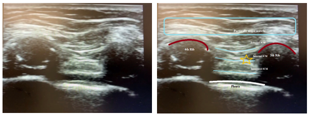
Intercostal Nerve Block, Sonogram. This sonogram illustrates the correct placement for administering a local anesthetic (yellow star) between the innermost intercostal muscle and the internal intercostal muscle during an intercostal nerve block. Also shown is the location of the parietal pleura.
Contributed by R Lopez-Rincon, MD
References
Feng LJ. Painless abdominoplasty: the efficacy of combined intercostal and pararectus blocks in reducing postoperative pain and recovery time. Plastic and reconstructive surgery. 2010 Nov:126(5):1723-1732. doi: 10.1097/PRS.0b013e3181ef8fe5. Epub [PubMed PMID: 21042130]
Hsieh MJ, Wang KC, Liu HP, Gonzalez-Rivas D, Wu CY, Liu YH, Wu YC, Chao YK, Wu CF. Management of acute postoperative pain with continuous intercostal nerve block after single port video-assisted thoracoscopic anatomic resection. Journal of thoracic disease. 2016 Dec:8(12):3563-3571. doi: 10.21037/jtd.2016.12.30. Epub [PubMed PMID: 28149550]
Elsy B. Clinically relevant anatomical variations in posterior intercostal neurovascular bundle. Folia morphologica. 2025:84(4):915-926. doi: 10.5603/fm.104771. Epub 2025 May 6 [PubMed PMID: 40326085]
Bhatia A, Gofeld M, Ganapathy S, Hanlon J, Johnson M. Comparison of anatomic landmarks and ultrasound guidance for intercostal nerve injections in cadavers. Regional anesthesia and pain medicine. 2013 Nov-Dec:38(6):503-7. doi: 10.1097/AAP.0000000000000006. Epub [PubMed PMID: 24121611]
Hwang EG, Lee Y. Effectiveness of intercostal nerve block for management of pain in rib fracture patients. Journal of exercise rehabilitation. 2014 Aug:10(4):241-4. doi: 10.12965/jer.140137. Epub 2014 Aug 31 [PubMed PMID: 25210700]
Shoeibi G, Babakhani B, Mohammadi SS. The efficacy of ilioinguinal-iliohypogastric and intercostal nerve co-blockade for postoperative pain relief in kidney recipients. Anesthesia and analgesia. 2009 Jan:108(1):330-3. doi: 10.1213/ane.0b013e31818c1b13. Epub [PubMed PMID: 19095869]
Level 1 (high-level) evidenceJaved S, Tabansi P. Use of intercostal nerve block for chest wall pain in a patient with CLOVES syndrome. Pain management. 2022 Sep:12(6):681-685. doi: 10.2217/pmt-2021-0120. Epub 2022 Jul 8 [PubMed PMID: 35801429]
Saby A, Swaminathan K, Pangarkar S, Tribuzio B. Alleviating Thoracotomy Pain With Intercostal Liposomal Bupivacaine: A Case Report. PM & R : the journal of injury, function, and rehabilitation. 2016 Nov:8(11):1119-1122. doi: 10.1016/j.pmrj.2016.06.003. Epub 2016 Jun 9 [PubMed PMID: 27292436]
Level 3 (low-level) evidenceElkhashab Y, Wang D. A Review of Techniques of Intercostal Nerve Blocks. Current pain and headache reports. 2021 Nov 4:25(10):67. doi: 10.1007/s11916-021-00975-y. Epub 2021 Nov 4 [PubMed PMID: 34738179]
Hui H, Miao H, Qiu F, Li H, Lin Y, Jiang B, Zhang Y. Comparison of analgesic effects of percutaneous and transthoracic intercostal nerve block in video-assisted thoracic surgery: a propensity score-matched study. Journal of cardiothoracic surgery. 2024 Jan 30:19(1):33. doi: 10.1186/s13019-024-02490-8. Epub 2024 Jan 30 [PubMed PMID: 38291461]
Level 2 (mid-level) evidenceChaudhri BB, Macfie A, Kirk AJ. Inadvertent total spinal anesthesia after intercostal nerve block placement during lung resection. The Annals of thoracic surgery. 2009 Jul:88(1):283-4. doi: 10.1016/j.athoracsur.2008.09.070. Epub [PubMed PMID: 19559248]
Level 3 (low-level) evidenceGadsden J, Kwofie K, Shastri U. Continuous intercostal versus paravertebral blockade for multiple fractured ribs. The journal of trauma and acute care surgery. 2012 Jul:73(1):293-4; author reply 294. doi: 10.1097/TA.0b013e31825aaeb5. Epub [PubMed PMID: 22743402]
Level 3 (low-level) evidenceGuerra-Londono CE, Privorotskiy A, Cozowicz C, Hicklen RS, Memtsoudis SG, Mariano ER, Cata JP. Assessment of Intercostal Nerve Block Analgesia for Thoracic Surgery: A Systematic Review and Meta-analysis. JAMA network open. 2021 Nov 1:4(11):e2133394. doi: 10.1001/jamanetworkopen.2021.33394. Epub 2021 Nov 1 [PubMed PMID: 34779845]
Level 1 (high-level) evidenceSpaans LN, Dijkgraaf MGW, Susa D, de Loos ER, Mourisse JMJ, Bouwman RA, Verhagen AFTM, van den Broek FJC, OPtriAL Study Group, Meijer P, Kuut M, Hanneman N, Bousema J, Franssen A, Brokx H, van Duyn E, Potters JW, van den Broek R, van Brakel T, Rijna H, Boom A, Noyez V, Hendriks JMH, Yogeswaran SK, Dickhoff C, van Dorp M. Intercostal or Paravertebral Block vs Thoracic Epidural in Lung Surgery: A Randomized Noninferiority Trial. JAMA surgery. 2025 Aug 1:160(8):855-864. doi: 10.1001/jamasurg.2025.1899. Epub [PubMed PMID: 40560556]
Level 1 (high-level) evidence