 Tools
Tools
 Marcus Gunn Pupil
Introduction
Marcus Gunn pupil, also known as a relative afferent pupillary defect (RAPD), is an important clinical sign indicating asymmetric dysfunction of the afferent visual pathway.[1] Normally, exposure to light results in constriction of both pupils through the direct and consensual pupillary light reflex.[2] In contrast, an MGP reflects weakness in the afferent limb of this reflex pathway, typically due to disease affecting the optic nerve, retina, or visual pathways. An MGP is characterized by constriction of the pupils of both eyes when the light stimulus is applied to the normal eye and dilation of both pupils when the light is subsequently transferred from the normal eye to the affected eye (see Image. Left Optic Nerve and Optic Tracts). This paradoxical dilation occurs because the affected eye transmits a weaker afferent signal, thereby reducing pupillomotor input.[3]
First described by Hirschberg and later associated with Scottish ophthalmologist Robert Marcus Gunn, an MGP is a hallmark sign of unilateral or asymmetric visual pathway dysfunction.[4] An MGP is commonly observed in conditions such as optic neuritis and neuropathy, glaucoma, retinal detachment, and central retinal artery or vein occlusion. Less commonly, damage to the optic chiasm, tracts, and midbrain may cause an RAPD.[5][6] Because an MGP may be present even when other visual findings are subtle, careful assessment of pupillary responses using techniques such as the swinging flashlight test is an essential component of the ophthalmic and neurologic examination. Early recognition of an MGP can help clinicians identify underlying pathology, guide diagnostic evaluation, and facilitate timely management to prevent vision loss and other complications.
Etiology
Register For Free And Read The Full Article
 Search engine and full access to all medical articles
- 10 free questions in your specialty
- Free CME/CE Activities
- Free daily question in your email
- Save favorite articles to your dashboard
- Emails offering discounts
Learn more about a Subscription to StatPearls Point-of-Care
Etiology
Lesions of the optic nerve are among the most common causes of an MGP. Conditions such as optic neuritis, ischemic optic neuropathy, traumatic optic neuropathy, and glaucoma can damage retinal ganglion cell axons and impair afferent signaling.[1] Retinal and posterior segment disorders may also lead to an MGP through loss of photoreceptors or viable retinal tissue. These conditions include retinal detachment,[3][7] central retinal vein occlusion,[8] central serous chorioretinopathy (may not be clinically appreciable),[9] severe macular disease,[10] retinitis pigmentosa,[11] and endophthalmitis. In such cases, the magnitude of the defect often correlates with the extent and asymmetry of retinal damage.
Certain conditions may produce an apparent or induced MGP. Dense cataracts can create a contralateral RAPD due to increased intraocular light scattering and altered retinal illumination.[12] For the same reason, anisocoria greater than 2 mm can cause an MGP of the eye with the smaller pupil.[13] A temporary, contralateral MGP may occur after patching one eye, due to dark adaptation and increased retinal sensitivity of the patched eye.[14] Other factors, such as amblyopia, may occasionally produce subtle or artifactual pupillary asymmetry.[13][15]
Lesions involving the optic chiasm, optic tract,[16][17] or pretectal region may also produce an MGP.[18] Lesions of the optic chiasm, while less common than those of the optic nerve, may damage more fibers from one eye compared to the other, resulting in an MGP.[19] Lesions of the optic tract can cause a contralateral MGP due to more nasal fibers crossing at the chiasm than temporal fibers that stay on the ipsilateral side.[20] Unilateral lesions of the olivary pretectal area or the brachium of the superior colliculus can cause a tectal MGP, which is contralateral in nature and can present without vision loss due to the pupillary fibers branching off before reaching the visually processing areas.[6] An MGP should not be confused with the Marcus Gunn phenomenon (jaw-winking syndrome), a separate condition in which eyelid movement occurs during chewing.[21]
Epidemiology
An MGP does not exhibit a specific age or sex predilection. Findings from a study using a binocular pupillometer showed that 42% of the normal population had an MGP between 0.08 and 0.22 log units, and 6% had an MGP between 0.23 and 0.39 log units.[22] The presence of an MGP was attributed either to measurement inaccuracy or to actual synaptic asymmetries in the visual pathway. Findings from one of the most recent population-based studies evaluating MGP in a Syrian cohort showed that clinically significant MGP was identified in 7.9% of individuals, with a higher prevalence observed among older individuals.[23] Furthermore, an MGP is noted in more than 90% of cases of acute unilateral cases of optic neuritis, up to 90% of cases of central retinal artery occlusion, 91% cases of ischemic central retinal vein occlusion (CRVO), more than 50% cases of retinal detachment involving the macula, and 23% cases of primary open-angle glaucoma.[3][8][24][25][26]
Pathophysiology
When a bright light shines on one eye, the pupil constricts. This pupillary constriction is known as a direct light reflex. Simultaneously, the pupil of the other eye also constricts. This response in the contralateral eye is known as a consensual light reflex. The light reflex is mediated by retinal photoreceptors and subserved by 4 neurons.[27]
- The sensory afferent neurons connect each retina with both pretectal nuclei located in the midbrain at the level of the superior colliculus. Impulses from the nasal half of the retina decussate in the optic chiasm and pass via the contralateral optic tract to reach the contralateral pretectal nucleus. Impulses from the temporal half of the retina travel via the ipsilateral optic tract to reach the ipsilateral pretectal nucleus. Estimates suggest that the nasal fibers decussate within the chiasm in a ratio of crossed (nasal) to uncrossed (temporal) fibers of 53:47.[28] According to a new model, temporal retinal fibers may partially cross at the optic chiasm, in addition to the crossing of nasal fibers, leading to greater input from one eye into the contralateral optic tract.[17]
- The internuncial neurons connect each pretectal nucleus to both Edinger-Westphal nuclei that activate both the ciliary and pupillary sphincter muscles. Thus, a uniocular light stimulus evokes bilateral crystalline lens accommodation and pupillary constriction. This consensual light reflex results in a similar degree of pupillary constriction because pupillomotor output to both eyes is the same.[29]
- The preganglionic motor neuron connects the Edinger-Westphal nucleus to the ciliary ganglion. These fibers pass through the inferior division of the oculomotor nerve.
- The postganglionic motor neurons innervate the sphincter pupillae via short ciliary nerves.
An MGP is observed in lesions of the afferent arm of the light pathway. When the light is shifted from the normal eye to the abnormal eye, the total pupillomotor input is reduced, and the amount of pupillary constriction is less when compared to stimulation of the normal eye.[30] This difference in pupillomotor input results in an MGP.
Marcus Gunn Pupil in Lesions of the Optic Nerve
Ischemic optic neuropathies, such as arteritic and nonarteritic ischemic optic neuropathy, cause an MGP via ischemia and infarction of the optic nerve, with secondary optic nerve edema. Other diseases causing such asymmetric or unilateral damage to the optic nerve resulting in an MGP include glaucoma, anterior and retrobulbar optic neuritis, and traumatic optic neuropathy.
Marcus Gunn Pupil in Lesions of the Optic Tract
The axons of the retinal ganglion cells from the nasal retina decussate in the optic chiasm, joining the temporal fibers of the other side. In addition, a partial crossing of temporal fibers may occur in the chiasm.[17] Thus, an asymmetry of pupillomotor input into the optic tracts occurs, with greater input from one eye to the contralateral tract. Consequently, individuals with unilateral optic tract lesions may show an MGP in the eye contralateral to the lesion, which generally has a temporal field defect.[16][17]
Marcus Gunn Pupil in Lesions of the Pretectum
The asymmetry of pupillomotor input extends from the optic tract to the pretectal nucleus. Consequently, lesions of the pretectum produce an MGP in the contralateral eye. The MGP in such situations is without the associated loss of visual acuity, color vision, or visual field, because just before fibers reach the lateral geniculate body (light pathway), a few fibers (pupillary reflex) branch off to the pretectum. Therefore, for lesions at the brachium of the superior colliculus adjacent to the pretectal nucleus, vision is not affected.[18]
Marcus Gunn Pupil in Glaucoma and Visual Field Defects
In conditions such as glaucoma, an MGP occurs secondary to damage to retinal ganglion cells and loss of the retinal nerve fiber layer.[31] At least 26% of retinal ganglion cells should be damaged to elicit an MGP.[32] An MGP is observed only when glaucomatous damage is asymmetrically severe. An MGP may be noted even before the appearance of diagnostic glaucomatous visual field defects.[33] Findings from studies showed that the central 30° of the visual field provides the most pupillomotor input, and asymmetrical visual field loss outside of 30° is much less likely to result in an MGP.[34]
Marcus Gunn Pupil in Retinal Disorders
An MGP occurs secondary to the loss of functional photoreceptors in the retina.[10] The central retina provides the most pupillomotor input, resulting in a greater pupillary response than the same stimulus applied to the peripheral retina.[35] Consequently, detachment of each peripheral quadrant of the retina causes an estimated 0.35 log unit light sensitivity loss, while detachment of the macula causes a 0.68 log unit loss.[7] Ischemic CRVO typically produces over a 0.9 log unit loss, whereas nonischemic CRVO produces less than a 0.6 log unit loss.[8] Most macular diseases do not cause an MGP unless they are very severe.[36] In macular disease, visual acuity declines more relative to the degree of MGP, whereas in optic neuropathy, vision loss may correlate more closely with MGP level.[9] Localized macular dystrophies and degenerations, as well as diffuse retinal dystrophies such as retinitis pigmentosa, usually do not show an MGP response unless the disease is asymmetric. Even in that case, the MGP may be subclinical. However, variants such as unilateral retinitis pigmentosa can show an MGP usually of under 0.3 log units.[11] Central serous chorioretinopathy may cause a mild MGP (usually ≤0.4 log units, but sometimes up to 0.9 log units), which improves with the resolution of subretinal fluid.[9]
Induced Marcus Gunn Pupil
Marcus Gunn pupil in cataract: The occurrence of an MGP in cataracts is controversial because an MGP usually indicates underlying damage to the afferent visual pathway (optic nerve or retina) rather than the cataract itself. Since Galen's time, an MGP has been noted to indicate poor visual prognosis after a cataract surgical procedure.[4] When a dense cataract causes an MGP in the contralateral eye, the MGP occurs by increasing the pupillomotor effectiveness of the stimulus light, likely because of increased intraocular light scattering by the cataract.[12] Peripheral photoreceptors may be stimulated by diffusely scattered light, and the light may bounce back again on the retina due to reflection by the posterior surface of the cataract.[12] Additionally, a dense cataract may induce dark adaptation of the affected eye.[12] A mature cataract can induce a mean of 0.44 log units (maximum 0.6 log units) defect in the contralateral eye, which disappears after cataract extraction.[12]
Marcus Gunn pupil in eye patching and dark adaptation: Ocular occlusion due to patching of the eye or eye disease, such as an eyelid mass or ptosis, increases retinal sensitivity via dark adaptation and can cause a false MGP of up to 1.50 log units in the unoccluded eye. This induced MGP disappears within 30 minutes of examination in an illuminated room after the eye occlusion has been removed.[14]
Marcus Gunn pupil in anisocoria: In anisocoria, the retina in one eye is shaded by the iris more than the other. This difference in retinal illumination can produce an RAPD in the eye with the smaller pupil. For every 1 mm difference in pupil size, a 0.10 log unit defect in RAPD is noted. Therefore, a 3 mm difference between the pupils can produce a clinically detectable RAPD. This artifactual pupillary response can be alleviated by placing neutral-density filters over the larger pupil at a rate of 0.10 log unit per millimeter of anisocoria.[13][37]
Miscellaneous
Marcus Gunn pupil in amblyopia: According to classic teaching, a structural abnormality of the retina or optic nerve is not present in amblyopia; consequently, an MGP should not be present. However, a study of individuals with amblyopic eyes found that an easily visible MGP (greater than 0.3 log units) was observed in 29 of 55 individuals (52.7%), and 45 of 55 individuals (81.8%) had a detectable MGP. Although most cases had an MGP of up to 0.6 log units, some showed a lower MGP of less than 0.3 log units, detected using a modified standard RAPD test.[15] The cause of MGP in amblyopic eyes is unknown,[38] but it is associated with impaired development of X ganglion cells in the fovea of the amblyopic eye. Studies have not revealed a correlation between amblyopia severity and MGP grade.[38]
History and Physical
Patients with an MGP can present with normal visual function or may have symptoms of an underlying disorder. For example, an MGP can persist after optic neuritis resolves and vision returns to normal.[18][39][40] Although this is often a point of confusion for medical trainees and staff, an MGP does not cause anisocoria.[41]
Ocular Examination
- Visual acuity: May range from 20/20 to light perception
- Ocular alignment: Patients with long-standing vision loss may manifest sensory strabismus
- Assessment of pupils:
- A weaker initial constriction with greater redilatation
- An initial stall with greater redilatation
- An initial and immediate dilatation, known as pupillary escape
- Pupillary escape: A specific and evident sign of MGP and is not to be confused with hippus, which is a natural rhythmic oscillation in the pupil diameter [42]
- Anterior segment examination: The presence of iris neovascularization should raise suspicion of ischemic CRVO
- Fundus evaluation: Helps to identify underlying causes such as optic neuropathy (eg, optic neuritis and ischemic optic neuropathy), retinal detachment, retinal vascular occlusion, glaucoma, and other pathologies
Supportive Tests
Supportive tests include optical coherence tomography of the retina and optic nerve, fluorescein angiography, fundus autofluorescence, visual field testing, color vision, contrast sensitivity, visual evoked potential, and MRI in cases of optic neuritis. Brightness acuity testing can be performed to rule out crystalline lens (cataract) or corneal (eg, Fuchs dystrophy) pathology. Please see StatPearls' companion resource, "Tests for Potential Vision," for further information.
Evaluation
An MGP typically indicates disease in the prechiasmal visual pathway.[4] The ability to detect and quantify an MGP is an essential part of the neuro-ophthalmic examination.
Tests to Identify and Quantify an MGP
- Gunn test
- Kestenbaum-Gunn test and modified Gunn test
- Swinging flashlight test
- Pupillography
The use of neutral density filters and cross-polarized filters aids in quantifying an MGP when used in conjunction with the swinging flashlight test or the Modified Gunn test.[37][43] RAPD severity correlates with retinal nerve fiber layer thinning and ganglion cell layer damage.[44]
Gunn test
The original description of a clinical test for an RAPD was reported by the Scottish physician Robert Marcus Gunn in 1902. In contrast, some literature suggests that the German ophthalmologist Julius Hirschberg had already noted the phenomenon of pupillary light reflex attenuation in unilateral optic nerve disease 20 years prior.[45] The original Gunn test involved covering and uncovering the eye in bright light and observing the pupillary response. A positive response was recorded when the pupil of the affected eye failed to constrict promptly compared with the unaffected eye.[46]
Modified Gunn test, Kestenbaum-Gunn test, and swinging flashlight test
In a 1946 publication, the Austrian physician Alfred Kestenbaum named RAPD testing and the pupillary response for Marcus Gunn. Occasionally, the test is named after both of them (Kestenbaum-Gunn test). In 1959, the American ophthalmologist Paul Levatin introduced and popularized the swinging flashlight test. His method was a modification of the original test in which, instead of occluding the eye, a bright light was alternately shone into each eye. The swinging flashlight test also standardized the illumination duration for each eye, ensuring accurate results. A study found that the original Gunn test correctly identified an MGP in 57% of cases, compared to 93% with the swinging flashlight test.[36][47][48][47]
Swinging flashlight test procedure:
- Perform the test in a semi-dark room and ask the patient to fixate on a distant letter or pattern (looking at a near object causes accommodative miosis).
- Shine a focused light source, such as a penlight or an indirect ophthalmoscope, into one eye from below the patient's eye at a distance of 5 to 10 cm.
- After a 3-second pause, quickly shift the light to the other eye.[49]
- Move the whole light from side to side, keeping the distance and angle equal. Holding the light in front of the nose and sequentially illuminating the 2 pupils by shifting the angle away from the central axis can stimulate the near response.
- Alternate the light between the 2 eyes, pausing 3 seconds on each eye.
- Look for a change in pupil size as the light is alternated.
- An MGP dilates when illuminated. The contralateral (unilluminated) pupil should dilate at the same time.
Clinical grading of RAPD based on the swinging flashlight test:
The grades are determined by averaging pupillary responses over at least 6 light swings:
- Grade 1: A weak initial constriction and greater redilatation
- Grade 2: An initial stall and greater redilatation
- Grade 3: An immediate dilatation
- Grade 4: An immediate pupillary dilatation (followed by secondary constriction) following prolonged illumination of a good eye for 6 seconds (bleaching of the normal eye)
- Grade 5: An immediate pupillary dilatation with no secondary constriction following prolonged illumination of a good eye for 6 seconds [49]
Grades 4 and 5 are elicited after bleaching the photoreceptors of the good eye to reduce its pupil power.
Reverse method RAPD testing:
When an efferent defect results in an unresponsive or fixed pupil, detecting an MGP remains possible by performing a modification of the swinging flashlight test.[36] When a pupil cannot react because of posterior synechiae, pharmacological immobilization, trauma, or other physical or neurological damage, clinicians should focus on the reactive pupil. The modification is as follows:
- Illuminate the reactive pupil from the side with a dim light that is bright enough to observe pupillary responses. Do not shine this light directly into the pupil.
- Complete the swinging flashlight test.
- If an MGP is present in the eye with an unresponsive pupil, when light is thrown onto the normal eye, its pupil constricts. When the light is moved to the abnormal eye, the pupil in the normal eye dilates or constricts less. Returning the light to the normal eye causes its pupil to constrict again.
- The unresponsive pupil may be larger or smaller than the responsive pupil, which can alter the relative illumination of the retina and affect the test result.
Quantification of Marcus Gunn Pupil Using Neutral-Density Filters
Quantification is performed by placing a neutral-density filter over the better eye and then performing a swinging flashlight test. The stimulus in the better eye is reduced by applying a neutral density filter (typically 0.3, 0.6, 0.9, or 1.2 log units), and the swinging flashlight test is performed with increasing filter densities. The endpoint of the test is achieved when the pupillary responses are equal. The filter density required to reach this point is recorded in log units to quantify the defect size. The smallest detectable defect is 0.3 log units. Clinicians should avoid light spill at the edge of the filters as the light is swung from one eye to the other. Because the neutral density filter blocks light, visualizing the pupil beyond a 1.2 log unit neutral density filter is difficult, necessitating the examiner to look around the filter to observe the pupillary reflex. During the first attempt, overshooting the endpoint and inducing a pseudo-RAPD in the better or filtered eye is preferable, after which the filter density can be reduced.[37][49]
Using Neutral-Density Filters to Test for a Subtle Marcus Gunn Pupil
To unmask a subtle MGP, a neutral density filter of 0.3 log units is placed in front of the eye with suspected optic neuropathy, and the swinging flashlight test is then performed. Placing the neutral density filter in front of the weak eye further decreases the pupillomotor input of that eye and heightens the intereye pupillomotor input difference, thereby unmasking the RAPD.[15][36][50]
The Kestenbaum Number
In individuals with unilateral optic nerve disease, when one eye is covered and the other is exposed to bright light, the pupil of the affected eye dilates to a larger diameter. This difference (in millimeters) in the size of the 2 pupils is a measure of the difference in pupillomotor input between the 2 eyes and roughly corresponds to the RAPD measured in log units of neutral density filter.[30]
Limitations
The swinging flashlight test is subject to various sources of inconsistency, including interindividual variability, the clinician's experience, ambient lighting conditions, patient cooperation, and the lack of precise quantification criteria. Measuring the Kestenbaum number or quantifying an MGP is difficult in children because of hippus, in older adults because of age-related miosis, and in any individual with reduced iris mobility.[30]
Pupillography
Modern pupillography uses a computerized device to simulate the swinging flashlight test, enabling more accurate detection and quantification of MGPs.[51] These are binocular pupillometers that provide information on pupil diameter, pupil diameter-time curves, and symmetry of pupillary responses. The data studied in pupillography include average pupillary constriction amplitude, constriction velocity, and constriction onset latency. Constriction amplitude is the best parameter for detecting MGPs and differentiating patients from normal subjects.[52] Recent advances, such as camera-based automated pupillometry and virtual reality–based eye-tracking systems, enable objective detection of RAPD and may facilitate its use in teleophthalmology.[53] Newer techniques, such as ultrasonography pupillometry, may detect subtle optic nerve dysfunction even when the clinical swinging flashlight test does not reveal an RAPD.[54]
Treatment / Management
MGP has no specific treatment. Treatment is directed toward the underlying disorder affecting the afferent visual pathway. Because an MGP most commonly indicates pathology of the optic nerve, retina, or visual pathways, prompt evaluation and treatment of the causative condition are essential to prevent vision loss and other complications. Management strategies depend on the underlying etiology. The most common causes of an MGP include:
- Optic neuritis: Diagnosis may include magnetic resonance imaging, optical coherence tomography, visual evoked potential testing, and various laboratory tests. Treatment may include corticosteroids.[55]
- Ischemic optic neuropathies: Diagnosis may include clinical observation, laboratory testing (including erythrocyte sedimentation rate, C-reactive protein, and platelet count), fluorescein angiography, and a temporal artery biopsy, and is managed by controlling risk factors and initiating immediate corticosteroid treatment when giant cell arteritis is suspected.[56][57]
- Asymmetric glaucoma: Glaucoma is evaluated using tests such as measurement of intraocular pressure, optical coherence tomography, visual field testing, pachymetry, and gonioscopy. Treatment requires reducing intraocular pressure with topical medications, laser therapy, or surgical procedures.[58]
- Optic nerve compression or trauma: Diagnosis is facilitated by computed tomography and magnetic resonance imaging, as well as visual function tests such as color vision and visual field testing. Management of orbital tumors, thyroid eye disease, or trauma to the optic nerve ranges from surgical decompression (acute trauma or thyroid eye disease), high-dose corticosteroids (inflammation or thyroid eye disease), or observation in some cases.[59][60]
- Extensive retinal damage: Severe, unilateral retinal disorders, such as retinal detachment, central retinal vein occlusion, or large retinal infections, require prompt ophthalmologic evaluation with B-scan ultrasonography, fundus photography, angiography, and optical coherence tomography. Treatment options include surgical repair and systemic and intravitreal pharmacologic therapy.[61][62] (B3)
Careful clinical assessment should precede pharmacologic pupil dilation, especially when a patient presents with vision loss. Early diagnosis, appropriate referral to ophthalmology or neurology, and coordinated follow-up are essential to preserve visual function and improve clinical outcomes.
Differential Diagnosis
Hippus
Continuous oscillations of the pupil are often observed under constant retinal illumination.[63]
Alternating Contraction Anisocoria
Alternating contraction anisocoria is an uncommon syndrome of the anterior midbrain in which light stimulation of one pupil causes it to constrict more than the other, causing the contralateral pupil to appear larger. As the light stimulus is moved to the other pupil, this pupil now constricts more, leaving the original pupil as the larger one. This condition appears to the clinician as alternating anisocoria and RAPD. The cause is hypothesized to be a lesion at the posterior commissure and one or both brachia of the superior colliculus because of damage to a combination of both first- and second-order neurons. Alternating contraction anisocoria can present in otherwise healthy individuals or result from viral encephalomyelitis, syphilis, or traumatic brain injury.[29]
Prognosis
The prognosis of MGP varies according to the cause:
- In optic neuropathy, the MGP magnitude often correlates with the degree of visual acuity loss, whereas in maculopathy, vision impairment exceeds that reflected by RAPD.[9]
- In glaucoma, an MGP is objective evidence of visual field loss,[25] and can signify the earliest onset of optic nerve damage in patients with ocular hypertension.[64] Combining MGP assessment with ocular coherence tomography angiography may help detect early asymmetric glaucoma by identifying both functional and microvascular changes.[44]
- In retinal vein occlusion, an MGP of over 0.9 log units is associated with extensive capillary nonperfusion and poor visual acuity that is typically worse than 20/400 (3/60).[8] A significant increase in MGP degree in a patient with nonischemic CRVO is an early indicator of conversion to ischemic CRVO.
- In retinal detachment, an MGP typically occurs in retinal detachment involving the macula, and postoperative visual acuity is typically lower in detachments involving the macula.[65]
- In cataract, if the patient has an MGP in the same eye, postoperative visual gain tends to be poor due to associated defects in the anterior visual pathway.[12] A poor visual prognosis should always be explained in this case.
Complications
Failure to detect this clinical sign results in complications related to the underlying etiology. Most of the pathology associated with an MGP represents advanced disease of the posterior segment (retina and optic nerve) and brain (optic chiasm, optic radiations, and pretectal area), which is more likely to sustain rapid, irreversible injury than disease of the anterior segment. In cases such as arteritic anterior optic neuropathy or a compressive malignant lesion, detecting an MGP can be both sight-saving and life-saving.
Consultations
Testing for an MGP is a standard part of pretesting performed during a comprehensive eye examination. Pupillary testing is also routinely performed by primary care clinicians, nurses, and emergency department clinicians. When an MGP is present, consultations can involve different ophthalmology specialists (eg, retina, glaucoma, neuro-ophthalmology), depending on the suspected etiology. When an MGP is suspected but uncertain, consultation with a pupillographer or a neuro-ophthalmologist may be indicated.
Deterrence and Patient Education
Measurement of an MGP has proven useful for early detection of visual field loss in certain optic nerve and retinal diseases.[24] An MGP is a very sensitive and specific finding for predicting ischemia and differentiating ischemic from nonischemic central retinal vein occlusion.[8] Thus, the presence of an MGP can be used to counsel patients on the urgent need for further ophthalmological and other testing to determine the underlying etiology.
Pearls and Other Issues
Diagnostic pearls include the following:
- An MGP with normal visual acuity, color vision, and without field defects increases suspicion for a higher-order lesion at the level of the pretectum.
- An MGP and a contralateral field defect, specifically temporal field defects, increase suspicion for an optic tract lesion.
- An MGP and neovascularization of the iris increase suspicion for ischemic CRVO.
- An MGP and anisocoria may result from unequal light adaptation due to unequal pupil size. Repeating the test after 30 minutes of light adaptation is recommended.
- A cataract in the same eye as the MGP increases the suspicion of posterior segment pathology.
- A dense cataract and an MGP in the contralateral eye can be caused by dark adaptation of the eye with the cataract, causing it to respond more strongly to a light stimulus. The cataract effect, which causes light scattering, may add to this phenomenon. Light adaptation may allow the clinician to correct for this phenomenon.
Enhancing Healthcare Team Outcomes
As MGP is an abnormal pupillary response indicating asymmetric dysfunction of the afferent limb of the pupillary light reflex, most commonly involving the prechiasmal visual pathway. Normally, illumination of one eye causes constriction of both pupils through direct and consensual reflexes. An MGP occurs when light is moved from a normal eye to an affected eye, and both pupils dilate because of reduced afferent input. An MGP is frequently associated with optic nerve disorders such as optic neuritis, ischemic optic neuropathy, and glaucoma, as well as retinal diseases, including retinal detachment and central retinal vein occlusion. Detection relies on careful pupillary examination, most commonly using the swinging flashlight test, and may be supported by quantitative techniques, such as neutral density filters or automated pupillometry. Clinicians should avoid dilating the pupils prematurely, as important diagnostic pupillary signs may be missed during early evaluation.
Interprofessional collaboration plays a critical role in early detection and appropriate treatment of an MGP and its underlying causes. Optometrists, ophthalmologists, and neurologists lead diagnostic evaluation and treatment planning, whereas primary care clinicians and advanced practice clinicians often identify abnormal pupillary findings and initiate timely referral. Nurses and trainees in inpatient wards or intensive care units frequently perform routine neurologic assessments and may detect abnormal pupillary reflexes, prompting urgent specialist consultation. Pharmacists support safe medication use for underlying conditions, whereas coordinated communication among care team members facilitates prompt diagnostic testing and follow-up. Careful interdisciplinary assessment may also reveal otherwise silent conditions, such as pituitary adenomas, which can be identified through timely referral for radiological imaging. In emergency settings, point-of-care ultrasonography has been explored as an adjunct to assess pupillary responses when direct examination is limited, particularly in ocular trauma.[66] Systematic evaluation and coordinated care improve diagnostic accuracy, support timely referral, and enhance patient outcomes.
Media
(Click Image to Enlarge)
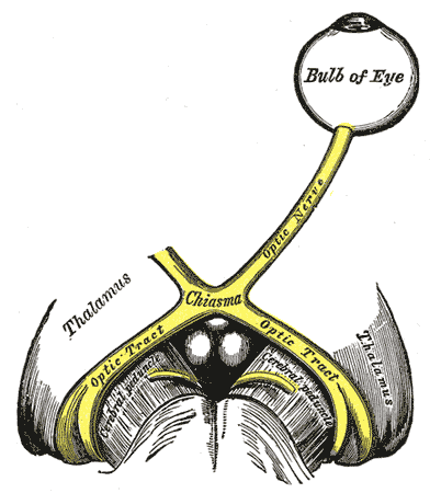
Left Optic Nerve and Optic Tracts. A Marcus Gunn pupil indicates an afferent defect, usually at the level of the retina or optic nerve. Moving a bright light from the unaffected eye to the affected eye would cause both eyes to dilate, because the ability to perceive the bright light is diminished.
Henry Vandyke Carter, Public Domain, via Wikimedia Commons

References
Kohn AN, Moss AP, Podos SM. Relative afferent pupillary defects in glaucoma without characteristic field loss. Archives of ophthalmology (Chicago, Ill. : 1960). 1979 Feb:97(2):294-6 [PubMed PMID: 550800]
Level 3 (low-level) evidenceEllis CJ. The pupillary light reflex in normal subjects. The British journal of ophthalmology. 1981 Nov:65(11):754-9 [PubMed PMID: 7326222]
Bovino JA, Burton TC. Measurement of the relative afferent pupillary defect in retinal detachment. American journal of ophthalmology. 1980 Jul:90(1):19-21 [PubMed PMID: 7395954]
Thompson HS. Afferent pupillary defects. Pupillary findings associated with defects of the afferent arm of the pupillary light reflex arc. American journal of ophthalmology. 1966 Nov:62(5):860-73 [PubMed PMID: 5928836]
Sharifi A, Sigireddi RR, Lyons LJ, Kini AT, Al Othman BA, Lee AG. Localizing Thalamomesencephalic Afferent and Efferent Pupillary Defects. Journal of neuro-ophthalmology : the official journal of the North American Neuro-Ophthalmology Society. 2021 Mar 1:41(1):e136-e138. doi: 10.1097/WNO.0000000000000903. Epub [PubMed PMID: 32028453]
Donaldson L, Rebello R, Rodriguez AR. Relative Afferent Pupillary Defect with Normal Vision: Unique Localisation to the Contralateral Brachium of the Superior Colliculus. Neuro-ophthalmology (Aeolus Press). 2020 Apr:44(2):128-130. doi: 10.1080/01658107.2019.1668432. Epub 2019 Dec 12 [PubMed PMID: 32395164]
Folk JC, Thompson HS, Farmer SG, O'Gorman TW, Dreyer RF. Relative afferent pupillary defect in eyes with retinal detachment. Ophthalmic surgery. 1987 Oct:18(10):757-9 [PubMed PMID: 3431805]
Servais GE, Thompson HS, Hayreh SS. Relative afferent pupillary defect in central retinal vein occlusion. Ophthalmology. 1986 Mar:93(3):301-3 [PubMed PMID: 2422618]
Han DP, Thompson HS, Folk JC. Differentiation between recently resolved optic neuritis and central serous retinopathy. Use of tests of visual function. Archives of ophthalmology (Chicago, Ill. : 1960). 1985 Mar:103(3):394-6 [PubMed PMID: 3977714]
Newsome DA, Milton RC, Gass JD. Afferent pupillary defect in macular degeneration. American journal of ophthalmology. 1981 Sep:92(3):396-402 [PubMed PMID: 7294099]
Level 3 (low-level) evidenceJiang MQ, Thompson HS. Pupillary defects in retinitis pigmentosa. American journal of ophthalmology. 1985 May 15:99(5):607-8 [PubMed PMID: 4003509]
Lam BL, Thompson HS. A unilateral cataract produces a relative afferent pupillary defect in the contralateral eye. Ophthalmology. 1990 Mar:97(3):334-8 [PubMed PMID: 2336271]
Level 3 (low-level) evidenceLam BL, Thompson HS. An anisocoria produces a small relative afferent pupillary defect in the eye with the smaller pupil. Journal of neuro-ophthalmology : the official journal of the North American Neuro-Ophthalmology Society. 1999 Sep:19(3):153-9 [PubMed PMID: 10494942]
DuBois LG, Sadun AA. Occlusion-induced contralateral afferent pupillary defect. American journal of ophthalmology. 1989 Mar 15:107(3):306-7 [PubMed PMID: 2923167]
Level 3 (low-level) evidencePortnoy JZ, Thompson HS, Lennarson L, Corbett JJ. Pupillary defects in amblyopia. American journal of ophthalmology. 1983 Nov:96(5):609-14 [PubMed PMID: 6638127]
Newman SA, Miller NR. Optic tract syndrome. Neuro-ophthalmologic considerations. Archives of ophthalmology (Chicago, Ill. : 1960). 1983 Aug:101(8):1241-50 [PubMed PMID: 6882255]
Level 3 (low-level) evidenceSchmid R, Wilhelm B, Wilhelm H. Naso-temporal asymmetry and contraction anisocoria in the pupillomotor system. Graefe's archive for clinical and experimental ophthalmology = Albrecht von Graefes Archiv fur klinische und experimentelle Ophthalmologie. 2000 Feb:238(2):123-8 [PubMed PMID: 10766280]
Forman S, Behrens MM, Odel JG, Spector RT, Hilal S. Relative afferent pupillary defect with normal visual function. Archives of ophthalmology (Chicago, Ill. : 1960). 1990 Aug:108(8):1074-5 [PubMed PMID: 2136343]
Level 3 (low-level) evidenceSy KJD, Cruz FMO. Ophthalmologic Findings of Parachiasmal Lesions in a Tertiary Philippine Hospital. Acta medica Philippina. 2025:59(7):67-73. doi: 10.47895/amp.vi0.9712. Epub 2025 Jun 13 [PubMed PMID: 40666743]
Chen NS, McDonald HM, Micieli J, Margolin E. Diagnosis and etiologic classification of optic tract lesions. Brain communications. 2025:7(5):fcaf354. doi: 10.1093/braincomms/fcaf354. Epub 2025 Sep 19 [PubMed PMID: 41030890]
Zhuang S, Qi Q, Lin M, Li J. Progress in MGJWS: A 74-Year Review of Marcus Gunn Jaw-Winking Syndrome. Journal of child neurology. 2025 Feb:40(2):132-147. doi: 10.1177/08830738241282698. Epub 2024 Dec 8 [PubMed PMID: 39648605]
Level 2 (mid-level) evidenceWilhelm H, Peters T, Lüdtke H, Wilhelm B. The prevalence of relative afferent pupillary defects in normal subjects. Journal of neuro-ophthalmology : the official journal of the North American Neuro-Ophthalmology Society. 2007 Dec:27(4):263-7 [PubMed PMID: 18090558]
Alsaydjamil S, Bdeiwi H, Aljundi R, Abu Ghedda S, Ibrahim M, Shawakh H, Sultan H. Examining pupil characteristics and relative afferent pupil defect prevalence in Syrian patients. Annals of medicine and surgery (2012). 2025 Jun:87(6):3194-3200. doi: 10.1097/MS9.0000000000003354. Epub 2025 May 12 [PubMed PMID: 40486633]
Cox TA, Thompson HS, Corbett JJ. Relative afferent pupillary defects in optic neuritis. American journal of ophthalmology. 1981 Nov:92(5):685-90 [PubMed PMID: 7304695]
Page CJ, Merritt JC, Evans B. Relative afferent pupillary defects in primary open-angle glaucoma--five years' experience. Journal of the National Medical Association. 1985 Dec:77(12):979-84 [PubMed PMID: 4078926]
Level 3 (low-level) evidenceLakkis T, Elshoura AMA, Soria Behr GA, Eduardo Mauricio MC, Sil-Zavaleta S, Cai LY, Rai M. Central Retinal Artery Occlusion in Acute Care: Current Practices and Emerging Therapies. Cureus. 2025 Sep:17(9):e92786. doi: 10.7759/cureus.92786. Epub 2025 Sep 20 [PubMed PMID: 41127733]
Bouffard MA. The Pupil. Continuum (Minneapolis, Minn.). 2019 Oct:25(5):1194-1214. doi: 10.1212/CON.0000000000000771. Epub [PubMed PMID: 31584534]
Kupfer C, Chumbley L, Downer JC. Quantitative histology of optic nerve, optic tract and lateral geniculate nucleus of man. Journal of anatomy. 1967 Jun:101(Pt 3):393-401 [PubMed PMID: 6051727]
LOWENSTEIN O. Alternating contraction anisocoria; a pupillary syndrome of the anterior midbrain. A.M.A. archives of neurology and psychiatry. 1954 Dec:72(6):742-57 [PubMed PMID: 13206493]
Jiang MQ, Thompson HS, Lam BL. Kestenbaum's number as an indicator of pupillomotor input asymmetry. American journal of ophthalmology. 1989 May 15:107(5):528-30 [PubMed PMID: 2712133]
Tatsumi Y, Nakamura M, Fujioka M, Nakanishi Y, Kusuhara A, Maeda H, Negi A. Quantification of retinal nerve fiber layer thickness reduction associated with a relative afferent pupillary defect in asymmetric glaucoma. The British journal of ophthalmology. 2007 May:91(5):633-7 [PubMed PMID: 17050576]
Kerrison JB, Buchanan K, Rosenberg ML, Clark R, Andreason K, Alfaro DV, Grossniklaus HE, Kerrigan-Baumrind LA, Kerrigan DF, Miller NR, Quigley HA. Quantification of optic nerve axon loss associated with a relative afferent pupillary defect in the monkey. Archives of ophthalmology (Chicago, Ill. : 1960). 2001 Sep:119(9):1333-41 [PubMed PMID: 11545640]
Level 3 (low-level) evidenceJohnson LN, Hill RA, Bartholomew MJ. Correlation of afferent pupillary defect with visual field loss on automated perimetry. Ophthalmology. 1988 Dec:95(12):1649-55 [PubMed PMID: 3068603]
Kardon RH, Haupert CL, Thompson HS. The relationship between static perimetry and the relative afferent pupillary defect. American journal of ophthalmology. 1993 Mar 15:115(3):351-6 [PubMed PMID: 8442495]
Kardon RH, Kirkali PA, Thompson HS. Automated pupil perimetry. Pupil field mapping in patients and normal subjects. Ophthalmology. 1991 Apr:98(4):485-95; discussion 495-6 [PubMed PMID: 2052302]
Broadway DC. How to test for a relative afferent pupillary defect (RAPD). Community eye health. 2012:25(79-80):58-9 [PubMed PMID: 23520419]
Thompson HS, Corbett JJ, Cox TA. How to measure the relative afferent pupillary defect. Survey of ophthalmology. 1981 Jul-Aug:26(1):39-42 [PubMed PMID: 7280994]
Level 3 (low-level) evidenceIkeda H. Visual acuity, its development and amblyopia. Journal of the Royal Society of Medicine. 1980 Aug:73(8):546-55 [PubMed PMID: 7230230]
Level 3 (low-level) evidenceGirkin CA, Perry JD, Miller NR. A relative afferent pupillary defect without any visual sensory deficit. Archives of ophthalmology (Chicago, Ill. : 1960). 1998 Nov:116(11):1544-5 [PubMed PMID: 9823369]
Level 3 (low-level) evidenceEllis CJ. The afferent pupillary defect in acute optic neuritis. Journal of neurology, neurosurgery, and psychiatry. 1979 Nov:42(11):1008-17 [PubMed PMID: 501365]
Kerr RG, Bacon AM, Baker LL, Gehrke JS, Hahn KD, Lillegraven CL, Renner CH, Spilman SK. Underestimation of Pupil Size by Critical Care and Neurosurgical Nurses. American journal of critical care : an official publication, American Association of Critical-Care Nurses. 2016 May:25(3):213-9. doi: 10.4037/ajcc2016554. Epub [PubMed PMID: 27134226]
Broadway DC. How to test for a relative afferent pupillary defect (RAPD). Community eye health. 2016:29(96):68-69 [PubMed PMID: 28381906]
Rosenberg ML, Oliva A. The use of crossed polarized filters in the measurement of the relative afferent pupillary defect. American journal of ophthalmology. 1990 Jul 15:110(1):62-5 [PubMed PMID: 2195895]
Nakamura M, Sakamoto M, Ueda K, Okuda M, Takano F, Yamada-Nakanishi Y. Detection of Relative Afferent Pupillary Defect and Its Correlation with Structural and Functional Asymmetry in Patients with Glaucoma Using Hitomiru, a Novel Hand-Held Pupillometer. Journal of clinical medicine. 2023 Jun 8:12(12):. doi: 10.3390/jcm12123936. Epub 2023 Jun 8 [PubMed PMID: 37373631]
Landau WM. Clinical neuromythology. I. The Marcus Gunn phenomenon: loose canon of neuro-ophthalmology. Neurology. 1988 Jul:38(7):1141-2 [PubMed PMID: 3386834]
Roper-Hall G. Historical Vignette: Robert Marcus Gunn (1850-1909): Scottish Ophthalmologist, Skilled Observer, and Gifted Teacher. The American orthoptic journal. 2015:65():121-7. doi: 10.3368/aoj.65.1.121. Epub [PubMed PMID: 26564938]
Enyedi LB, Dev S, Cox TA. A comparison of the Marcus Gunn and alternating light tests for afferent pupillary defects. Ophthalmology. 1998 May:105(5):871-3 [PubMed PMID: 9593390]
Level 1 (high-level) evidenceLEVATIN P. Pupillary escape in disease of the retina or optic nerve. Archives of ophthalmology (Chicago, Ill. : 1960). 1959 Nov:62():768-79 [PubMed PMID: 14416133]
Bell RA, Waggoner PM, Boyd WM, Akers RE, Yee CE. Clinical grading of relative afferent pupillary defects. Archives of ophthalmology (Chicago, Ill. : 1960). 1993 Jul:111(7):938-42 [PubMed PMID: 8328935]
Thompson HS, Corbett JJ. Asymmetry of pupillomotor input. Eye (London, England). 1991:5 ( Pt 1)():36-9 [PubMed PMID: 2060668]
Pillai MR, Sinha S, Aggarwal P, Ravindran RD, Privitera CM. Quantification of RAPD by an automated pupillometer in asymmetric glaucoma and its correlation with manual pupillary assessment. Indian journal of ophthalmology. 2019 Feb:67(2):227-232. doi: 10.4103/ijo.IJO_648_18. Epub [PubMed PMID: 30672475]
Cohen LM, Rosenberg MA, Tanna AP, Volpe NJ. A Novel Computerized Portable Pupillometer Detects and Quantifies Relative Afferent Pupillary Defects. Current eye research. 2015:40(11):1120-7. doi: 10.3109/02713683.2014.980007. Epub 2015 Feb 6 [PubMed PMID: 25658805]
Bruegger D, Grabe HM, Vicini R, Dysli M, Lussi D, Abegg M. Detection of Relative Afferent Pupillary Defects Using Eye Tracking and a VR Headset. Translational vision science & technology. 2023 Jun 1:12(6):22. doi: 10.1167/tvst.12.6.22. Epub [PubMed PMID: 37367721]
Siebald F, Grittner U, Otto C, Bereuter C, Zimmermann HG, Harms L, Klonner J, Schreiber SJ, Paul F, Ruprecht K, Schmidt FA. Ultrasound pupillometry for the detection of a relative afferent pupillary defect (RAPD): Systematic evaluation in patients with optic neuritis and comparison with infrared video pupillometry. PloS one. 2025:20(1):e0315712. doi: 10.1371/journal.pone.0315712. Epub 2025 Jan 10 [PubMed PMID: 39792831]
Level 1 (high-level) evidenceAlchaki AR. Updates on the Acute and Maintenance Management of Optic Neuritis Including Multiple Sclerosis, Neuromyelitis Optica Spectrum Disorder, and Myelin Oligodendrocyte Glycoprotein Antibody Disease. Neurologic clinics. 2026 May:44(2):149-168. doi: 10.1016/j.ncl.2025.12.003. Epub 2026 Feb 6 [PubMed PMID: 41922028]
Ababneh OH, Al-Asmar RK, Fashho ER, Qatawneh AT. Nonarteritic Anterior Ischemic Optic Neuropathy: Review and Update. Neurologic clinics. 2026 May:44(2):169-186. doi: 10.1016/j.ncl.2025.11.001. Epub 2026 Jan 13 [PubMed PMID: 41922029]
von der Emde L, Petzinna SM, Herwig-Carl MC, Adamson MS, Bauer CJ, Esser J, Isaak A, Wall K, Terheyden JH, Holz FG, Schäfer VS, Ach T. Advances in diagnosing and treating giant cell arteritis: New hope for arteritic anterior ischemic optic neuropathy. Survey of ophthalmology. 2026 Mar-Apr:71(2):483-497. doi: 10.1016/j.survophthal.2025.06.009. Epub 2025 Jun 18 [PubMed PMID: 40541842]
Level 3 (low-level) evidenceChuang LH, Tsai DH, Chan YH, Shao SC, Lai CC, Lai EC. Trends in First-Line Glaucoma Treatment from 2013 to 2024: A Multi-institutional and Multinational Cohort Study. Ophthalmology and therapy. 2026 Mar:15(3):1083-1094. doi: 10.1007/s40123-026-01327-y. Epub 2026 Feb 4 [PubMed PMID: 41636973]
Alryalat SA, Alasheh A, Salim QA, Yaaqba N. Updates on Treatment in Thyroid Eye Disease for the Neurologist. Neurologic clinics. 2026 May:44(2):321-335. doi: 10.1016/j.ncl.2025.12.002. Epub 2026 Jan 19 [PubMed PMID: 41922040]
De Lott LB. Optic Neuropathies. Continuum (Minneapolis, Minn.). 2025 Apr 1:31(2):381-406 [PubMed PMID: 40179401]
Echegaray JJ, Vanner EA, Zhang L, Fortun JA, Albini TA, Berrocal AM, Smiddy WE, Flynn HW Jr, Sridhar J, Gregori NZ, Townsend JH, Davis JL, Haddock LJ. Outcomes of Pars Plana Vitrectomy Alone versus Combined Scleral Buckling plus Pars Plana Vitrectomy for Primary Retinal Detachment. Ophthalmology. Retina. 2021 Feb:5(2):169-175. doi: 10.1016/j.oret.2020.09.013. Epub 2020 Sep 25 [PubMed PMID: 32980532]
Darabuş DM, Dărăbuş RG, Munteanu M. The Diagnosis and Treatment of Branch Retinal Vein Occlusions: An Update. Biomedicines. 2025 Jan 5:13(1):. doi: 10.3390/biomedicines13010105. Epub 2025 Jan 5 [PubMed PMID: 39857689]
Bouma H, Baghuis LC. Hippus of the pupil: periods of slow oscillations of unknown origin. Vision research. 1971 Nov:11(11):1345-51 [PubMed PMID: 5148578]
Kaback MB, Burde RM, Becker B. Relative afferent pupillary defect in glaucoma. American journal of ophthalmology. 1976 Apr:81(4):462-8 [PubMed PMID: 1266925]
Level 3 (low-level) evidenceBurton TC. Recovery of visual acuity after retinal detachment involving the macula. Transactions of the American Ophthalmological Society. 1982:80():475-97 [PubMed PMID: 6763802]
Ramamoorthy T, Manu Ayyan S, Deb AK. Diagnostic Value of Point-of-Care Ultrasound-Guided Assessment of Relative Afferent Pupillary Defect in Adult Ocular Trauma Patients Presenting to the Emergency Department: A Prospective Cohort Study. Journal of ultrasound in medicine : official journal of the American Institute of Ultrasound in Medicine. 2024 Jul:43(7):1343-1351. doi: 10.1002/jum.16458. Epub 2024 Apr 6 [PubMed PMID: 38581178]