 Tools
Tools
 Mallory-Weiss Syndrome
Introduction
Mallory-Weiss syndrome (MWS) is recognized as one of the causes of nonvariceal upper gastrointestinal haemorrhage, accounting for 3% to 10% of cases.[1][2][3][4][5] First described in 1929 by Kenneth Mallory and Soma Weiss, MWS is defined by superficial, longitudinal mucosal lacerations at the esophagogastric junction.[6] These tears result from a sudden increase in intra-abdominal pressure, typically induced by recurrent vomiting, forceful retching, or severe coughing. Although traditionally associated with alcohol use, MWS may also occur in the setting of pregnancy, eating disorders, or endoscopic procedures.
The diagnosis of MWS should be considered in patients with upper gastrointestinal bleeding preceded by vomiting of clear fluid or bile, followed by hematemesis. Confirmation is achieved by esophagogastroduodenoscopy (EGD), which enables direct visualization of the mucosal tear and therapeutic hemostasis when indicated. Most Mallory-Weiss tears, reported in up to 90% of cases, resolve spontaneously without endoscopic or other intervention. The rebleeding rate after successful hemostasis is approximately 7%, typically occurring within 24 hours of the initial procedure.[7][8] Optimal care requires an interprofessional approach, with timely recognition of risk factors and coordinated management essential for reducing recurrence and preventing complications.
Etiology
Register For Free And Read The Full Article
 Search engine and full access to all medical articles
- 10 free questions in your specialty
- Free CME/CE Activities
- Free daily question in your email
- Save favorite articles to your dashboard
- Emails offering discounts
Learn more about a Subscription to StatPearls Point-of-Care
Etiology
Excessive and chronic alcohol use is the most common risk factor for MWS, identified in 50% to 70% of cases.[9] Alcohol promotes vomiting, damages the gastrointestinal mucosa, and alters esophageal motility. Hemorrhage is typically more severe in the setting of portal hypertension and esophageal varices.[10] Additional factors such as gastroesophageal reflux and gastroparesis compromise mucosal integrity and increase intragastric pressure, thereby predisposing to mucosal tears. In a 10-year analysis of MWS cases in Germany, reflux esophagitis was the most frequent comorbidity (23.6%), followed by hiatal hernia (19.7%), although the latter does not appear to confer independent risk.[11][12] Approximately 20% of patients present without an identifiable risk factor.[13]
Mallory-Weiss tears are most often preceded by vomiting, which may be associated with metabolic disorders, including diabetic ketoacidosis and renal failure, hyperemesis gravidarum, eating disorders such as anorexia nervosa and bulimia nervosa, gastroenteritis, upper gastrointestinal luminal stenosis, trichobezoars, chemotherapy, and intracranial hypertension.[14][15][16] Other precipitating factors include coughing, lifting, straining, seizures, and iatrogenic causes such as nasogastric tube placement.
MWS may occur as a complication of endoscopic gastrointestinal procedures, with an incidence of 5.4%, or following transesophageal echocardiography, with reported rates of 0.07% to 0.49%.[17][18] The frequency of endoscopy-associated tears appears reduced when deep sedation or general anesthesia is employed. Although rare, MWS has been reported after Roux-en-Y gastric bypass, where diagnosis is often delayed because of limited endoscopic access to the excluded stomach.[19] Marked increases in intra-abdominal pressure, such as those occurring during cardiopulmonary resuscitation, may also precipitate MWS.[20] The use of nonsteroidal anti-inflammatory drugs (NSAIDs) and anticoagulants does not increase the risk of tear formation but exacerbates bleeding severity and complicates hemostatic management.[21]
Epidemiology
The incidence of MWS varies by geographic region and diagnostic methods. The condition accounts for 3% to 10% of nonvariceal upper gastrointestinal hemorrhage and is more prevalent in populations with high alcohol consumption. Younger individuals are affected more frequently, with a higher incidence in male patients than in female patients. In the U.S., MWS is most often identified in hospitalized adults between 40 and 60 years of age, with a mean age of 56 years. Approximately 72% of cases occur in men.
Pathophysiology
The precise mechanism of MWS remains incompletely defined but reflects a complex interplay of mechanical and mucosal factors.[22] A sudden rise in intra-abdominal and intragastric pressure, triggered by forceful vomiting or persistent singultus, places stress on the esophageal mucosa. Opposing muscle contractions generate shear forces, and gastric contents may be propelled into the esophagus, producing lacerations.
MWS most often presents with a single longitudinal mucosal tear, although multiple or circumferential lesions have been described. Most lacerations remain confined to the mucosal layer, but extension into deeper tissues may disrupt blood vessels and cause upper gastrointestinal bleeding. Chronic gastroesophageal reflux disease and alcohol-induced mucosal injury reduce tissue resilience and increase susceptibility to tearing.
History and Physical
MWS most often develops after a sudden rise in intra-abdominal pressure due to violent and repeated vomiting, coughing, prolonged singultus, or medical interventions such as cardiopulmonary resuscitation and therapeutic endoscopy. The syndrome typically presents as nonhemorrhagic vomiting followed by hematemesis, indicating mucosal tearing, although bleeding may occur at the onset. Clinical manifestations include hematemesis, melena in prolonged or mild bleeding, hematochezia in severe or persistent hemorrhage, and features of anemia such as pallor, tachycardia, and orthostatic hypotension. Pain is generally absent unless complications develop. Clinical examination is essential for assessing bleeding severity and detecting signs of shock in critical presentations.
Evaluation
MWS should be considered in patients presenting with upper gastrointestinal bleeding, particularly after repeated or forceful vomiting. Initial evaluation must prioritize hemodynamic status. In hemodynamically stable patients, endoscopy should be performed within 12 hours of hospital admission. Unstable patients require resuscitation before undergoing endoscopy, which should ideally be performed within 24 hours of stabilization.[23][24][25]
The characteristic endoscopic finding in MWS is a longitudinal mucosal tear at or near the esophagogastric junction, most often in the cardia along the lesser curvature of the stomach, or less commonly on the posterolateral right wall of the distal esophagus.[26] Tears typically measure 1 to 2 cm and remain confined to the mucosa or submucosa. A solitary lesion is identified in approximately 90% of cases, although multiple or more extensive lacerations may occur (see Image. Mallory-Weiss Syndrome on Esophagoscopy).[27] EGD also excludes alternative sources of upper gastrointestinal bleeding, such as gastroduodenal ulcers and esophageal varices, identifies stigmata of recent or active hemorrhage, and determines the need for hemostatic intervention.
Computed tomography angiography may be warranted when complications are suspected or endoscopy is contraindicated. Laboratory evaluation is also essential, including a complete blood count to assess the degree of blood loss, coagulation studies to determine bleeding risk from underlying disease or anticoagulant use, and renal and liver function tests to evaluate for comorbidities.
Treatment / Management
Therapeutic strategies for MWS depend on the presence of active bleeding or hemodynamic stability. Hemorrhage is mild and self-limited in most cases, making conservative medical management sufficient. Patients with ongoing bleeding, endoscopic stigmata such as a visible vessel or adherent clot, or comorbidities including cirrhosis or cardiovascular disease require endoscopic or other hemostatic intervention.
When EGD reveals no active hemorrhage, high-dose proton pump inhibitors (PPIs) should be initiated to promote mucosal healing and reduce recurrence risk. Endoscopic therapy is not indicated in this setting. Antiemetics such as ondansetron or promethazine may be administered to control vomiting.
In cases of active hemorrhage, management should begin at admission with rapid assessment of clinical severity and hemodynamic status. Shock requires immediate fluid resuscitation, with volume replacement and blood transfusion indicated when hemoglobin is below 8 g/dL. Intravenous PPIs should be administered as an 80-mg bolus followed by continuous infusion. Upper gastrointestinal endoscopy should be performed within 12 to 24 hours.
Endoscopic hemostatic therapy is the recommended 1st-line intervention for active bleeding. Hemostasis is achieved in more than 90% of cases, with a significant reduction in rebleeding risk compared to medical therapy and a low complication rate. Techniques used alone or in combination include epinephrine injection, multipolar electrocoagulation, argon plasma coagulation, hemostatic clipping, and endoscopic band ligation (see Image. Endoscopic View of Hemoclip Application for Bleeding Control).[28] Mechanical methods such as clipping and band ligation demonstrate greater efficacy than injection alone for achieving initial hemostasis and preventing rebleeding.[29][30] Hemostatic powder spray may be employed as a salvage measure when conventional therapies fail.[31]
Postendoscopic management parallels that of other causes of upper gastrointestinal bleeding.[32] In the absence of endoscopic stigmata of recent bleeding (eg, active bleeding, adherent clot, or nonbleeding visible vessel), oral PPI therapy should be initiated and antiemetics administered upon recovery from sedation. Such patients may be discharged with instructions to seek immediate care in the event of recurrent bleeding. Mild-to-moderate active bleeding warrants hospitalization.
Patients with risk factors for rebleeding (eg, portal gastropathy, coagulopathy, anticoagulant use), severe bleeding at presentation, advanced age, or comorbidities in which rebleeding poses a high risk of life-threatening complications (eg, chronic obstructive pulmonary disease, coronary artery disease, cerebrovascular disease, chronic kidney disease) are generally admitted for observation and given an intravenous PPI infusion for up to 72 hours. Both discharged and hospitalized patients may begin a clear liquid diet, advancing to a full diet if stable for 24 hours. Repeat endoscopy for MWS after successful hemostasis is indicated only in the event of recurrent bleeding.
If endoscopic therapy fails, arterial embolization is an effective alternative for the localization and control of active bleeding. Surgical oversewing of the mucosal tear is rarely required and is seldom performed, given the effectiveness of contemporary endoscopic techniques.[33] Patient monitoring and education remain essential, particularly in severe or high-risk cases, with targeted management of comorbidities and modifiable factors such as alcohol use, NSAID exposure, and recurrent vomiting (see Image. Diagnostic and Therapeutic Algorithm for Mallory-Weiss Syndrome).
Differential Diagnosis
Other causes of upper gastrointestinal bleeding must be systematically excluded, such as the following:
- Esophageal varices associated with portal hypertension, often causing hemorrhage without preceding vomiting and potentially coexisting with MWS in alcoholic cirrhosis [34]
- Boerhaave syndrome, a transmural esophageal rupture that may progress to mediastinitis
- Bleeding peptic ulcer
- Pill-induced erosive esophagitis
- Infectious esophagitis (eg, due to Candida, cytomegalovirus, or herpes virus)
- Lye or other caustic injury
- Esophagogastric tumors [35]
- Vascular lesions, including Dieulafoy lesions and angiodysplasias
Systematic exclusion of differential diagnoses is critical, as management strategies vary considerably. Integration of clinical history, EGD findings, and laboratory data provides the foundation for accurate diagnosis and effective therapy.
Prognosis
The prognosis of MWS is generally favorable. However, the risk of complications and mortality increases in the presence of advanced age, initial hypovolemic shock, persistent or recurrent bleeding, coagulopathy, acute kidney injury, or chronic liver disease.[36]
Complications
Serious complications, such as massive or recurrent hemorrhage and perforation, are uncommon. In rare instances, mucosal tears may progress to transmural rupture, necessitating emergency surgical intervention.[37]
Deterrence and Patient Education
Bleeding in MWS is generally self-limited or readily managed with endoscopic interventions, although recurrence may occur if predisposing factors are not adequately addressed. Patients should be informed of contributing factors and interventions that reduce the risk of recurrence. Support for alcohol withdrawal should be provided, with referral to addiction specialists when appropriate. Use of NSAIDs or anticoagulants should be reassessed, with PPIs prescribed for gastric protection if discontinuation is not feasible. Patients with risk factors for recurrent vomiting, including pregnancy and chemotherapy, should receive prompt antiemetic treatment to minimize mucosal stress. Regular follow-up should be ensured for individuals with comorbidities, such as coagulation disorders or chronic liver disease, which are associated with increased recurrence risk.
Enhancing Healthcare Team Outcomes
Effective management of MWS requires structured coordination supported by standardized interprofessional protocols and clear communication among healthcare providers. Management should be based on unified guidelines that delineate the roles of emergency medicine physicians, gastroenterologists, nurses, and endoscopy technicians. Surgeons and interventional radiologists may be involved when therapeutic endoscopy does not achieve definitive hemostasis.
A coordinated approach facilitates rapid intervention, enables timely endoscopy, shortens hospitalization, and reduces the risk of recurrence. Interprofessional collaboration improves clinical performance and outcomes in gastrointestinal hemorrhage management.[38][39] Continuous training is critical to reinforce integrated education, enhance team coordination, and optimize patient care.
Media
(Click Image to Enlarge)
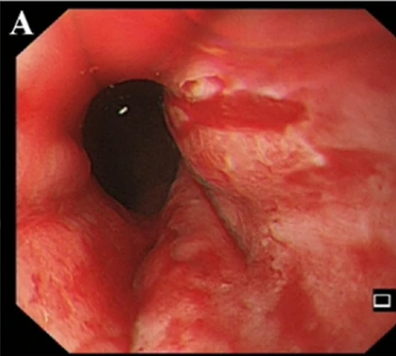
Mallory-Weiss Syndrome on Esophagoscopy. This image, captured via esophagoscopy, shows the distal esophagus. The visible tissue is ulcerated and torn. This finding is consistent with Mallory-Weiss Syndrome, which is characterized by tears in the esophageal lining caused by forceful vomiting or retching.
Shi S-S et al 2023.
Licensed under the Creative Commons Attribution 4.0 International License (CC BY 4.0)
.
.
(Click Image to Enlarge)
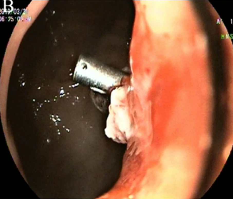
Endoscopic View of Hemoclip Application for Bleeding Control. This endoscopic image shows a medical procedure in progress. A hemoclip has been precisely applied to an area of tissue that appears to be actively bleeding or at risk of bleeding. The clip is deployed to achieve hemostasis. This intervention is commonly used to stop gastrointestinal bleeding.
Shi S-S, et al 2023.
Licensed under the Creative Commons Attribution 4.0 International License (CC BY 4.0)
.
(Click Image to Enlarge)
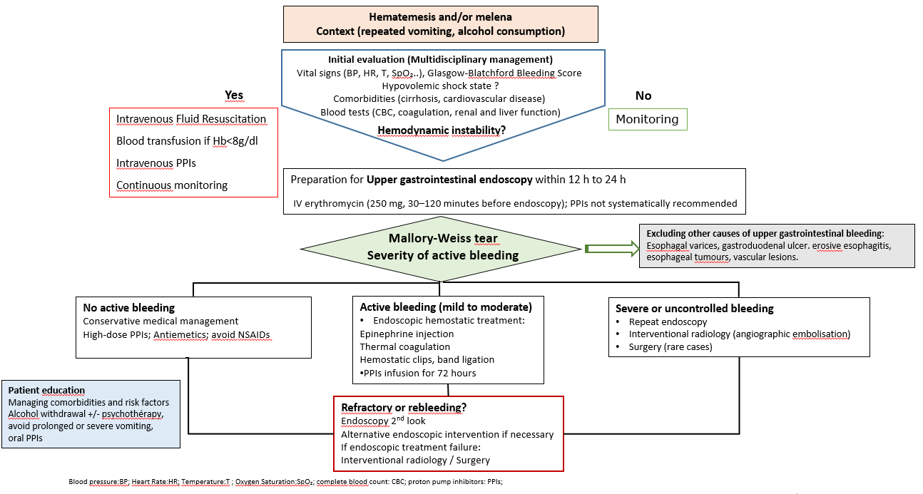
Diagnostic and Therapeutic Algorithm for Mallory-Weiss Syndrome. This flowchart outlines the clinical management of hematemesis or melena. The initial evaluation is detailed, including an assessment of hemodynamic stability. The algorithm provides guidance on the management of a Mallory-Weiss tear based on the severity of bleeding, from conservative care to surgical intervention. Patient education is also included in the chart.
By Zakia Kassama.
References
Enestvedt BK, Gralnek IM, Mattek N, Lieberman DA, Eisen G. An evaluation of endoscopic indications and findings related to nonvariceal upper-GI hemorrhage in a large multicenter consortium. Gastrointestinal endoscopy. 2008 Mar:67(3):422-9. doi: 10.1016/j.gie.2007.09.024. Epub 2008 Jan 18 [PubMed PMID: 18206878]
Gibson JA, Odze RD. Pathology of diseases that cause upper gastrointestinal tract bleeding. Gastrointestinal endoscopy clinics of North America. 2011 Oct:21(4):583-96. doi: 10.1016/j.giec.2011.07.006. Epub 2011 Aug 10 [PubMed PMID: 21944412]
Yin A, Li Y, Jiang Y, Liu J, Luo H. Mallory-Weiss syndrome: clinical and endoscopic characteristics. European journal of internal medicine. 2012 Jun:23(4):e92-6. doi: 10.1016/j.ejim.2012.02.005. Epub 2012 Mar 4 [PubMed PMID: 22560400]
Nojkov B, Cappell MS. Distinctive aspects of peptic ulcer disease, Dieulafoy's lesion, and Mallory-Weiss syndrome in patients with advanced alcoholic liver disease or cirrhosis. World journal of gastroenterology. 2016 Jan 7:22(1):446-66. doi: 10.3748/wjg.v22.i1.446. Epub [PubMed PMID: 26755890]
Bakhshipour A, Rafaiee R. Upper and Lower Gastrointestinal Bleeding: A Retrospective Study on 10 Years Experiences in Southeastern Iran. Middle East journal of digestive diseases. 2023 Apr:15(2):116-120. doi: 10.34172/mejdd.2023.329. Epub 2023 Apr 30 [PubMed PMID: 37546509]
Level 2 (mid-level) evidenceDECKER JP, ZAMCHECK N, MALLORY GK. Mallory-Weiss syndrome: hemorrhage from gastroesophageal lacerations at the cardiac orifice of the stomach. The New England journal of medicine. 1953 Dec 10:249(24):957-63 [PubMed PMID: 13111391]
Sonnenberg A, Yoo ER, Bakis G. Illusion of endoscopic success in instances of spontaneous disease resolution. Gastrointestinal endoscopy. 2023 Sep:98(3):437-440. doi: 10.1016/j.gie.2023.05.001. Epub 2023 May 5 [PubMed PMID: 37150415]
Bharucha AE, Gostout CJ, Balm RK. Clinical and endoscopic risk factors in the Mallory-Weiss syndrome. The American journal of gastroenterology. 1997 May:92(5):805-8 [PubMed PMID: 9149189]
Cherednikov EF, Kunin AA, Cherednikov EE, Moiseeva NS. The role of etiopathogenetic aspects in prediction and prevention of discontinuous-hemorrhagic (Mallory-Weiss) syndrome. The EPMA journal. 2016:7(1):7. doi: 10.1186/s13167-016-0056-4. Epub 2016 Mar 20 [PubMed PMID: 26998186]
Schuman BM, Threadgill ST. The influence of liver disease and portal hypertension on bleeding in Mallory-Weiss syndrome. Journal of clinical gastroenterology. 1994 Jan:18(1):10-2 [PubMed PMID: 8113576]
Mertens A, Essing T, Roderburg C, Luedde T, Kandler J, Loosen SH. A Systematic Analysis of Incidence, Therapeutic Strategies, and In-hospital Mortality of Mallory-Weiss Syndrome in Germany. Journal of clinical gastroenterology. 2024 Aug 1:58(7):640-649. doi: 10.1097/MCG.0000000000001918. Epub 2024 Aug 1 [PubMed PMID: 37668412]
Level 1 (high-level) evidenceCorral JE, Keihanian T, Kröner PT, Dauer R, Lukens FJ, Sussman DA. Mallory Weiss syndrome is not associated with hiatal hernia: a matched case-control study. Scandinavian journal of gastroenterology. 2017 Apr:52(4):462-464. doi: 10.1080/00365521.2016.1267793. Epub 2016 Dec 22 [PubMed PMID: 28007004]
Level 2 (mid-level) evidenceKortas DY, Haas LS, Simpson WG, Nickl NJ 3rd, Gates LK Jr. Mallory-Weiss tear: predisposing factors and predictors of a complicated course. The American journal of gastroenterology. 2001 Oct:96(10):2863-5 [PubMed PMID: 11693318]
Shi SS, Yang XZ, Zhang XY, Huang L, Guo HD, Li SF, Zhang W, Zhang YQ. Mallory-Weiss syndrome in four hemodialysis patients: a case study. BMC nephrology. 2023 Jun 27:24(1):188. doi: 10.1186/s12882-023-03250-x. Epub 2023 Jun 27 [PubMed PMID: 37365498]
Level 3 (low-level) evidenceSuzuki T, Wagata M, Konno H, Ito T, Torii Y, Murakoshi T. Case with a Nonreassuring Fetal Status Induced by Massive Hematemesis due to Mallory-Weiss Tear That Required Emergency Cesarean Section at 38 Weeks' Gestation. Case reports in obstetrics and gynecology. 2015:2015():762463. doi: 10.1155/2015/762463. Epub 2015 Dec 31 [PubMed PMID: 26881157]
Level 3 (low-level) evidenceLieto E, Auricchio A, Belfiore MP, Del Sorbo G, De Sena G, Napolitano V, Ruggiero A, Galizia G, Cardella F. Mallory-Weiss syndrome from giant gastric trichobezoar: A case report. World journal of gastrointestinal surgery. 2023 May 27:15(5):972-977. doi: 10.4240/wjgs.v15.i5.972. Epub [PubMed PMID: 37342849]
Level 3 (low-level) evidenceChen W, Zhu XN, Wang J, Zhu LL, Gan T, Yang JL. Risk factors for Mallory-Weiss Tear during endoscopic submucosal dissection of superficial esophageal neoplasms. World journal of gastroenterology. 2019 Sep 14:25(34):5174-5184. doi: 10.3748/wjg.v25.i34.5174. Epub [PubMed PMID: 31558865]
Cappell MS, Dass K, Manickam P. Characterization of the syndrome of UGI bleeding from a Mallory-Weiss tear associated with transesophageal echocardiography. Digestive diseases and sciences. 2014 Oct:59(10):2381-9. doi: 10.1007/s10620-014-3195-2. Epub 2014 May 18 [PubMed PMID: 24838497]
Zhang Y,Bian S,Li M,Zhang M,Zhang P,Liu Y,Zhang Z, Upper gastrointestinal bleeding after Roux-en-Y gastric bypass: Mallory-Weiss syndrome with marginal ulcer. Gastroenterology report. 2025; [PubMed PMID: 40703586]
Tang MM, Fang DF, Liu B. Upper gastrointestinal bleeding from a Mallory-Weiss tear associated with transesophageal echocardiography during successful cardiopulmonary resuscitation: A case report. World journal of clinical cases. 2022 Mar 26:10(9):2954-2960. doi: 10.12998/wjcc.v10.i9.2954. Epub [PubMed PMID: 35434104]
Level 3 (low-level) evidenceOba J, Usuda D, Tsuge S, Sakurai R, Kawai K, Matsubara S, Tanaka R, Suzuki M, Takano H, Shimozawa S, Hotchi Y, Usami K, Tokunaga S, Osugi I, Katou R, Ito S, Mishima K, Kondo A, Mizuno K, Takami H, Komatsu T, Nomura T, Sugita M. Hemorrhagic shock due to submucosal esophageal hematoma along with mallory-weiss syndrome: A case report. World journal of clinical cases. 2022 Sep 26:10(27):9911-9920. doi: 10.12998/wjcc.v10.i27.9911. Epub [PubMed PMID: 36186194]
Level 3 (low-level) evidenceHu Z, Liu D. Mallory-Weiss Syndrome. The New England journal of medicine. 2024 May 30:390(20):e49. doi: 10.1056/NEJMicm2310882. Epub [PubMed PMID: 38810187]
Gralnek IM, Stanley AJ, Morris AJ, Camus M, Lau J, Lanas A, Laursen SB, Radaelli F, Papanikolaou IS, Cúrdia Gonçalves T, Dinis-Ribeiro M, Awadie H, Braun G, de Groot N, Udd M, Sanchez-Yague A, Neeman Z, van Hooft JE. Endoscopic diagnosis and management of nonvariceal upper gastrointestinal hemorrhage (NVUGIH): European Society of Gastrointestinal Endoscopy (ESGE) Guideline - Update 2021. Endoscopy. 2021 Mar:53(3):300-332. doi: 10.1055/a-1369-5274. Epub 2021 Feb 10 [PubMed PMID: 33567467]
Laine L, Barkun AN, Saltzman JR, Martel M, Leontiadis GI. ACG Clinical Guideline: Upper Gastrointestinal and Ulcer Bleeding. The American journal of gastroenterology. 2021 May 1:116(5):899-917. doi: 10.14309/ajg.0000000000001245. Epub [PubMed PMID: 33929377]
Alali AA, Barkun AN. An update on the management of non-variceal upper gastrointestinal bleeding. Gastroenterology report. 2023:11():goad011. doi: 10.1093/gastro/goad011. Epub 2023 Mar 20 [PubMed PMID: 36949934]
Expert Panels on Vascular and Gastrointestinal Imaging, Nagpal P, Dane B, Aghayev A, Fowler KJ, Hedgire SS, Bartel TB, Cash BD, Collins JD, Kirsch DS, Lo HS, Pietryga JA, Ripley B, Santillan CS, Kim DH, Steigner ML. ACR Appropriateness Criteria® Nonvariceal Upper Gastrointestinal Bleeding: 2024 Update. Journal of the American College of Radiology : JACR. 2024 Nov:21(11S):S433-S447. doi: 10.1016/j.jacr.2024.08.021. Epub [PubMed PMID: 39488353]
Lecleire S, Antonietti M, Ducrotté P. [Mallory-Weiss syndrome: diagnosis and treatment]. Presse medicale (Paris, France : 1983). 2010 Jun:39(6):640-4. doi: 10.1016/j.lpm.2009.09.019. Epub [PubMed PMID: 19931377]
Wilkins T, Wheeler B, Carpenter M. Upper Gastrointestinal Bleeding in Adults: Evaluation and Management. American family physician. 2020 Mar 1:101(5):294-300 [PubMed PMID: 32109037]
Lee S, Ahn JY, Jung HY, Jung KW, Lee JH, Kim DH, Choi KD, Song HJ, Lee GH, Kim JH, Kim SO. Effective endoscopic treatment of Mallory-Weiss syndrome using Glasgow-Blatchford score and Forrest classification. Journal of digestive diseases. 2016 Oct:17(10):676-684. doi: 10.1111/1751-2980.12409. Epub [PubMed PMID: 27624697]
Lecleire S, Antonietti M, Iwanicki-Caron I, Duclos A, Ramirez S, Ben-Soussan E, Hervé S, Ducrotté P. Endoscopic band ligation could decrease recurrent bleeding in Mallory-Weiss syndrome as compared to haemostasis by hemoclips plus epinephrine. Alimentary pharmacology & therapeutics. 2009 Aug 15:30(4):399-405. doi: 10.1111/j.1365-2036.2009.04051.x. Epub 2009 May 26 [PubMed PMID: 19485979]
Saltzman JR. Hemostatic Spray for the Management of Gastrointestinal Bleeding. Gastroenterology & hepatology. 2019 Jan:15(1):40-43 [PubMed PMID: 30899208]
Shung DL, Laine L. Review article: Upper gastrointestinal bleeding - review of current evidence and implications for management. Alimentary pharmacology & therapeutics. 2024 May:59(9):1062-1081. doi: 10.1111/apt.17949. Epub 2024 Mar 22 [PubMed PMID: 38517201]
Morales P, Baum AE. Therapeutic Alternatives for the Mallory-Weiss Tear. Current treatment options in gastroenterology. 2003 Feb:6(1):75-83 [PubMed PMID: 12521575]
Gralnek IM, Camus Duboc M, Garcia-Pagan JC, Fuccio L, Karstensen JG, Hucl T, Jovanovic I, Awadie H, Hernandez-Gea V, Tantau M, Ebigbo A, Ibrahim M, Vlachogiannakos J, Burgmans MC, Rosasco R, Triantafyllou K. Endoscopic diagnosis and management of esophagogastric variceal hemorrhage: European Society of Gastrointestinal Endoscopy (ESGE) Guideline. Endoscopy. 2022 Nov:54(11):1094-1120. doi: 10.1055/a-1939-4887. Epub 2022 Sep 29 [PubMed PMID: 36174643]
Okamoto T, Fukuda K. Adenocarcinoma of the Gastroesophageal Junction Masquerading as Mallory-Weiss Syndrome. Case reports in gastroenterology. 2022 Jan-Apr:16(1):8-13. doi: 10.1159/000520627. Epub 2022 Jan 17 [PubMed PMID: 35221882]
Level 3 (low-level) evidenceFujisawa N, Inamori M, Sekino Y, Akimoto K, Iida H, Takahata A, Endo H, Hosono K, Sakamoto Y, Akiyama T, Koide T, Tokoro C, Takahashi H, Saito K, Abe Y, Nakamura A, Kubota K, Saito S, Koyama S, Nakajima A. Risk factors for mortality in patients with Mallory-Weiss syndrome. Hepato-gastroenterology. 2011 Mar-Apr:58(106):417-20 [PubMed PMID: 21661406]
Cuccì M, Caputo F, Fraternali Orcioni G, Roncallo A, Ventura F. Transition of a Mallory-Weiss syndrome to a Boerhaave syndrome confirmed by anamnestic, necroscopic, and autopsy data: A case report. Medicine. 2018 Dec:97(49):e13191. doi: 10.1097/MD.0000000000013191. Epub [PubMed PMID: 30544378]
Level 3 (low-level) evidenceLoftus TJ, Go KL, Hughes SJ, Croft CA, Smith RS, Efron PA, Moore FA, Brakenridge SC, Mohr AM, Jordan JR. Improved outcomes following implementation of an acute gastrointestinal bleeding multidisciplinary protocol. The journal of trauma and acute care surgery. 2017 Jul:83(1):41-46. doi: 10.1097/TA.0000000000001295. Epub [PubMed PMID: 27779592]
Papadinas A, Butt J. Outcomes in patients with acute upper gastrointestinal bleeding following changes to management protocols at an Australian hospital. JGH open : an open access journal of gastroenterology and hepatology. 2020 Aug:4(4):617-623. doi: 10.1002/jgh3.12303. Epub 2020 Feb 5 [PubMed PMID: 32782947]