 Tools
Tools
 Malar Rash
Introduction
Malar rash, commonly referred to as a butterfly rash, is a distinctive erythematous eruption involving the cheeks and nasal bridge that typically spares the nasolabial folds.[1][2] It is most classically associated with SLE. According to the American College of Rheumatology, malar rash is one of the classification criteria for SLE; it also may occur in other forms of lupus, including discoid lupus and subacute cutaneous lupus.[3] The differential diagnosis for malar rash is broad and includes inflammatory, infectious, and dermatologic conditions such as SLE, dermatomyositis, contact dermatitis (including photoallergic dermatitis), drug-induced photosensitivity, erysipelas, cellulitis, erythema infectiosum, rosacea, seborrheic dermatitis, and pemphigus erythematosus. Accurate clinical recognition is essential to distinguish benign cutaneous conditions from those associated with systemic disease.[4][5][6][7][8][9][10][11] Management begins with careful evaluation to identify the underlying etiology and assess for systemic involvement, followed by sun protection (eg, protective clothing, sunscreen use, and behavioral modification) and targeted treatment of the underlying condition when identified.
Etiology
Register For Free And Read The Full Article
 Search engine and full access to all medical articles
- 10 free questions in your specialty
- Free CME/CE Activities
- Free daily question in your email
- Save favorite articles to your dashboard
- Emails offering discounts
Learn more about a Subscription to StatPearls Point-of-Care
Etiology
Common causes of malar rash include erysipelas, cellulitis, lupus erythematosus, rosacea, seborrheic dermatitis, dermatomyositis, contact dermatitis, and erythema infectiosum. Erysipelas is an acute bacterial skin infection involving the superficial lymphatic vessels and upper dermis, most commonly caused by group A streptococci. It typically presents as a painful, rapidly spreading, well-demarcated erythematous plaque with a shiny surface, often accompanied by marked edema and a peau d’orange appearance. Systemic symptoms, including fever, chills, and malaise, are common. Although the lower extremities are most frequently affected, the face is the second most common site of involvement.[12][13][14]
In the early twentieth century, the face was a common site of cellulitis; however, the incidence of facial involvement has declined over time. Cellulitis is a bacterial skin infection affecting the deeper dermis and subcutaneous tissue, most commonly caused by streptococci and Staphylococcus aureus. In contrast to erysipelas, cellulitis is typically less well demarcated, with minimal surface change and less prominent edema. Systemic symptoms are less common and, when present, the clinical course is generally more gradual and indolent.[15][16] Systemic lupus erythematosus is an autoimmune disease involving multiple organ systems in which the immune system produces numerous autoantibodies that target tissues such as the kidneys, joints, skin, brain, and heart. Clinical manifestations vary widely, ranging from mild rashes and arthritis to systemic symptoms such as fever, fatigue, and joint pain, as well as severe organ damage and life-threatening disease.[17]
Rosacea is a chronic inflammatory skin disorder characterized by facial erythema, telangiectasia, and inflammatory papules or pustules in the absence of comedones. It classically involves the cheeks, nose, chin, and forehead. Four clinical subtypes of rosacea have been described, with the erythematotelangiectatic subtype most closely resembling the malar rash seen in SLE. An important distinguishing feature is involvement of the nasolabial folds, which are typically spared in lupus-associated malar rash.[18] Rosacea may also be associated with recurrent episodes of facial flushing triggered by consumption of hot beverages or alcohol, emotional stress, and temperature changes. Both genetic predisposition and environmental factors contribute to disease pathogenesis.[4]
Malar rash is a common, chronic, relapsing inflammatory skin disorder that predominantly affects sebaceous-rich areas, including the scalp, face, chest, and back. Ill-defined erythematous patches with overlying yellow, greasy scale characterize malar rash. The etiology is multifactorial and includes increased sebaceous gland activity, immune dysregulation, skin barrier dysfunction, and overgrowth of Malassezia species.[19]
Dermatomyositis can present with facial erythema involving the malar region, but it has distinguishing clinical features. The rash is typically violaceous rather than bright erythematous and often includes a heliotrope rash with periorbital edema. Facial involvement may extend beyond the malar area to other photosensitive regions, such as the V-sign and shawl sign, and pruritus is common.[7][20]
According to the North American Contact Dermatitis Group, the face is one of the most common sites for primary dermatitis.[21] The face is exposed to a wide range of cosmetic and personal care products that may contain susceptible allergens. The most common allergens reported to cause facial dermatitis include fragrances, preservatives (particularly methylisothiazolinone and methylchloroisothiazolinone), nickel, and balsam of Peru.[22] Photoallergic contact dermatitis results from a delayed hypersensitivity reaction to a photosensitizing agent following UV exposure. It typically affects sun-exposed areas such as the face, the anterior neck in a V-shaped distribution, the dorsal hands, and the forearms, while sparing photoprotected sites. Chemical sunscreen agents are the most common triggers, whereas physical blockers such as titanium dioxide and zinc oxide are not known causes.[23]
Erythema infectiosum, also known as fifth disease, is caused by parvovirus B19 and most commonly affects children. It typically begins with a mild viral prodrome followed by the characteristic erythematous slapped-cheek facial rash. The facial eruption may be succeeded by a lacy reticular rash on the extremities, which resolves over several weeks but can recur with sun exposure, heat, or stress.[24]
Less common causes of malar rash include pemphigus erythematosus and drug-induced photosensitivity. Pemphigus erythematosus is a localized variant of pemphigus foliaceus mediated by IgG autoantibodies directed against desmoglein 1, which results in subcorneal blistering due to acantholysis. The condition predominantly affects photodistributed areas, including the face, and may mimic cutaneous lupus erythematosus with a malar distribution. Its etiology involves autoimmune mechanisms, with UV light exposure acting as a trigger that promotes cleavage of desmoglein 1 and immune complex deposition.[25][26]
Photosensitizing drug reactions are cutaneous eruptions triggered by UV radiation exposure in patients taking photosensitizing medications. These reactions are classified as phototoxic or photoallergic, with phototoxic reactions being more common. Numerous medications are implicated, most frequently amiodarone, chlorpromazine, doxycycline, hydrochlorothiazide, nonsteroidal anti-inflammatory drugs (naproxen, piroxicam, ketoprofen, ibuprofen), and certain antifungal (voriconazole) and oncologic (vemurafenib) agents. Most photosensitizing drugs act within the UVA spectrum. Clinical distinction between phototoxic and photoallergic reactions may be challenging due to overlapping features.[6][27]
Epidemiology
Cellulitis and Erysipelas
Because cellulitis and erysipelas are not reportable disorders, the exact prevalence is not clear; however, it is a common infection involving all racial and ethnic groups. Both conditions can occur at any age, and facial involvement, while historically less frequent than involvement of the extremities, is clinically important due to potential complications. Awareness of its presentation is essential for timely recognition and treatment.
Systemic Lupus Erythematosus
The Lupus Foundation of America has estimated that at least 1.5 million individuals in the United States have SLE. Worldwide, the highest prevalence and incidence rates are reported in North America, with a prevalence of 241 per 100,000 population and an incidence of 23.2 per 100,000 person-years. Lower incidence rates have been observed in Ukraine and Africa (0.3 per 100,000 person-years), and the lowest prevalence was reported in Northern Australia (0 cases in 847 people). Women are affected more frequently than men across all age and ethnic groups. Among different racial and ethnic populations, individuals of African descent have the highest prevalence and incidence, whereas White populations have the lowest. Historical data from the 1970s through the 2000s indicate that annual incidence ranged from approximately 1 to 10 per 100,000 population, with prevalence ranging from 5.8 to 130 per 100,000 population.[28]
Rosacea
Rosacea is a chronic inflammatory facial disorder with a worldwide prevalence of 5.46%.[29] White individuals with sun-sensitive skin have the highest risk. The prevalence in this population is estimated at 2% to 22%. Rosacea can affect adults of any age, and its chronic nature can lead to persistent erythema, telangiectasia, and occasional inflammatory papules or pustules.[30][31]
Seborrheic Dermatitis
Seborrheic dermatitis follows a bimodal distribution, presenting most commonly in early infancy (2 weeks to 12 months) and later in adolescence and adulthood. Globally, it affects an estimated 4% of the population and is seen more frequently in adults than in infants or children.[32] Severity varies from mild scaling to more extensive erythematous plaques, and recurrence is common in patients with neurologic or immunologic comorbidities.
Dermatomyositis
The incidence of dermatomyositis is estimated at 9.63 cases per million people. The disease has a bimodal age distribution, with one peak in the 5 to 15 age range and another in the 45 to 60 age range.[33] Both children and adults may present with characteristic cutaneous findings and muscle involvement, with a slight female predominance.
Contact Dermatitis
The exact prevalence of facial contact dermatitis has not been well established; however, available data suggest it accounts for approximately 26% to 27% of contact dermatitis cases referred for patch testing.[22][34] Among patients with facial dermatitis, allergic contact dermatitis is diagnosed in an estimated 45% to 82% of cases, with the remainder attributed to irritant contact dermatitis or other etiologies.[34][23] Female patients are disproportionately affected, with female-to-male ratios reported as high as 9:1 in some studies. The mean age at presentation is typically in the mid-to-late 30s, with an increased risk in individuals over 40.[34]
Erythema Infectosum
Erythema infectiosum (fifth disease) most commonly affects school-aged children and often occurs in epidemic patterns, with increased incidence in late winter and spring.[35] Caused by parvovirus B19, the disease shows a distinct age distribution, with most cases in children aged 5 to 15 years. Transmission occurs primarily via respiratory droplets, facilitating spread in community settings such as schools, where outbreaks are frequently observed.[35]
Pemphigus Erythematosus
Pemphigus erythematosus is a rare autoimmune blistering disorder that represents a clinical subtype of pemphigus foliaceus, characterized by lesions predominantly in photodistributed areas.[25] The disease is mediated by IgG autoantibodies against desmoglein 1, resulting in subcorneal blistering. It may mimic cutaneous lupus erythematosus and often requires histopathologic and immunofluorescence studies for diagnosis
Drug-Induced Photosensitization
Drug-induced photosensitivity accounts for approximately 5% to 6% of patients evaluated for photosensitivity at specialized photoinvestigation centers, although the true prevalence remains uncertain due to underreporting and geographic variability.[36][37] Affected individuals are typically in the sixth decade of life, with a reported median age ranging from 52 to 62 years, and there is a slight female predominance (approximately 55%–56%).[36][38] The majority of cases occur in patients with lighter skin phototypes, with phototypes I–III accounting for roughly 80% of reported cases.[36] Clinical manifestations range from mild erythema to severe phototoxic reactions, and management often requires avoidance of the offending medication or modification of sun exposure.
Histopathology
Cellulitis
Histopathologic findings of cellulitis include vascular and lymphatic dilatation with marked edema in the dermal and subepidermal areas. Dense and diffuse neutrophilic infiltration is usually observed beneath the edema. These features reflect the acute inflammatory response characteristic of bacterial infection.[39]
Erysipelas
In the acute phase, erysipelas demonstrates a dense, predominantly neutrophilic inflammatory infiltrate in the dermis, with less involvement of the hypodermis. Prominent fibrin-rich dermal edema can lead to subepidermal blister formation, and the process may be associated with pustules, abscesses, or focal necrosis. Dilated lymphatics contain neutrophils and macrophages, with neutrophilic involvement of venule walls and occasional thrombosis.[40]
Dermatomyositis and Systemic Lupus Erythematosus
The main histopathologic findings of dermatomyositis and SLE include epidermal thinning, hydropic degeneration of the basal layer, disruption of the dermo-epidermal junction, upper dermal edema, sparse lymphocytic infiltration in the dermis, and fibrinoid degeneration of connective tissue. Direct immunofluorescence microscopy in SLE reveals deposition of IgG, IgM, and C3 at the basement membrane in both involved and uninvolved skin, which is not observed in dermatomyositis.[7]
Rosacea
Histopathologic features of rosacea include dilated vessels, lymphohistiocytic infiltration in the perifollicular and perivascular areas, neutrophils in the intrafollicular region, and dermal edema. Sebaceous gland hyperplasia, connective tissue expansion, and elastosis are frequently observed.[41]
Seborrheic Dermatitis
Seborrheic dermatitis is characterized by spongiosis, psoriasiform epidermal hyperplasia, and parakeratosis concentrated around follicular openings, often described as shoulder parakeratosis. Neutrophils may be present within scale crusts at follicular ostia. Chronic lesions show prominent psoriasiform hyperplasia and parakeratosis, closely resembling psoriasis histologically.[42]
Contact Dermatitis
Histopathologic findings in contact dermatitis typically include epidermal acanthosis with associated hyperkeratosis and parakeratosis, accompanied by a predominantly T-lymphocyte–mediated inflammatory infiltrate in the dermis.[43][44] A relatively specific feature of allergic contact dermatitis is the presence of spongiotic epidermal collections of Langerhans cells, reflecting the underlying delayed-type hypersensitivity reaction.[45]
Erythema Infectiosum
Erythema infectiosum, also called fifth disease, is caused by parvovirus B19. Its histopathology is relatively nonspecific, showing features common to viral exanthems, including a superficial perivascular lymphocytic infiltrate and variable interface changes. Epidermal changes are usually mild, and histologic findings should be interpreted in the context of the clinical presentation and laboratory confirmation of parvovirus B19 infection.[46]
Pemphigus Erythematosus
Pemphigus erythematosus demonstrates subcorneal acantholysis, consistent with pemphigus foliaceus, along with a characteristic immunopathologic pattern. Direct immunofluorescence reveals intraepidermal IgG deposition and granular-to-linear IgG deposits along the basement membrane zone, producing the classic lupus band appearance.[26]
Drug-Induced Photosensitivity
Histopathologic findings in phototoxic reactions reflect direct cellular injury, with keratinocyte necrosis, vacuolar interface changes at the dermal–epidermal junction, and minimal inflammatory infiltrate. The epidermis may show sunburn-like changes with scattered necrotic keratinocytes, and in severe cases, full-thickness epidermal necrosis may be present. Dermal melanophages are often present, indicating pigmentary incontinence due to melanin leakage from damaged epidermal cells into the dermis.[47][48] Photoallergic reactions demonstrate features of a delayed-type hypersensitivity response, with a more prominent inflammatory infiltrate than phototoxic reactions. Histologic findings typically show a spongiotic and/or lichenoid pattern with a predominantly T-lymphocytic infiltrate, as demonstrated by CD3 immunohistochemical staining.[47]
History and Physical
The initial step in narrowing the differential diagnosis of a malar rash or facial erythema is a thorough patient history combined with a comprehensive skin examination. Careful attention to associated symptoms, triggers, temporal patterns, and subtle morphologic features is critical for accurate diagnosis. A full skin examination is essential, as additional cutaneous findings may indicate an underlying cutaneous or systemic disorder.
It is important to ascertain the onset and duration of symptoms, distinguishing between acute, subacute, chronic, recurrent, or transient presentations. Identification of triggers and exacerbating factors can provide valuable diagnostic clues. Photosensitivity or recent sun exposure may suggest cutaneous lupus erythematosus, dermatomyositis, lupus tumidus erythematosus, or pemphigus erythematosus. A recent viral illness, particularly in children, may point toward parvovirus B19 infection. New medications, including photosensitizing agents, as well as the use of topical products or cosmetics, should also be explored.
Associated symptoms such as pruritus may indicate allergic contact dermatitis, atopic dermatitis, or photoallergic reactions, whereas pain or burning may reflect sunburn, irritant contact dermatitis, phototoxic reactions, erysipelas, or rosacea. Blistering, erosions, or crusting can suggest pemphigus erythematosus, and constitutional or systemic symptoms such as fever, fatigue, arthralgias, or myalgias may indicate lupus erythematosus, dermatomyositis, drug eruption, or viral infection. A detailed medication and exposure history, including supplements, is essential. Additionally, noting distribution beyond the face can provide clues: truncal or extremity involvement may be seen in viral exanthems, seborrheic dermatitis, or drug eruptions. Past and family history of autoimmune blistering diseases, connective tissue disorders, or recent exposure to infected contacts should also be elicited.
Physical examination should evaluate both the morphology and distribution of lesions. Diffuse symmetrical erythema without scale is characteristic of flushing or a ruddy complexion, whereas diffuse erythema with scale may indicate irritant or airborne allergic contact dermatitis or infantile atopic dermatitis. Symmetrical central facial erythema without scale can be seen in rosacea or erythema infectiosum due to parvovirus B19, while the presence of scale or crust suggests seborrheic dermatitis, atopic dermatitis, psoriasis, or pemphigus erythematosus. Photodistributed erythema or plaques may point to cutaneous lupus erythematosus, dermatomyositis, lupus tumidus erythematosus, pemphigus erythematosus, or phototoxic/photoallergic reactions. Localized plaques or patches can be seen in erysipelas, lupus tumidus erythematosus, cutaneous lymphoid hyperplasia, Jessner lymphocytic infiltrate, or granuloma faciale. Superficial erosions, crusting, or blistering favor a diagnosis of pemphigus erythematosus. Sparing of the nasolabial folds supports lupus-related malar rash, and telangiectasias are often present in rosacea or photodamage.
Examination should also include mucosal assessment, as oral erosions may be present in pemphigus erythematosus, although less commonly than in pemphigus vulgaris. Hair and scalp findings, including alopecia or scaling, may also be observed in pemphigus erythematosus or cutaneous lupus. Systemic findings, such as arthralgias or arthritis, may accompany parvovirus B19 infection or lupus erythematosus, and lymphadenopathy or fever may indicate viral infection. Finally, other cutaneous findings, such as a lacy or reticular rash on the extremities following facial erythema, are characteristic of parvovirus B19 infection and can help confirm the diagnosis.
Evaluation
Uncomplicated cases of erysipelas and cellulitis generally do not require laboratory evaluation. In patients who appear toxic, laboratory testing—including complete blood count with differential, erythrocyte sedimentation rate, and C-reactive protein—is recommended to assess the severity of infection. Malar rash in SLE may precede other systemic manifestations by months or years or may appear concurrently with acute disease. Laboratory findings in patients with isolated malar rash may mirror those seen in SLE, including positive antinuclear antibody test, elevated ESR, and elevated CRP. Histopathologic examination of lesional skin can provide additional diagnostic support. In patients presenting with malar rash without clinical signs of muscle disease, dermatomyositis should be considered. Skin biopsy is the primary diagnostic tool, and patients should be evaluated for subclinical muscle involvement, with periodic assessment of serum creatine kinase and aldolase every 2 to 3 months, along with formal muscle examination.
Contact dermatitis presenting as a malar rash should be evaluated with a detailed history of exposure to cosmetics, personal care products, and occupational allergens, with attention to the timing of symptom onset relative to the introduction of new products. The pattern of facial involvement—periorbital, lateral, central, or generalized—provides important diagnostic clues. Patch testing remains the gold standard for confirming allergic contact dermatitis.[23][49][50] Rosacea is diagnosed clinically based on characteristic facial erythema, telangiectasias, and absence of systemic involvement. Similarly, seborrheic dermatitis is diagnosed clinically by identifying erythematous patches with greasy scales in sebaceous-rich areas, including the scalp, face, and upper trunk.
Parvovirus B19 infection, also known as erythema infectiosum or fifth disease, should be suspected in immunocompetent patients presenting with fever, rash, and/or arthralgias. In children with classic disease, the diagnosis is usually clinical and does not require laboratory confirmation. Serologic testing for parvovirus B19–specific IgM may be considered in atypical presentations or in patients with unexplained new-onset arthropathy.
Evaluation of pemphigus erythematosus requires correlation of clinical findings with immunopathologic, serologic, and histologic studies. Direct immunofluorescence of perilesional skin is the diagnostic gold standard, demonstrating intercellular deposition of IgG and/or C3 within the epidermis. Serologic testing via enzyme-linked immunosorbent assay for anti–desmoglein 1 and 3 antibodies is highly sensitive and specific, helps distinguish pemphigus subtypes, and can monitor disease activity. Isolated desmoglein 1 positivity supports pemphigus foliaceus–type disease. Histopathology of lesional skin typically shows subcorneal acantholysis.[25][51]
Drug-induced photosensitivity should be evaluated with a focused history of medication exposure, timing of symptom onset, and photodistribution of lesions. Phototoxic reactions resemble severe sunburn, whereas photoallergic reactions present as eczematous dermatitis. Phototesting and photopatch testing may assist in diagnosing suspected photoallergic reactions. Drug rechallenge can confirm the diagnosis but should be performed cautiously due to the risk of recurrence.[6][27][36][52]
Treatment / Management
Management of malar rash depends on the underlying cause. For cellulitis and erysipelas, the mainstay of treatment is oral or intravenous antibiotic therapy, with the choice of agent guided by severity, patient comorbidities, and local resistance patterns. Supportive measures, including rest, limb elevation when applicable, and hydration, may also improve outcomes and reduce complications.
In patients with SLE or dermatomyositis, initial management focuses on sun protection through appropriate clothing, sunscreen use, and behavioral modification to prevent exacerbation of lesions. Additional treatment may include topical or intralesional corticosteroids for localized disease, antimalarial medications, such as hydroxychloroquine, for systemic involvement, and immunosuppressive agents for severe or refractory cases. Regular monitoring is essential to assess treatment response and minimize adverse effects.
Rosacea management may involve topical gels or creams to repair the skin and reduce telangiectasia, papules, and pustules, along with antibiotic therapy to control inflammation. For patients with phymatous changes, CO2 laser therapy or other procedural interventions may be considered to improve cosmesis. Patient education regarding triggers such as sun exposure, heat, and alcohol can help reduce flares and improve long-term disease control.
Seborrheic dermatitis is treated with topical antifungal agents, low-potency corticosteroids, topical calcineurin inhibitors, or topical phosphodiesterase 4 inhibitors. In refractory cases, short courses of systemic antifungal therapy may be indicated, particularly in adults with extensive involvement. Regular follow-up allows clinicians to adjust therapy based on response and minimize long-term side effects.
Facial contact dermatitis is managed primarily through strict avoidance of identified allergens or irritants, with short courses of low- to medium-potency topical corticosteroids typically limited to 1 to 2 weeks due to the increased risk of adverse effects on facial skin. Patch testing and careful history-taking are essential in persistent or unclear cases to identify causative agents, which commonly include cosmetics, personal care products, and airborne contactants. Topical calcineurin inhibitors may be used as steroid-sparing alternatives for sensitive facial areas or when longer-term therapy is required, providing effective control while reducing the risk of steroid-related side effects.[8][23]
Erythema infectiosum in immunocompetent patients is self-limiting, and treatment is primarily symptomatic, including antipyretics or analgesics for fever and joint discomfort. In most cases, lesions resolve without intervention within 1 to 3 weeks. Education of caregivers regarding the benign nature of the disease and measures to prevent transmission in school or household settings is recommended.
Pemphigus erythematosus is treated similarly to pemphigus foliaceus. Initial therapy typically involves lower doses of prednisolone (0.5 mg/kg/day) to control disease activity while minimizing adverse effects. Second-line options include oral immunosuppressive agents such as azathioprine, mycophenolate mofetil, and mycophenolic acid, while third-line treatments may include rituximab, intravenous immunoglobulin, dapsone, cyclophosphamide, or methotrexate for refractory or severe disease.[53] Close monitoring is necessary to adjust therapy and prevent complications.
Management of drug-induced photosensitivity centers on discontinuation of the offending medication when possible, combined with symptomatic treatment using topical corticosteroids. Strict sun protection is essential, particularly for patients who require continued use of the causative drug, and education on photoprotection strategies can reduce recurrence. Phototesting or photopatch testing may be considered in complex cases to identify causative agents and guide further management.[54][27]
Differential Diagnosis
Common differential diagnoses for malar rash include cellulitis, erysipelas, SLE, dermatomyositis, seborrheic dermatitis, rosacea, parvovirus B19 infection (erythema infectiosum), contact dermatitis, and drug-induced photosensitivity.
Prognosis
Cellulitis
The prognosis of cellulitis is generally favorable with prompt diagnosis and appropriate antimicrobial therapy. Most patients recover completely, although recurrence may occur, particularly in those with underlying risk factors such as lymphedema, venous insufficiency, or diabetes. Delayed treatment can lead to complications, including abscess formation, necrotizing infection, or systemic involvement.
Erysipelas
Erysipelas typically has an excellent prognosis when treated early with appropriate antibiotics. Symptoms often resolve rapidly, although recurrence is common, especially in patients with chronic edema, venous disease, or lymphatic dysfunction. Severe cases may result in systemic complications if treatment is delayed.
Systemic Lupus Erythematosus
The prognosis of SLE has improved substantially with advances in diagnosis and treatment. Outcomes depend on the extent of organ involvement, particularly renal and central nervous system disease. Many patients experience a relapsing-remitting course, and long-term survival is favorable with appropriate management.
Dermatomyositis
Prognosis in dermatomyositis varies depending on disease severity, response to therapy, and presence of associated malignancy or interstitial lung disease. With early diagnosis and immunosuppressive treatment, many patients achieve disease control, although relapses are common. Long-term morbidity may result from muscle weakness, calcinosis, or systemic complications.
Seborrheic Dermatitis
Seborrheic dermatitis is a chronic relapsing condition with a benign course. Symptoms can usually be well controlled with topical therapies, although complete remission is uncommon, and periodic flares are expected. Long-term prognosis is favorable with appropriate maintenance treatment.
Rosacea
Rosacea is a chronic inflammatory disorder with a variable course and a tendency to progress without treatment. Although not life-threatening, the disease may, if left untreated, lead to persistent erythema, phymatous changes, or ocular involvement. Long-term management can substantially improve symptoms and quality of life.
Parvovirus B19
Parvovirus B19 infection is typically self-limited in immunocompetent individuals, with complete recovery expected. Children with erythema infectiosum usually have an excellent prognosis without long-term sequelae. Complications may occur in immunocompromised patients or those with underlying hematologic disorders.
Contact Dermatitis
The prognosis of contact dermatitis is excellent with identification and avoidance of the offending allergen or irritant. Acute episodes usually resolve with appropriate topical therapy, although chronic or recurrent disease may occur if exposure persists. Long-term outcomes are favorable with effective allergen avoidance strategies.
Drug-Induced Photosensitivity
Drug-induced photosensitivity generally resolves after discontinuation of the offending medication and implementation of sun-protective measures. Most patients recover without permanent sequelae, although hyperpigmentation may persist in some cases. Prognosis is excellent when the condition is recognized early and managed appropriately.
Complications
Complications of facial dermatologic conditions vary depending on the underlying disorder. Cellulitis and erysipelas can lead to serious infectious sequelae, including abscess formation, bacteremia, endocarditis, septic arthritis or osteomyelitis, metastatic infection, sepsis, and toxic shock syndrome.[55] Dermatomyositis may cause life-threatening complications affecting the skin, lungs, muscles, and other organ systems.[20] Systemic lupus erythematosus can involve multiple organ systems, with cardiovascular disease, infections, and chronic kidney disease representing the leading causes of mortality.[56]
Seborrheic dermatitis is typically benign but may be complicated by secondary infection, postinflammatory hyperpigmentation, or severe pruritus that affects quality of life, particularly in infants or immunocompromised patients. Rosacea may result in persistent facial erythema, telangiectasias, inflammatory papules and pustules, and phymatous changes such as rhinophyma. Ocular involvement can lead to blepharitis, keratitis, or visual impairment if untreated. Parvovirus B19 infection is usually self-limited in immunocompetent individuals, but transient arthralgias or arthritis may occur in adults, and those with underlying hemolytic disorders may develop transient aplastic crisis. In immunocompromised patients or during pregnancy, infection can cause chronic anemia, fetal anemia, hydrops fetalis, or fetal loss.
Contact dermatitis may lead to secondary bacterial infection, postinflammatory hyperpigmentation or hypopigmentation, and chronic lichenification with persistent allergen exposure. Unrecognized triggers can result in recurrent or chronic dermatitis and reduced quality of life. Drug-induced photosensitivity can cause severe sunburn-like reactions, blistering, and secondary skin infection, with persistent postinflammatory hyperpigmentation or dyspigmentation. Repeated exposure without discontinuation of the offending medication may result in chronic photosensitivity or photodamage.
Consultations
Consultation with dermatology, rheumatology, or infectious disease may be appropriate.
Deterrence and Patient Education
Treatment and management of malar rash depend on the underlying condition. Patients with cellulitis or erysipelas generally respond well to appropriate antimicrobial therapy. Rosacea and seborrheic dermatitis often follow a relapsing-remitting course, and ongoing dermatology follow-up may be required to maintain symptom control. Patients with systemic lupus erythematosus or dermatomyositis typically require management and monitoring by a rheumatologist, with treatment tailored to disease activity and systemic involvement.
Enhancing Healthcare Team Outcomes
Malar rash is an erythematous eruption involving the cheeks and nasal bridge that typically spares the nasolabial folds and is most classically associated with systemic lupus erythematosus, though it may occur in other inflammatory, infectious, or dermatologic conditions. Its pathophysiology varies by etiology, ranging from autoimmune-mediated inflammation in lupus to infectious or irritant processes. Clinical presentation requires careful assessment of morphology, distribution, triggers, and associated systemic features. Evaluation emphasizes thorough history, physical examination, and targeted testing, including autoimmune serologies, biopsy, or patch testing when indicated. Management is directed at the underlying cause and commonly includes sun protection, topical therapies, or systemic treatment for autoimmune or infectious conditions.
Interprofessional collaboration is essential to optimize diagnostic accuracy and patient outcomes. Physicians and advanced practitioners lead evaluation and management, while primary care clinicians coordinate longitudinal care and identify early or systemic features. Nurses support patient education and monitoring, particularly regarding medication adherence and skin care. Pharmacists assist with medication selection and safety, especially for immunosuppressive or photosensitizing agents. Dermatologists, rheumatologists, and infectious disease specialists contribute expertise for complex or refractory cases. Effective communication, shared decision-making, and timely referral support risk reduction, prevent complications, and ensure coordinated, patient-centered care across settings.
Media
(Click Image to Enlarge)
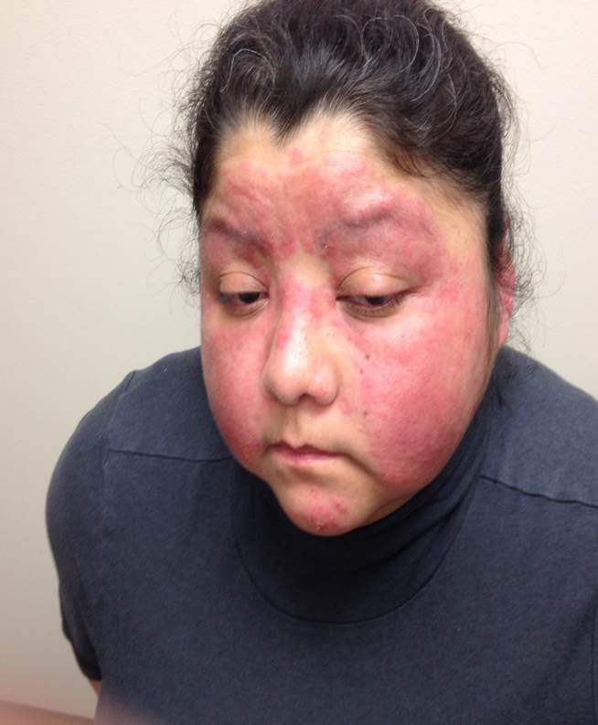
Rash, Face. Rash on the face presents on bilateral cheeks with sparing of the nasolabial folds.
Contributed by S Jones, MD
(Click Image to Enlarge)
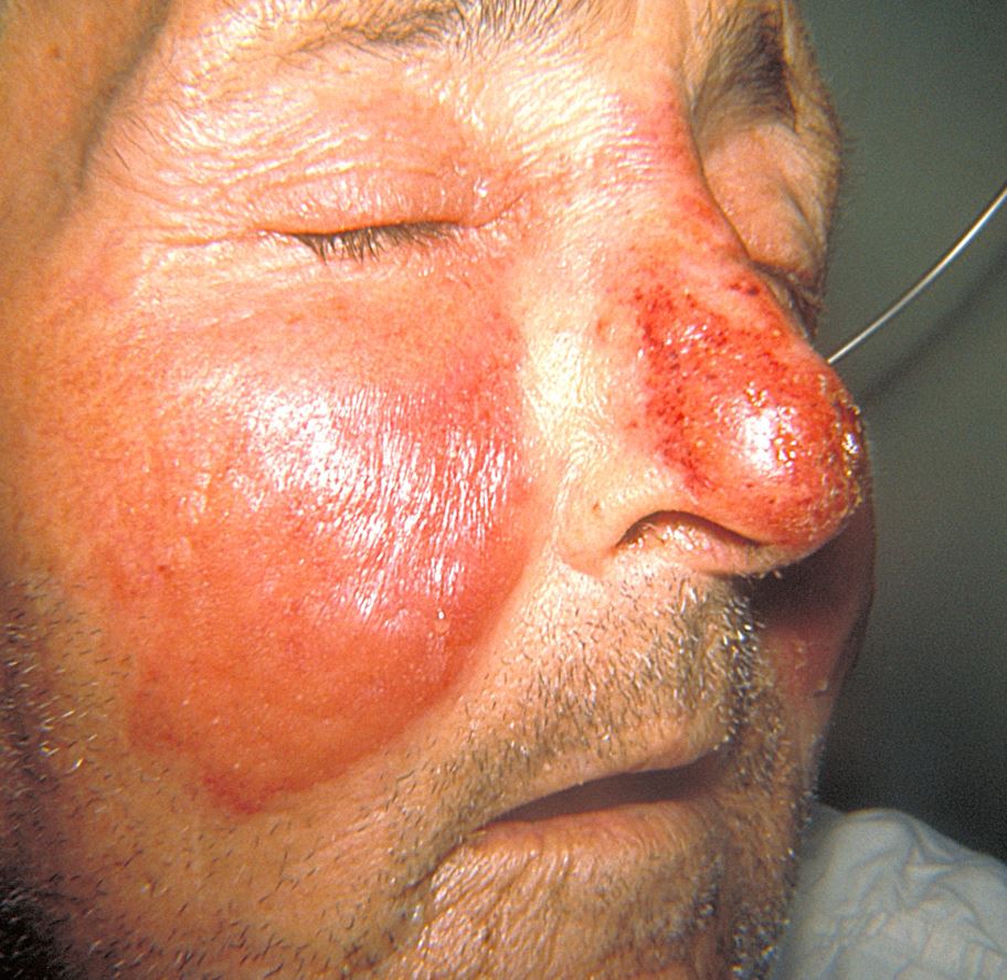
Erysipelas Rash.
Thomas F Sellers, MD; Public Health Image Library, Public Domain, Centers for Disease Control and Prevention
(Click Image to Enlarge)
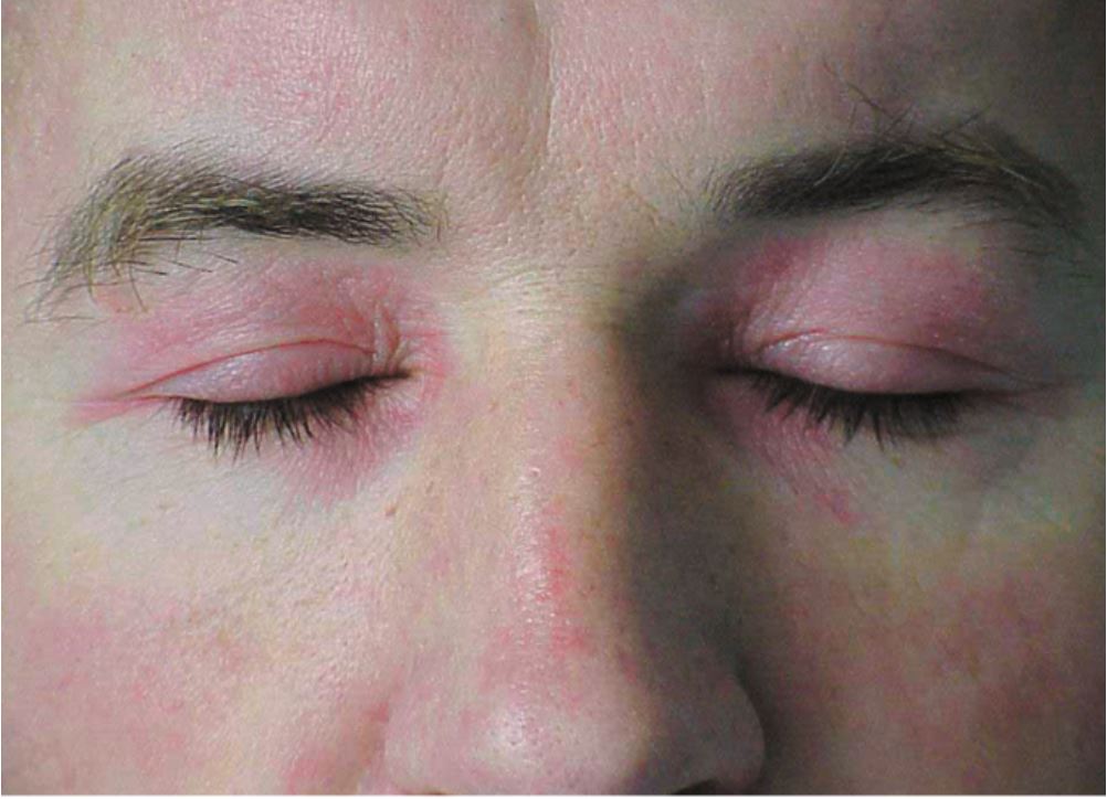
Heliotrope Rash. A violaceous eruption with mild periorbital edema is characteristic of the heliotrope rash in dermatomyositis.
Contributed by M Tarnopolsky, MD, PhD
References
Hoi A, Igel T, Mok CC, Arnaud L. Systemic lupus erythematosus. Lancet (London, England). 2024 May 25:403(10441):2326-2338. doi: 10.1016/S0140-6736(24)00398-2. Epub 2024 Apr 17 [PubMed PMID: 38642569]
Siegel CH, Sammaritano LR. Systemic Lupus Erythematosus: A Review. JAMA. 2024 May 7:331(17):1480-1491. doi: 10.1001/jama.2024.2315. Epub [PubMed PMID: 38587826]
Uva L, Miguel D, Pinheiro C, Freitas JP, Marques Gomes M, Filipe P. Cutaneous manifestations of systemic lupus erythematosus. Autoimmune diseases. 2012:2012():834291. doi: 10.1155/2012/834291. Epub 2012 Jul 25 [PubMed PMID: 22888407]
van Zuuren EJ. Rosacea. The New England journal of medicine. 2017 Nov 2:377(18):1754-1764. doi: 10.1056/NEJMcp1506630. Epub [PubMed PMID: 29091565]
Adalsteinsson JA, Kaushik S, Muzumdar S, Guttman-Yassky E, Ungar J. An update on the microbiology, immunology and genetics of seborrheic dermatitis. Experimental dermatology. 2020 May:29(5):481-489. doi: 10.1111/exd.14091. Epub 2020 Mar 16 [PubMed PMID: 32125725]
Montgomery S, Worswick S. Photosensitizing drug reactions. Clinics in dermatology. 2022 Jan-Feb:40(1):57-63. doi: 10.1016/j.clindermatol.2021.08.014. Epub 2021 Aug 8 [PubMed PMID: 35190066]
Kazandjieva J, Tsankov N, Pramatarov K. The red face revisited: connective tissue disorders. Clinics in dermatology. 2014 Jan-Feb:32(1):153-8. doi: 10.1016/j.clindermatol.2013.05.037. Epub [PubMed PMID: 24314389]
Li Y, Li L. Contact Dermatitis: Classifications and Management. Clinical reviews in allergy & immunology. 2021 Dec:61(3):245-281. doi: 10.1007/s12016-021-08875-0. Epub 2021 Jul 15 [PubMed PMID: 34264448]
Ndulue JK, Ndulue CJ. Rapidly Progressing Facial Rash. American family physician. 2025 Jul:112(1):83-84 [PubMed PMID: 40736498]
Miller JM, Binnicker MJ, Campbell S, Carroll KC, Chapin KC, Gonzalez MD, Harrington A, Jerris RC, Kehl SC, Leal SM Jr, Patel R, Pritt BS, Richter SS, Robinson-Dunn B, Snyder JW, Telford S 3rd, Theel ES, Thomson RB Jr, Weinstein MP, Yao JD. Guide to Utilization of the Microbiology Laboratory for Diagnosis of Infectious Diseases: 2024 Update by the Infectious Diseases Society of America (IDSA) and the American Society for Microbiology (ASM). Clinical infectious diseases : an official publication of the Infectious Diseases Society of America. 2024 Mar 5:():. pii: ciae104. doi: 10.1093/cid/ciae104. Epub 2024 Mar 5 [PubMed PMID: 38442248]
Malik AM, Tupchong S, Huang S, Are A, Hsu S, Motaparthi K. An Updated Review of Pemphigus Diseases. Medicina (Kaunas, Lithuania). 2021 Oct 9:57(10):. doi: 10.3390/medicina57101080. Epub 2021 Oct 9 [PubMed PMID: 34684117]
Celestin R, Brown J, Kihiczak G, Schwartz RA. Erysipelas: a common potentially dangerous infection. Acta dermatovenerologica Alpina, Pannonica, et Adriatica. 2007 Sep:16(3):123-7 [PubMed PMID: 17994173]
Stulberg DL, Penrod MA, Blatny RA. Common bacterial skin infections. American family physician. 2002 Jul 1:66(1):119-24 [PubMed PMID: 12126026]
Krasagakis K, Valachis A, Maniatakis P, Krüger-Krasagakis S, Samonis G, Tosca AD. Analysis of epidemiology, clinical features and management of erysipelas. International journal of dermatology. 2010 Sep:49(9):1012-7 [PubMed PMID: 20931671]
O'Connor K, Paauw D. Erysipelas: rare but important cause of malar rash. The American journal of medicine. 2010 May:123(5):414-6. doi: 10.1016/j.amjmed.2009.09.036. Epub [PubMed PMID: 20399315]
Level 3 (low-level) evidenceRath E, Skrede S, Mylvaganam H, Bruun T. Aetiology and clinical features of facial cellulitis: a prospective study. Infectious diseases (London, England). 2018 Jan:50(1):27-34. doi: 10.1080/23744235.2017.1354130. Epub 2017 Aug 2 [PubMed PMID: 28768452]
Askanase A, Shum K, Mitnick H. Systemic lupus erythematosus: an overview. Social work in health care. 2012:51(7):576-86. doi: 10.1080/00981389.2012.683369. Epub [PubMed PMID: 22905974]
Level 3 (low-level) evidenceFrazier W, Zemtsov RK, Ge Y. Rosacea: Common Questions and Answers. American family physician. 2024 Jun:109(6):533-542 [PubMed PMID: 38905551]
Navarro Triviño FJ, Velasco Amador JP, Rivera Ruiz I. Seborrheic Dermatitis Revisited: Pathophysiology, Diagnosis, and Emerging Therapies-A Narrative Review. Biomedicines. 2025 Oct 9:13(10):. doi: 10.3390/biomedicines13102458. Epub 2025 Oct 9 [PubMed PMID: 41153741]
Level 3 (low-level) evidenceCallen JP. Dermatomyositis. Lancet (London, England). 2000 Jan 1:355(9197):53-7 [PubMed PMID: 10615903]
Warshaw EM, Buonomo M, DeKoven JG, Pratt MD, Reeder MJ, Silverberg JI, Belsito DV, Maibach HI, Atwater AR, Houle MC, Taylor JS, Zug KA, DeLeo VA, Dunnick CA. Importance of Supplemental Patch Testing Beyond a Screening Series for Patients With Dermatitis: The North American Contact Dermatitis Group Experience. JAMA dermatology. 2021 Dec 1:157(12):1456-1465. doi: 10.1001/jamadermatol.2021.4314. Epub [PubMed PMID: 34730775]
Bruusgaard-Mouritsen MA, Garvey LH, Johansen JD. Facial contact dermatitis caused by cosmetic-relevant allergens. Contact dermatitis. 2021 Dec:85(6):650-659. doi: 10.1111/cod.13966. Epub 2021 Sep 24 [PubMed PMID: 34482547]
Fonacier L, Bernstein DI, Pacheco K, Holness DL, Blessing-Moore J, Khan D, Lang D, Nicklas R, Oppenheimer J, Portnoy J, Randolph C, Schuller D, Spector S, Tilles S, Wallace D, American Academy of Allergy, Asthma & Immunology, American College of Allergy, Asthma & Immunology, Joint Council of Allergy, Asthma & Immunology. Contact dermatitis: a practice parameter-update 2015. The journal of allergy and clinical immunology. In practice. 2015 May-Jun:3(3 Suppl):S1-39. doi: 10.1016/j.jaip.2015.02.009. Epub [PubMed PMID: 25965350]
Allmon A, Deane K, Martin KL. Common Skin Rashes in Children. American family physician. 2015 Aug 1:92(3):211-6 [PubMed PMID: 26280141]
Schmidt E, Kasperkiewicz M, Joly P. Pemphigus. Lancet (London, England). 2019 Sep 7:394(10201):882-894. doi: 10.1016/S0140-6736(19)31778-7. Epub [PubMed PMID: 31498102]
Oktarina DA, Poot AM, Kramer D, Diercks GF, Jonkman MF, Pas HH. The IgG "lupus-band" deposition pattern of pemphigus erythematosus: association with the desmoglein 1 ectodomain as revealed by 3 cases. Archives of dermatology. 2012 Oct:148(10):1173-8 [PubMed PMID: 22801864]
Level 3 (low-level) evidenceBlakely KM, Drucker AM, Rosen CF. Drug-Induced Photosensitivity-An Update: Culprit Drugs, Prevention and Management. Drug safety. 2019 Jul:42(7):827-847. doi: 10.1007/s40264-019-00806-5. Epub [PubMed PMID: 30888626]
Rees F, Doherty M, Grainge MJ, Lanyon P, Zhang W. The worldwide incidence and prevalence of systemic lupus erythematosus: a systematic review of epidemiological studies. Rheumatology (Oxford, England). 2017 Nov 1:56(11):1945-1961. doi: 10.1093/rheumatology/kex260. Epub [PubMed PMID: 28968809]
Level 2 (mid-level) evidenceSpoendlin J, Voegel JJ, Jick SS, Meier CR. A study on the epidemiology of rosacea in the U.K. The British journal of dermatology. 2012 Sep:167(3):598-605. doi: 10.1111/j.1365-2133.2012.11037.x. Epub 2012 Jul 23 [PubMed PMID: 22564022]
Level 2 (mid-level) evidenceRainer BM, Fischer AH, Luz Felipe da Silva D, Kang S, Chien AL. Rosacea is associated with chronic systemic diseases in a skin severity-dependent manner: results of a case-control study. Journal of the American Academy of Dermatology. 2015 Oct:73(4):604-8. doi: 10.1016/j.jaad.2015.07.009. Epub 2015 Aug 6 [PubMed PMID: 26256428]
Level 2 (mid-level) evidenceTan J, Berg M. Rosacea: current state of epidemiology. Journal of the American Academy of Dermatology. 2013 Dec:69(6 Suppl 1):S27-35. doi: 10.1016/j.jaad.2013.04.043. Epub [PubMed PMID: 24229634]
Level 3 (low-level) evidencePolaskey MT, Chang CH, Daftary K, Fakhraie S, Miller CH, Chovatiya R. The Global Prevalence of Seborrheic Dermatitis: A Systematic Review and Meta-Analysis. JAMA dermatology. 2024 Aug 1:160(8):846-855. doi: 10.1001/jamadermatol.2024.1987. Epub [PubMed PMID: 38958996]
Level 1 (high-level) evidenceBendewald MJ, Wetter DA, Li X, Davis MD. Incidence of dermatomyositis and clinically amyopathic dermatomyositis: a population-based study in Olmsted County, Minnesota. Archives of dermatology. 2010 Jan:146(1):26-30. doi: 10.1001/archdermatol.2009.328. Epub [PubMed PMID: 20083689]
Level 2 (mid-level) evidenceKasemsarn P, Iamphonrat T, Boonchai W. Risk factors and common contact allergens in facial allergic contact dermatitis patients. International journal of dermatology. 2016 Apr:55(4):417-24. doi: 10.1111/ijd.12880. Epub 2015 Sep 4 [PubMed PMID: 26341231]
Galvon FA. Erythema infectiosum: an epidemic that mimics rubeola. Canadian family physician Medecin de famille canadien. 1982 Dec:28():2218-20 [PubMed PMID: 20469391]
Alrashidi A, Rhodes LE, Sharif JCH, Kreeshan FC, Farrar MD, Ahad T. Systemic drug photosensitivity-Culprits, impact and investigation in 122 patients. Photodermatology, photoimmunology & photomedicine. 2020 Nov:36(6):441-451. doi: 10.1111/phpp.12583. Epub 2020 Jul 5 [PubMed PMID: 32564400]
Hofmann GA, Weber B. Drug-induced photosensitivity: culprit drugs, potential mechanisms and clinical consequences. Journal der Deutschen Dermatologischen Gesellschaft = Journal of the German Society of Dermatology : JDDG. 2021 Jan:19(1):19-29. doi: 10.1111/ddg.14314. Epub [PubMed PMID: 33491908]
Ge L, Huang X, Dong Z, Zhong T. Causes of drug-induced photosensitivity: an analysis using FDA adverse event reporting system database. Scientific reports. 2025 May 24:15(1):18102. doi: 10.1038/s41598-025-03114-4. Epub 2025 May 24 [PubMed PMID: 40413262]
Drucker AM, Piguet V. Hot Stuff: Thermal Imaging Aids in Cellulitis Diagnosis. The Journal of investigative dermatology. 2018 Mar:138(3):482-484. doi: 10.1016/j.jid.2017.10.008. Epub [PubMed PMID: 29477190]
Bonnetblanc JM, Bédane C. Erysipelas: recognition and management. American journal of clinical dermatology. 2003:4(3):157-63 [PubMed PMID: 12627991]
Lee WJ, Jung JM, Lee YJ, Won CH, Chang SE, Choi JH, Moon KC, Lee MW. Histopathological Analysis of 226 Patients With Rosacea According to Rosacea Subtype and Severity. The American Journal of dermatopathology. 2016 May:38(5):347-52. doi: 10.1097/DAD.0000000000000454. Epub [PubMed PMID: 26460622]
Ito A, Sugita K, Goto H, Yamamoto O. Implication of Perifollicular Clusters and Folliculotropic Distribution of Dendritic Cells in the Pathogenesis of Seborrhoeic Dermatitis. Acta dermato-venereologica. 2020 Jun 15:100(13):adv00183. doi: 10.2340/00015555-3519. Epub 2020 Jun 15 [PubMed PMID: 32421200]
Frings VG, Böer-Auer A, Breuer K. Histomorphology and Immunophenotype of Eczematous Skin Lesions Revisited-Skin Biopsies Are Not Reliable in Differentiating Allergic Contact Dermatitis, Irritant Contact Dermatitis, and Atopic Dermatitis. The American Journal of dermatopathology. 2018 Jan:40(1):7-16. doi: 10.1097/DAD.0000000000000842. Epub [PubMed PMID: 28296664]
Wildemore JK, Junkins-Hopkins JM, James WD. Evaluation of the histologic characteristics of patch test confirmed allergic contact dermatitis. Journal of the American Academy of Dermatology. 2003 Aug:49(2):243-8 [PubMed PMID: 12894072]
Wu PA, Wu J, Liu R, Sullivan S, Keller O, Caro-Chang L, Pemba Y, Fung MA. Epidermal spongiotic Langerhans cell collections, but not eosinophils, are a clue to the diagnosis of allergic contact dermatitis: A series of 170 clinically- and patch test-confirmed cases. Journal of the American Academy of Dermatology. 2025 Apr:92(4):853-860. doi: 10.1016/j.jaad.2024.11.062. Epub 2024 Dec 19 [PubMed PMID: 39709083]
Level 3 (low-level) evidenceYoung NS, Brown KE. Parvovirus B19. The New England journal of medicine. 2004 Feb 5:350(6):586-97 [PubMed PMID: 14762186]
Stein KR, Scheinfeld NS. Drug-induced photoallergic and phototoxic reactions. Expert opinion on drug safety. 2007 Jul:6(4):431-43 [PubMed PMID: 17688387]
Level 3 (low-level) evidenceGaghan LJ, Coates MM, Crouse LN, Miedema J, Mervak JE, Ziemer CM. Photodistributed Toxic Epidermal Necrolysis: Case Report and Review of Current Literature. JAMA dermatology. 2022 Jul 1:158(7):787-790. doi: 10.1001/jamadermatol.2022.1090. Epub [PubMed PMID: 35507359]
Level 3 (low-level) evidenceZirwas MJ. Contact Dermatitis to Cosmetics. Clinical reviews in allergy & immunology. 2019 Feb:56(1):119-128. doi: 10.1007/s12016-018-8717-9. Epub [PubMed PMID: 30421329]
Usatine RP, Riojas M. Diagnosis and management of contact dermatitis. American family physician. 2010 Aug 1:82(3):249-55 [PubMed PMID: 20672788]
Jiao D, Bystryn JC. Sensitivity of indirect immunofluorescence, substrate specificity, and immunoblotting in the diagnosis of pemphigus. Journal of the American Academy of Dermatology. 1997 Aug:37(2 Pt 1):211-6 [PubMed PMID: 9270506]
Banerji A, Solensky R, Phillips EJ, Khan DA. Drug Allergy Practice Parameter Updates to Incorporate Into Your Clinical Practice. The journal of allergy and clinical immunology. In practice. 2023 Feb:11(2):356-368.e5. doi: 10.1016/j.jaip.2022.12.002. Epub 2022 Dec 20 [PubMed PMID: 36563781]
Hertl M, Jedlickova H, Karpati S, Marinovic B, Uzun S, Yayli S, Mimouni D, Borradori L, Feliciani C, Ioannides D, Joly P, Kowalewski C, Zambruno G, Zillikens D, Jonkman MF. Pemphigus. S2 Guideline for diagnosis and treatment--guided by the European Dermatology Forum (EDF) in cooperation with the European Academy of Dermatology and Venereology (EADV). Journal of the European Academy of Dermatology and Venereology : JEADV. 2015 Mar:29(3):405-14. doi: 10.1111/jdv.12772. Epub 2014 Oct 22 [PubMed PMID: 25338479]
Drucker AM, Rosen CF. Drug-induced photosensitivity: culprit drugs, management and prevention. Drug safety. 2011 Oct 1:34(10):821-37. doi: 10.2165/11592780-000000000-00000. Epub [PubMed PMID: 21879777]
Raff AB, Kroshinsky D. Cellulitis: A Review. JAMA. 2016 Jul 19:316(3):325-37. doi: 10.1001/jama.2016.8825. Epub [PubMed PMID: 27434444]
Muñoz-Grajales C, Yilmaz EB, Svenungsson E, Touma Z. Systemic lupus erythematosus and damage: What has changed over the past 20 years? Best practice & research. Clinical rheumatology. 2023 Dec:37(4):101893. doi: 10.1016/j.berh.2023.101893. Epub 2023 Nov 22 [PubMed PMID: 37993371]