 Tools
Tools
 Malakoplakia
Introduction
Malakoplakia is a rare, chronic granulomatous inflammatory disorder that most often occurs in immunocompromised individuals. First described by Michaelis and Gutmann in 1902, malakoplakia is derived from the Greek words malakos ("soft") and plakes ("plaques"), reflecting the characteristic gross appearance of the lesions.[1][2] The condition most frequently involves the urinary tract; however, extra-genitourinary involvement has been well documented and includes the gastrointestinal tract, lungs, brain, lymph nodes, adrenal glands, tonsils, conjunctiva, skin, bone, abdominal wall, pancreas, retroperitoneum, and female genital tract.
Malakoplakia is commonly associated with conditions characterized by immune dysregulation or chronic illness, including solid organ transplantation, tuberculosis, sarcoidosis, allergic disorders, cytotoxic chemotherapy, acquired immunodeficiency syndrome, malignancy, corticosteroid use, alcohol abuse, poorly controlled diabetes mellitus, ulcerative colitis, and malnutrition.[3] Malakoplakia may arise in the setting of both infectious and noninfectious etiologies. Among infectious causes, Escherichia coli is the most frequently associated pathogen, although a variety of other bacterial organisms have also been reported. Management strategies range from antimicrobial therapy to surgical resection, depending on the extent of the disease and the clinical context.[1]
Etiology
Register For Free And Read The Full Article
 Search engine and full access to all medical articles
- 10 free questions in your specialty
- Free CME/CE Activities
- Free daily question in your email
- Save favorite articles to your dashboard
- Emails offering discounts
Learn more about a Subscription to StatPearls Point-of-Care
Etiology
The precise pathogenesis of malakoplakia remains incompletely understood; however, substantial evidence indicates that the condition arises from an acquired defect in macrophage bactericidal function. β-Glucuronidase, a lysosomal hydrolase, plays a key role in the degradation of bacterial cell wall components. In contrast, cyclic guanosine monophosphate (cGMP) serves as an essential second messenger that regulates microtubule assembly and vesicular trafficking within the phagolysosomal system.
Deficiencies in these enzymatic and signaling pathways create a permissive intracellular environment in which pathogenic organisms persist within phagolysosomes despite successful phagocytic uptake. As a result, partially degraded bacterial remnants accumulate within the macrophage cytoplasm due to impaired microbial clearance. The characteristic Michaelis-Gutmann bodies—calcified intracytoplasmic inclusions pathognomonic for malakoplakia—are thought to represent phagolysosomes that have failed to undergo normal exocytosis.[3]
Malakoplakia is most commonly associated with infections caused by enteric gram-negative bacilli, particularly E coli and Klebsiella pneumoniae, which are the predominant etiologic agents reported. However, a broad spectrum of additional microorganisms has been implicated, underscoring the disease's opportunistic and occasionally polymicrobial nature. Other gram-negative organisms associated with malakoplakia include Klebsiella, Proteus, Pseudomonas, Acinetobacter, Aerobacter, Salmonella, and Burkholderia species.
Gram-positive bacteria have also been reported, including Staphylococcus, Streptococcus, Enterococcus, Corynebacterium, and Rhodococcus species. Acid-fast bacilli, most notably Mycobacterium species, have been linked to malakoplakia in select cases. In addition to bacterial pathogens, rare associations with fungal organisms, such as Paracoccidioides brasiliensis, as well as viral agents, have been described in isolated case reports, highlighting the wide range of potential infectious triggers.[3][4][5][6][7]
Clinically, malakoplakia occurs with increased frequency in individuals with underlying immune impairment or dysregulation. A wide range of predisposing conditions has been described, encompassing both systemic immunosuppression and chronic inflammatory or debilitating illnesses. These conditions include solid organ transplantation requiring iatrogenic immunosuppression, chronic pulmonary tuberculosis, HIV/AIDS, and exposure to cytotoxic chemotherapy. Malakoplakia has also been reported in association with malignant neoplasms, poorly controlled diabetes mellitus, chronic alcohol abuse, and malnutrition, all of which can impair host defenses and phagocytic function. In addition, autoimmune and inflammatory disorders, such as ulcerative colitis and certain allergic conditions, have been documented in affected patients, suggesting that both quantitative and qualitative immune defects may contribute to disease pathogenesis.[3][6][7]
Epidemiology
Epidemiologic characterization of malakoplakia is limited by the rarity of the condition. Nevertheless, accumulated case series and retrospective analyses have identified several distinct demographic patterns. A notable sex predilection exists, although this varies by anatomic site. Women appear to be disproportionately affected by genitourinary tract involvement, with the urinary bladder representing the most commonly involved visceral organ. In contrast, cutaneous malakoplakia demonstrates a pronounced male predominance, occurring approximately twice as often in men as in women.[7]
Analysis of age distribution indicates that malakoplakia predominantly affects adults in the fifth decade of life and older, with the highest incidence observed among middle-aged and older individuals. This age-related pattern aligns with the established association between malakoplakia and immunocompromised states, as advancing age is often accompanied by immune senescence and an increased burden of comorbid conditions that impair macrophage function. Nevertheless, the disease spectrum includes rare pediatric cases, with documented occurrences in infants and young children. The lack of comprehensive epidemiologic data remains a significant limitation to fully understanding this condition, as existing knowledge is largely derived from single-institution case series and literature reviews.[7][8]
Pathophysiology
The pathogenesis of malakoplakia remains incompletely understood; however, existing evidence supports a central role for impaired macrophage-mediated bacterial clearance. In early or naturally occurring infections, disease chronicity appears to result from the failure of either the host immune response or the offending pathogen to achieve effective microbial eradication. This persistent antigenic stimulus promotes an ongoing inflammatory response, culminating in the characteristic accumulation of intracytoplasmic basophilic-to-eosinophilic inclusions known as Michaelis-Gutmann bodies.[4]
In vitro studies using human monocyte-derived cell lines have provided important mechanistic insights into this process. Following microbial exposure, extracellular bacteria are internalized into phagosomes, which subsequently mature and fuse with endolysosomal compartments. Effective bacterial clearance depends on efficient lysosomal degradation; however, experimental findings demonstrate significant heterogeneity in macrophage responses. Although many phagocytosed organisms are successfully lysed within 48 hours without triggering host cell apoptosis, a subset of macrophages retains substantial amounts of undigested bacterial material. This observation implicates lysosomal dysfunction—specifically inadequate release of β-glucuronidase—as a key pathogenic factor.[9]
The impaired degradative process appears to stem from the interdependent roles of β-glucuronidase and cGMP in preserving normal microtubule dynamics and phagolysosomal function. Dysfunction of either component disrupts the efficient processing of internalized microorganisms, leading to the progressive accumulation of partially digested bacterial material within the macrophage cytoplasm. This retained intracellular debris is thought to serve as the nidus for Michaelis-Gutmann body formation, establishing a direct mechanistic link between defective bacterial degradation and the granulomatous inflammation characteristic of malakoplakia. In addition, activation of dimethylarginine dimethylaminohydrolase has been associated with aberrant calcium and iron deposition within affected tissues, further amplifying the inflammatory response and contributing to progressive structural damage.[9]
Clinical validation of these in vitro findings was provided by van Crevel et al (1998). The authors demonstrated profound monocyte dysfunction in patients with malakoplakia, characterized by impaired bactericidal activity, defective phagolysosomal fusion, and reduced oxidative burst capacity. The consistent presence of Michaelis-Gutmann bodies in this clinical setting further supports defective intracellular bacterial killing as a central feature of disease pathogenesis and emphasizes the importance of comprehensive immune evaluation in patients presenting with chronic or recurrent bacterial infections.[9] Brownstein et al describe the rare coexistence of sarcoidosis and malakoplakia, raising important diagnostic and pathophysiologic considerations. This unusual association suggests the possibility of shared macrophage-centered immunopathogenic mechanisms underlying these 2 distinct granulomatous disorders.[9]
Histopathology
Histologic examination reveals a dense inflammatory infiltrate composed predominantly of lymphocytes, plasma cells, histiocytes, and associated nuclear debris. The histiocytes are characteristically enlarged and foamy, often displaying eccentric nuclei. The cytoplasm of histiocytes contains variably sized, round granular inclusions, many of which exhibit concentric lamination with a central basophilic core surrounded by a pale halo. These inclusions stain positively with periodic acid-Schiff, Prussian blue, and von Kossa stains, consistent with Michaelis-Gutmann bodies and confirming the diagnosis of malakoplakia (see Image. Photomicrograph of Malakoplakia). Among these stains, the von Kossa stain is considered the most sensitive for highlighting Michaelis-Gutmann bodies.[4][9]
History and Physical
The clinical presentation of malakoplakia is highly variable and depends on the organ system involved. Reported manifestations span the gastrointestinal, genitourinary, hepatobiliary, cutaneous, soft-tissue, and head-and-neck regions, frequently presenting as mass-forming lesions that can mimic malignancy on imaging. The genitourinary system is most frequently affected by malakoplakia.
Patients commonly present with flank pain, urinary frequency, urgency, and intermittent gross hematuria. Imaging studies may demonstrate mass-like lesions within the urinary tract or renal parenchymal involvement.[3] Prostatic malakoplakia typically presents with lower urinary tract symptoms, including frequency, urgency, and dysuria, and may be accompanied by an elevated prostate-specific antigen level, with imaging revealing a prostate mass.[10]
Gastrointestinal tract involvement typically presents with chronic lower abdominal pain, altered bowel habits, tenesmus, intermittent loose stools mixed with mucus and blood, rectal bleeding, or symptoms suggestive of bowel obstruction.[11] Appendiceal and cecal malakoplakia have been clinically misdiagnosed as acute appendicitis or cecal carcinoma. Affected patients may present with nausea, vomiting, anorexia, unexplained weight loss, intermittent high-grade fevers, and right-sided abdominal pain radiating to the lower back and right anterior thigh.
Imaging in such cases often reveals a mass-like lesion in the right iliac fossa accompanied by multiple enlarged nodules.[9][12] Gallbladder involvement presents with right upper quadrant abdominal pain.[13] Pancreatic involvement may present with fever, chills, dyspnea, and abdominal pain.
Imaging studies can reveal an acute necrotic collection within the pancreas.[14] Esophageal malakoplakia associated with Barrett's esophagus has presented with anemia and melena.[9] Perianal and perineal disease may present as nonhealing, complex fistulous disease refractory to surgical interventions.[5]
Hepatic and hepatobiliary malakoplakia typically presents with fever, chills, and abdominal pain, with imaging studies revealing complex mass-like lesions or microabscesses.[15] Pelvic malakoplakia may present with chronic, poorly localized abdominal pain accompanied by lower urinary tract symptoms and recurrent urinary tract infections. Imaging studies can demonstrate mass-like lesions involving the adrenal glands, presacral soft tissues, and ovaries.[2][4]
Uterine or endometrial involvement typically presents with postmenopausal bleeding and associated endometrial thickening.[16] Cervical malakoplakia presents as a cervical mass.[17] Head and neck involvement may manifest as flesh-colored, exophytic lesions along the gingiva or as vocal hoarseness and cough when the larynx is affected. Periorbital involvement has been described as a gradually enlarging, painless, firm fusiform swelling of the eyelid.[7][18][19]
Malakoplakia may also present incidentally as a retroperitoneal mass on surveillance imaging, reflecting deep-seated disease in the absence of an overt external lesion.[20] Cutaneous involvement is rare. Clinically, lesions may present as plaques, nodules, cobblestoned or ulcerated masses, and frequently mimic neoplastic or other granulomatous conditions.[21][22]
Collectively, malakoplakia is characterized by nonspecific systemic and organ-specific manifestations, including fever, abdominal pain, diarrhea, rectal bleeding, dysuria, hematuria, weight loss, and focal swelling, depending on the organ system involved. Imaging findings often reveal mass-like lesions, confluent microabscesses, or poorly defined areas of soft tissue infiltration. Owing to this broad, frequently tumor-mimicking clinical and radiologic spectrum, malakoplakia is commonly misdiagnosed as malignancy or other inflammatory conditions until definitive histopathologic evaluation is performed.
Evaluation
The diagnostic evaluation of malakoplakia begins with a comprehensive clinical history and physical examination. Cross-sectional imaging may help narrow the differential diagnosis; however, definitive diagnosis requires histopathologic confirmation through biopsy of the suspected lesion to both establish the diagnosis of malakoplakia and exclude neoplastic, inflammatory, or infectious processes. Identification of Michaelis-Gutmann bodies is the hallmark histologic finding. In addition, microbiologic cultures obtained from lesional tissue can provide valuable adjunctive information to guide targeted antimicrobial therapy.[9]
Treatment / Management
Management of malakoplakia primarily relies on prolonged antimicrobial therapy targeting intracellular pathogens implicated in disease pathogenesis. Fluoroquinolones, such as ciprofloxacin and ofloxacin; trimethoprim-sulfamethoxazole; tetracyclines; and rifaximin are favored because of their ability to achieve high intracellular concentrations within macrophages and exert effective bactericidal activity. Reported cure rates with fluoroquinolone-based regimens range from approximately 66% to 100%.
Initial empiric therapy may include agents such as amoxicillin-clavulanate or azithromycin, with subsequent modification guided by microbiologic culture results and antimicrobial susceptibility testing.[9][23] In patients receiving immunosuppressive therapy, consideration should be given to dose reduction when clinically appropriate.[24] Adjunctive therapies, including bethanechol chloride, budesonide, and ascorbic acid, have been utilized to increase intracellular cGMP levels, thereby enhancing lysosomal function and improving macrophage bactericidal activity.[25](B2)
For certain sites of involvement, surgical intervention may represent the only definitive treatment. For example, appendectomy is the treatment of choice for appendiceal malakoplakia, particularly when conservative management fails or when a mass-forming lesion is present.[26] More extensive surgical resection is reserved for cases of refractory disease, the development of obstructive symptoms, or situations in which malignancy cannot be reliably excluded on clinical or radiologic grounds.[12] (B3)
For urinary tract involvement, semiannual radiologic and endoscopic surveillance is recommended to assess disease progression and monitor therapeutic response.[27] Increasing clinician awareness and fostering multicenter collaborative studies at both national and international levels are essential to addressing the diagnostic and therapeutic challenges posed by this rare condition. As an uncommon yet clinically important differential diagnosis in patients with chronic infections or mass-forming lesions—particularly among immunocompromised individuals—malakoplakia requires timely histopathologic identification and early initiation of appropriate antimicrobial therapy to reduce complications and prevent unnecessary surgical intervention.[9](B3)
Differential Diagnosis
The differential diagnosis includes other infectious diseases, neoplastic and reactive processes, such as:
Prognosis
Malakoplakia is generally regarded as a condition with low mortality; however, delayed or incorrect diagnosis may result in substantial morbidity, including organ dysfunction and sepsis. Consequently, the true clinical burden of malakoplakia is likely underestimated, partly due to its diagnostic complexity and the limited use of histopathologic evaluation for lesions initially presumed to be neoplastic.[30] Disease recurrence is relatively common, particularly among immunocompromised patients.
In the absence of timely and appropriate therapy, malakoplakia can progress to irreversible organ damage, especially when vital organs such as the kidneys are involved. Although mortality is rare, fatal outcomes have been reported in association with severe complications, most notably sepsis and renal failure.[27] The clinical course is influenced by the site and extent of disease involvement, the presence of comorbid conditions, and the patient's response to medical or surgical management.[9]
The prognosis of malakoplakia is heterogeneous, with some patients achieving favorable outcomes with medical therapy alone and others requiring surgical intervention, often undertaken because the lesions clinically or radiologically mimic malignancy.[31] Early and accurate diagnosis, along with individualized management strategies, is therefore essential to minimizing complications and preventing unnecessary or overly aggressive surgical intervention.[32] In conclusion, malakoplakia is generally associated with a low risk of mortality; however, in immunocompromised patients and in cases involving visceral organs, such as the kidneys, lungs, or gastrointestinal tract, delayed diagnosis can lead to serious complications, including sepsis and organ failure. Prompt recognition of the condition and early initiation of appropriate antimicrobial therapy are therefore essential to preventing adverse outcomes.[9]
Complications
Disease recurrence is not uncommon, particularly among immunocompromised individuals. In cases of delayed or suboptimal therapy, malakoplakia may progress to irreversible end-organ damage, especially when the kidneys are involved. Although overall mortality remains low, fatal outcomes have been reported, most often in association with severe complications including sepsis and renal failure. The clinical course is largely determined by the site and extent of disease involvement, the burden of underlying comorbidities, and the patient's response to medical or surgical interventions.[9][23]
Deterrence and Patient Education
Patients with a history of immunosuppression, whether iatrogenic or related to an underlying medical condition, should be counseled regarding potential complications or sequelae, including malakoplakia. These patients should be advised to promptly notify their healthcare provider of any persistent, enlarging, or ulcerating lesions, as well as symptoms that fail to resolve despite appropriate antimicrobial therapy.
Enhancing Healthcare Team Outcomes
Malakoplakia is a rare inflammatory disorder that most often occurs in immunocompromised individuals. Case reports in the literature describe successful treatment with antimicrobial therapy, surgical excision, or a combination of both; however, large randomized controlled trials evaluating specific treatment strategies are lacking. Because malakoplakia can involve nearly any organ system, optimal management requires an interprofessional team–based approach. This team may include clinicians responsible for managing underlying immunosuppressive conditions or therapies, such as infectious disease specialists, rheumatologists, primary care clinicians, oncologists, and transplant specialists; dermatologists; surgeons; radiologists; endoscopists; and pharmacists.
The prescribing clinician may coordinate with a pharmacist to optimize immunosuppressive regimens. When malakoplakia is suspected, consultation with dermatology, surgery, radiology, or endoscopy may be necessary to obtain a tissue biopsy for definitive diagnosis. Depending on the disease's location and complexity, the coordinated involvement of infectious disease specialists, dermatologists, and surgeons may be required to achieve optimal outcomes. Early identification of at-risk patients and inclusion of malakoplakia in the differential diagnosis can help reduce morbidity and prevent unnecessary interventions.
Media
(Click Image to Enlarge)
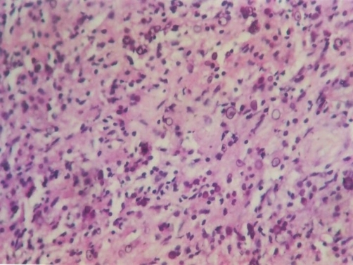
Photomicrograph of Malakoplakia. This specimen reveals intracellular and extracellular Michaelis-Gutmann bodies (hematoxylin-eosin, original magnification ×400).
Patnayak R, Reddy MK, Subramanian S, Jena A, Ravisankar G, Dandu RS. An unusual case of bilateral hydroureteronephrosis caused by uretero-vesico malakoplakia in a young male: a case report and review of the literature. Cases J. 2009;2:7527. doi: 10.1186/1757-1626-2-7527.
References
Triozzi JL, Rodriguez JV, Velagapudi R, Fallahzadeh MK, Binari LA, Paueksakon P, Fogo AB, Concepcion BP. Malakoplakia of the Kidney Transplant. Kidney international reports. 2023 Mar:8(3):680-684. doi: 10.1016/j.ekir.2022.12.016. Epub 2022 Dec 29 [PubMed PMID: 36938075]
Carbone R, Bonometti A, Cassani C, Lucato E, Rossi M, Spinillo A, Paulli M, Cesari S. Bilateral Ovarian Malakoplakia: Case Report and Review of the Literature With Clinical and Diagnostic Considerations. International journal of gynecological pathology : official journal of the International Society of Gynecological Pathologists. 2021 Jan:40(1):60-64. doi: 10.1097/PGP.0000000000000659. Epub [PubMed PMID: 31851062]
Level 2 (mid-level) evidenceXia Z, Du D, Zhang Z, Hu Z, Liu Z. Malakoplakia of urinary bladde: 3 cases reports and review. International urology and nephrology. 2024 Jun:56(6):1779-1784. doi: 10.1007/s11255-023-03920-7. Epub 2024 Jan 18 [PubMed PMID: 38236373]
Level 3 (low-level) evidenceZhang L, Zhang J, Wen J, Yang H, Sun L, Huang T, Sun Y, Jiang W, Tan C. Malakoplakia mimicking malignant rectal carcinoma with bone erosion in a SLE patient: diagnosis insights from a case report and literature review. Frontiers in immunology. 2025:16():1542777. doi: 10.3389/fimmu.2025.1542777. Epub 2025 May 21 [PubMed PMID: 40469298]
Level 3 (low-level) evidenceProthe C, Haouari MA, Adam J, de Parades V. Pelviperineal Malakoplakia. The American journal of gastroenterology. 2026 Jun 1:121(6):1306. doi: 10.14309/ajg.0000000000003860. Epub 2025 Nov 21 [PubMed PMID: 41432498]
Grewal HS, Virk RK, Carroll ME, Benvenuto LJ, Robbins H, Shah L, D'Ovidio F, Arcasoy SM. Malakoplakia Presenting as a Lung Mass in a Lung Transplant Recipient: Case Report. Transplantation proceedings. 2022 Jan-Feb:54(1):173-175. doi: 10.1016/j.transproceed.2021.11.013. Epub 2021 Dec 29 [PubMed PMID: 34973840]
Level 3 (low-level) evidenceRoark SC, Bruett CT, Dominger MG, Freedman PD, Reich RF. Malakoplakia Involving the Maxilla: A Case Report and a Review of the Literature. Head and neck pathology. 2024 Jun 25:18(1):57. doi: 10.1007/s12105-024-01668-9. Epub 2024 Jun 25 [PubMed PMID: 38916759]
Level 3 (low-level) evidenceGerard A, Mesa H, Danziger-Isakov L, Barros KL, Alali M. Successful treatment of malakoplakia of the liver and skin in a pediatric liver transplant patient. Pediatric transplantation. 2023 Aug:27(5):e14492. doi: 10.1111/petr.14492. Epub 2023 Mar 1 [PubMed PMID: 36860187]
Barbu LA, Vasile L, Mogoş GFR, Şurlin V, Vîlcea ID, Cercelaru L, Mogoantă SŞ, Mărgăritescu ND, Ţenea-Cojan TŞ. Appendiceal malakoplakia: a review of the literature and case presentation. Romanian journal of morphology and embryology = Revue roumaine de morphologie et embryologie. 2025 Apr-Jun:66(2):313-321. doi: 10.47162/RJME.66.2.04. Epub [PubMed PMID: 40851241]
Level 3 (low-level) evidenceGuo S, Liu Y, Wang W, Tao J. Prostatic Malakoplakia: A case report. Asian journal of surgery. 2024 Sep 11:():. pii: S1015-9584(24)01976-6. doi: 10.1016/j.asjsur.2024.08.236. Epub 2024 Sep 11 [PubMed PMID: 39266357]
Level 3 (low-level) evidencePrakash A, Noor N, Marchant A, Ghori M, Molani RA. COLONIC MALAKOPLAKIA. Journal of Ayub Medical College, Abbottabad : JAMC. 2024 Apr-Jun:36(2):451-453. doi: 10.55519/JAMC-02-12643. Epub [PubMed PMID: 39610001]
Li Voon Chong J, Ali N. Caecal malakoplakia: a rare mimic of malignancy. BMJ case reports. 2024 Jan 23:17(1):. doi: 10.1136/bcr-2023-257130. Epub 2024 Jan 23 [PubMed PMID: 38262716]
Level 3 (low-level) evidenceSajjanar AB, Vagha S. Malakoplakia of the Gallbladder: A Case Report. Cureus. 2023 May:15(5):e38912. doi: 10.7759/cureus.38912. Epub 2023 May 11 [PubMed PMID: 37313104]
Level 3 (low-level) evidenceAfzal RM, Rangaswamy B, Landau M, John I, Singh H, Phillips AE. Pancreatic Malakoplakia: A Rare Pathology Associated With Acute Pancreatitis. ACG case reports journal. 2023 Apr:10(4):e01042. doi: 10.14309/crj.0000000000001042. Epub 2023 Apr 19 [PubMed PMID: 37091205]
Level 3 (low-level) evidenceAnsari Z, Ahmad A, Bejarano PA, Pinna A, Zervos X. Malakoplakia in a Transplanted Liver. ACG case reports journal. 2024 Sep:11(9):e01486. doi: 10.14309/crj.0000000000001486. Epub 2024 Sep 11 [PubMed PMID: 39262618]
Level 3 (low-level) evidenceWu KY, Chen TC, Wang CJ, Weng CH, Chao A. Uterine Malakoplakia Identified by Diagnostic Hysteroscopy. Journal of minimally invasive gynecology. 2021 May:28(5):924-926. doi: 10.1016/j.jmig.2020.11.009. Epub 2020 Nov 22 [PubMed PMID: 33238208]
Li J, Mi J, Wang J, Zhuo Z. Case report: A rare case of malacoplakia resembling a malignant tumor of the cervix: a case report and review of the literature. Frontiers in medicine. 2024:11():1409239. doi: 10.3389/fmed.2024.1409239. Epub 2024 Jun 4 [PubMed PMID: 38895183]
Level 3 (low-level) evidenceThrishulamurthy CJ, Ahmed HS, Sohail J M, Biligi DS. Clinical Evaluation and Surgical Management of Ocular Malakoplakia of the Caruncle and Periorbital Region: A Report of Two Case and Literature Review. Ophthalmic plastic and reconstructive surgery. 2024 Jul-Aug 01:40(4):e121-e125. doi: 10.1097/IOP.0000000000002643. Epub 2024 Mar 21 [PubMed PMID: 38534076]
Level 3 (low-level) evidenceZeng S, Li S, Yang Q, Tang Q, Tang X, Li M, Huang P. Malakoplakia of Larynx: A Case Report and Literature Review of Localized Malakoplakia of Larynx. Journal of voice : official journal of the Voice Foundation. 2025 Jan:39(1):289.e9-289.e13. doi: 10.1016/j.jvoice.2022.08.011. Epub 2022 Sep 9 [PubMed PMID: 36089557]
Level 3 (low-level) evidenceDominguez D, Eichhorn J, Palot Manzil FF. Retroperitoneal Malakoplakia Mimicking Metastatic Disease. Clinical nuclear medicine. 2025 Jan 1:50(1):75-76. doi: 10.1097/RLU.0000000000005536. Epub 2024 Oct 30 [PubMed PMID: 39480213]
Marchalik R, Marchitto MC, Jedrych J. Cutaneous Malakoplakia of the Right Cheek in an Immunocompromised Man. The American Journal of dermatopathology. 2022 Oct 1:44(10):756-759. doi: 10.1097/DAD.0000000000002256. Epub 2022 Jul 6 [PubMed PMID: 36122335]
Whittington CP, Saleh JS, Schadler E, Bitar C, Wang F, Andea AA. Cutaneous malakoplakia arising on the ankle of a patient with pyoderma gangrenosum. Journal of cutaneous pathology. 2023 Nov:50(11):942-946. doi: 10.1111/cup.14519. Epub 2023 Aug 24 [PubMed PMID: 37615213]
Abbasi AB, Gamino G, Zambeli-Ljepović A, Whelan AM, Roll GR, Altshuler PJ. Malakoplakia among kidney transplant recipients: case series and literature review. Frontiers in immunology. 2025:16():1605146. doi: 10.3389/fimmu.2025.1605146. Epub 2025 May 23 [PubMed PMID: 40486516]
Level 2 (mid-level) evidenceVishwajeet V, Nalwa A, Jangid MK, Choudhary GR, Khera P, Bajpai N, Elhence PA. Renal Allograft Malakoplakia Presenting as a Pseudotumoral Lesion. Saudi journal of kidney diseases and transplantation : an official publication of the Saudi Center for Organ Transplantation, Saudi Arabia. 2023 Jul 1:34(4):371-377. doi: 10.4103/1319-2442.395454. Epub 2024 Feb 12 [PubMed PMID: 38345593]
Meredith T, Dharan N, Killen L, Anandabaskaran S, Kim A, Yang T, Marriott D, Hayward C, Macdonald P. Colonic malakoplakia in a dual stem cell and cardiac transplant recipient: A case report and literature review. Transplant infectious disease : an official journal of the Transplantation Society. 2021 Apr:23(2):e13488. doi: 10.1111/tid.13488. Epub 2020 Oct 22 [PubMed PMID: 33034139]
Level 3 (low-level) evidencePastena D, Giambusso M, Castri F, Potenza AE, Caprino P, Sacchetti F, Sofo L. Malakoplakia of the appendix in a young healthy adult: a case report and literature review. Journal of surgical case reports. 2022 Aug:2022(8):rjac357. doi: 10.1093/jscr/rjac357. Epub 2022 Aug 4 [PubMed PMID: 35935467]
Level 3 (low-level) evidencePolisini G, Delle Fave RF, Capretti C, Marronaro A, Costa AM, Quaresima L, Mazzaferro D, Galosi AB. Malakoplakia of the urinary bladder: A review of the literature. Archivio italiano di urologia, andrologia : organo ufficiale [di] Societa italiana di ecografia urologica e nefrologica. 2022 Sep 27:94(3):350-354. doi: 10.4081/aiua.2022.3.350. Epub 2022 Sep 27 [PubMed PMID: 36165484]
Saleem A, Hussain M, Schechter S, Sturm MB. Colonic malakoplakia. BMJ case reports. 2025 Mar 25:18(3):. pii: e265174. doi: 10.1136/bcr-2025-265174. Epub 2025 Mar 25 [PubMed PMID: 40132942]
Level 3 (low-level) evidenceJeong JW, Noh JH, Kang JH, Park JH, Lee JH. Cecal malakoplakia: A case report. Korean journal of clinical oncology. 2021 Jun:17(1):44-47. doi: 10.14216/kjco.21007. Epub 2021 Jun 30 [PubMed PMID: 36945214]
Level 3 (low-level) evidenceWang Z, Ren J. Clinical Analysis of Renal Failure Caused by Malakoplakia: A Case Report and Literature Review. Frontiers in medicine. 2022:9():770731. doi: 10.3389/fmed.2022.770731. Epub 2022 Mar 3 [PubMed PMID: 35308499]
Level 3 (low-level) evidenceBoualaoui I, El Aboudi A, Mikou MA, El Abidi H, Ouskri S, Mesbahi S, Ibrahimi A, Hachem ES, Yassine N. Malignant masquerade: A case report of urachal malakoplakia disguised as urachal tumor. Urology case reports. 2025 May:60():103012. doi: 10.1016/j.eucr.2025.103012. Epub 2025 Mar 18 [PubMed PMID: 40276710]
Level 3 (low-level) evidenceKinsella PM, Smibert OC, Whitlam JB, Steven M, Masia R, Gandhi RG, Kotton CN, Holmes NE. Successful use of azithromycin for Escherichia coli-associated renal allograft malakoplakia: a report of two cases. European journal of clinical microbiology & infectious diseases : official publication of the European Society of Clinical Microbiology. 2021 Dec:40(12):2627-2631. doi: 10.1007/s10096-021-04270-x. Epub 2021 May 14 [PubMed PMID: 33990891]
Level 3 (low-level) evidence