 Tools
Tools
 Discoid Lupus Erythematosus
Introduction
Lupus erythematosus is a multisystem disorder that predominantly affects the skin. There are many types of lupus, including systemic lupus erythematosus (SLE), drug-induced lupus erythematosus, neonatal lupus erythematosus, and cutaneous lupus erythematosus. The most common types of cutaneous lupus erythematosus include acute cutaneous lupus erythematosus, subacute cutaneous lupus erythematosus, and chronic cutaneous lupus erythematosus; discoid lupus erythematosus is a subtype of chronic cutaneous lupus erythematosus. Dr James Gilliam described the most commonly used classification of cutaneous lesions in lupus erythematosus. He distinguished lesions as either specific or nonspecific, based on the presence of interface dermatitis on histopathologic examination. Within the category of specific cutaneous lesions, he subdivided these into acute cutaneous lupus erythematosus, subacute cutaneous lupus erythematosus, and chronic cutaneous erythematosus.[1][2]
Discoid lupus erythematosus is the most common subset of chronic cutaneous lupus erythematosus. Patients may or may not report photosensitivity, but lesions are frequently photodistributed and often exhibit secondary atrophy or scarring. Most patients with discoid lupus erythematosus do not have significant systemic disease. Discoid lupus erythematosus can also occur as a manifestation of SLE in approximately 20% of patients. Other less common variants of chronic cutaneous lupus erythematosus include hypertrophic lupus erythematosus, tumid lupus erythematosus, lupus erythematosus panniculitis (also known as lupus profundus), chilblain lupus erythematosus, oral discoid lupus erythematosus, and discoid lupus erythematosus lesions on the palms and soles.[3][4]
Etiology
Register For Free And Read The Full Article
 Search engine and full access to all medical articles
- 10 free questions in your specialty
- Free CME/CE Activities
- Free daily question in your email
- Save favorite articles to your dashboard
- Emails offering discounts
Learn more about a Subscription to StatPearls Point-of-Care
Etiology
Lupus erythematosus is an inflammatory connective tissue disease characterized by generalized autoimmunity, pathogenic autoantibodies, and immune complexes, attributable to loss of immune tolerance. For discoid lupus erythematosus without associated SLE (chronic discoid lupus erythematosus), the evidence does not show whether circulating inflammatory cells and autoantibodies are involved in the pathogenesis. Still, it is evident that the cutaneous inflammatory infiltrates are dominated by Th1, rather than Th17, cells, in contrast to SLE.[5][6]
Epidemiology
Lupus can occur in all age groups, but discoid lupus erythematosus occurs more frequently in women in their fourth and fifth decades of life. Approximately 25% of patients with SLE may develop typical discoid lesions at some point during their illness, and 1% to 5% of patients with discoid lupus may develop SLE.
Ethnicity is also a major risk factor for developing LE, and its effect in some populations is almost as strong as that of gender. SLE prevalence is 4-fold higher in African-American women than in White American women (4 in 1000 versus 1 in 1000). In addition, African Americans tend to develop the disease at an earlier age and have a higher mortality rate.[7][8]
Pathophysiology
The pathogenesis of cutaneous lupus erythematosus is multifactorial, with an interplay between genetic and environmental factors. Some contributing environmental factors include UV radiation, medications, cigarette smoking, and, possibly, viruses. The interaction among these multiple factors triggers an inflammatory cascade of cytokines, chemokines, and inflammatory cell responses. Genes previously associated with SLE, including TYK2, IRF5, and CTLA4, confer an increased risk of developing discoid lupus erythematosus.
An analysis of 405 patients by Bockle et al found that smoking is highly associated with discoid lupus erythematosus. Bockle et al hypothesized that smoking may play a pathogenic role in cutaneous lupus erythematosus variants, including discoid lupus erythematosus and tumid lupus, by inducing apoptosis, stimulating T-cell proliferation, and increasing photosensitivity. Another explanation is that smoking induces DNA damage, leading to the formation of DNA adducts and the production of double-stranded DNA antibodies. Keratinocytes may also contribute to lupus skin damage by increasing the apoptotic rate and the production of proinflammatory cytokines, such as interferon-alpha and interleukin-6 for SLE and interferon-lambda for discoid lupus erythematosus.[9][10]
Histopathology
The histopathologic findings in cutaneous lupus vary by subtype. Overlap is evident in the histologic findings across the various clinical phenotypes, particularly acute cutaneous lupus erythematosus, subacute cutaneous lupus erythematosus, and discoid lesions. In cutaneous lupus erythematosus, basal cell damage (also referred to as vacuolar degeneration, hydropic change, or interface dermatitis) and lymphohistiocytic inflammatory infiltrates are commonly observed. In discoid lupus lesions, periadnexal inflammation, follicular plugging, and hyperkeratosis are typically observed in addition to interface dermatitis.[11] Dermal mucinosis, along with a thickened basement membrane, is also frequently noted.
Direct immunofluorescence (DIF) is the examination of the skin for deposits of immunoreactants. DIF of lesional skin can be useful for establishing a diagnosis of cutaneous lupus erythematosus when routine histopathology is equivocal. DIF does not replace routine histologic staining as the method of choice for establishing a diagnosis. In active lesions of discoid lupus erythematosus, DIF of lesional skin is positive in most cases. Antibody deposition at the dermal-epidermal junction and around hair follicles is the most characteristic DIF finding in cutaneous lupus erythematosus. These deposits are typically granular and composed primarily of immunoglobulin G and immunoglobulin M.[12][13]
The lupus band test (LBT) is a diagnostic procedure used to detect immunoglobulin and complement deposits along the dermo-epidermal junction in patients with lupus erythematosus. LBT can help distinguish systemic lupus erythematosus from chronic lupus erythematosus because, in patients with SLE, the LBT is frequently positive in both uninvolved and involved skin. In contrast, in patients with cutaneous lupus erythematosus, only the involved skin is positive.[14][15] The LBT is positive on the lesional skin in 75% of patients. Ideal lesions for LBT for discoid lupus erythematosus are on the head and neck that have been present for at least a few months.
History and Physical
Discoid lupus erythematosus is the most common form of chronic cutaneous erythematosus and can occur as a localized form (80%) with lesions on the face, ears, and scalp or as disseminated discoid lupus erythematosus (20%) with lesions above and below the neck. The disseminated form of discoid lupus erythematosus, especially when involving the trunk, is associated with an increased risk of progression to SLE up to 28%.[16]
Discoid lesions rarely present below the neck without concurrent involvement above the neck. Occasionally, discoid lesions develop on mucosal surfaces, including the lips, nasal mucosa, conjunctivae, and genital mucosa.[5] Some patients with discoid lesions exhibit a photodistribution. Sun exposure appears to play a role in lesion development. However, patients can have discoid lesions on the sun-protected skin, and there is no clear association between sun exposure and their development.
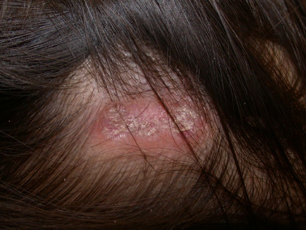
The first morphological sign of discoid lupus erythematosus is a well-defined, annular erythematous patch or plaque of varying size, followed by follicular hyperkeratosis, which is adherent to the skin. By removing the adherent scale, follicle-sized keratotic spikes similar to carpet tacks can be observed (carpet tack sign).[17] The lesions slowly expand with active inflammation and hyperpigmentation at the periphery, leaving depressed central atrophy, scarring, telangiectasia, and hypopigmentation.[18] Discoid lupus erythematosus can progress to irreversible scarring alopecia on the scalp. Although uncommon, a squamous cell carcinoma can develop in a longstanding discoid lesion at 2% to 3% and is often associated with a poor prognosis.[19]
Patients who present with discoid lesions may have associated arthralgias, but, over time, only approximately 10% to 20% of these patients eventually meet the classification criteria for SLE. Hyperkeratotic, verrucous plaques are noted in hypertrophic discoid lupus erythematosus, a rare entity of discoid lupus erythematosus.[20]
Evaluation
In evaluating discoid lupus erythematosus, the dermatologist should obtain a detailed history and perform a thorough cutaneous examination, looking for signs of systemic disease. The diagnosis of discoid lupus erythematosus is primarily based on clinical features, although histological examination may be necessary to confirm the diagnosis.[21][22]
The initial step in diagnosis involves an antinuclear antibody titer, which is highly sensitive (95%-100%) but not specific. Autoantibodies to SSA/Ro, SSB/La, U1RNP, histones, and ssDNA are common in patients with SLE but are not disease-specific. No other specific autoantibodies are routinely used in practice to differentiate the subtypes of cutaneous lupus erythematosus. A further possible target of auto-antibodies is annexin 1, which has been suggested to play a crucial role in preventing autoimmune diseases. A recent study found a significantly higher level of anti-annexin 1 antibodies in patients with discoid lupus erythematosus, suggesting that anti-annexin 1 antibodies may be a new diagnostic marker for discoid lupus erythematosus. Anti-annexin 1 antibody levels in the serum did not correlate with disease activity.[23]
Treatment / Management
Early treatment of discoid lupus erythematosus lesions may result in complete clearance, whereas treatment failure results in permanent scarring. Hair loss, depressed scars, and pigmentary changes are often disfiguring, particularly in darker-skinned people. Some general measures, such as sun avoidance, avoidance of photosensitizing medications, and liberal application of sunscreen, are encouraged because cutaneous lesions are known to be exacerbated by sunlight.[24] Smoking cessation is encouraged, as smoking can increase discoid lupus erythematosus disease activity. Studies demonstrate a statistically significant decrease in the efficacy of antimalarial medication in individuals who have currently or ever smoked.[25][26][27]
Current first-line treatment for discoid lupus erythematosus consists of photoprotection in conjunction with topical or intralesional corticosteroids and topical calcineurin inhibitors.[28] Chronic discoid lupus erythematosus lesions that are not responsive to topical therapy or have a widespread disease involvement are candidates for systemic therapy. When discoid lupus erythematosus is refractory to these measures, other agents with varying degrees of proven efficacy are used. Currently, no medications have been approved specifically, and many of the drugs described in the literature were developed for use in other autoimmune disorders.(A1)
Acute exacerbations of discoid lupus erythematosus are treated with the application of a super-high or high-potency topical corticosteroid.[29] Clinical improvement is typically observed within 2 weeks of therapy. Treatment can be stopped when lesions are free of erythema or scale, indicating disease inactivity. Cutaneous atrophy is the major adverse effect associated with chronic use of topical corticosteroids. If lesions are refractory to high-potency topical corticosteroid use for 2 to 4 weeks, alternative therapy with intralesional corticosteroids or topical calcineurin inhibitors should be initiated. Topical calcineurin inhibitors such as tacrolimus 0.1% or 0.03% ointment and pimecrolimus 1% cream are most commonly used for facial lesions, as they do not cause atrophy.[30] However, these inhibitors are more expensive and have a slow onset of action. Clinical improvement is typically noted within 4 weeks of therapy. Low-potency topical corticosteroids or topical calcineurin inhibitors are typically used to maintain the discoid lupus erythematosus lesions in remission once the acute flare resolves.[31] Burning sensation after application that resolves within 1 to 2 weeks is the most common adverse effect of topical calcineurin inhibitors. Topical lesions that do not respond to the above therapy are treated with intralesional triamcinolone acetonide at a dose of 0.1 mL (3-5 mg/mL). Multiple injections into the lesion 1 cm apart should be given. Injections can be repeated every 3 to 4 weeks. If there is no clinical improvement after 2 to 3 injections, alternative treatment should be initiated. Common adverse effects of intralesional corticosteroids include dyspigmentation and skin atrophy.(A1)
Antimalarials are immunotherapeutic and are considered first-line systemic therapy for cutaneous lupus erythematosus. Hydroxychloroquine and chloroquine with or without quinacrine are currently used in the treatment of discoid lupus erythematosus. Hydroxychloroquine is preferred over chloroquine due to the lower risk of adverse effects, specifically retinal toxicity. Quinacrine is known to cause hematological abnormalities. Hydroxychloroquine is typically started at 200 mg/d and increased to 200 mg/d twice within 1 to 2 weeks if there are no adverse effects. The maximum dose of hydroxychloroquine should not exceed 5 mg/kg of real body weight per day, given its association with retinal toxicity.[32] Around 6 to 8 weeks of therapy are required to assess the effectiveness. Quinacrine at a dose of 100 mg/d is added to hydroxychloroquine if no clinical improvement is noted with hydroxychloroquine alone.[33] The chloroquine dose should not exceed 2.3 mg/kg of real body weight per day due to the risk of retinopathy.[32] Hydroxychloroquine and chloroquine should not be used together due to cumulative retinal toxicity. Patients should undergo baseline and periodic eye examinations during therapy. Blue-gray discoloration of the face, shins, and palate, and bleaching of light-colored hair may occur. Quinacrine use may lead to generalized yellow pigmentation of the skin, secretions, and sclera. (B2)
Other therapeutic options, such as retinoids, vitamin A analogs with anti-keratinizing and anti-inflammatory effects, are sometimes used in cutaneous lupus erythematosus, but documentation in the literature is limited. Topical retinoids such as tretinoin 0.05% cream or tazarotene 0.05% gel are used to treat hypertrophic discoid lupus erythematosus.[34] Topical retinoids are pregnancy category X drugs, and cutaneous irritation is a commonly observed adverse effect. Immunomodulators and Immunosuppressive agents such as methotrexate, systemic retinoids, dapsone, mycophenolate mofetil, azathioprine, intravenous immune globulin (IVIG), cyclophosphamide, and cyclosporine have also been used in the management of discoid lupus erythematosus. These therapies are generally considered second-line options for patients whose disease is refractory to first-line treatments.[35](B3)
Methotrexate at a dose range of 10 to 25 mg per week is used to treat discoid lupus erythematosus lesions. Clinical improvement is noted within 2 to 4 weeks of use, with the resolution of the lesions in about 6 to 8 weeks.[36] A dose of 5 to 10 mg of methotrexate is given as a test dose to assess if the patient can tolerate the drug. Liver function tests and a complete blood count are performed 5 to 6 days after drug administration to assess for adverse effects or intolerance to the medication. The most common adverse effects include gastrointestinal upset, hepatotoxicity, nephrotoxicity, teratogenicity, pulmonary fibrosis, bone marrow suppression, and oral ulcers. Daily folic acid supplementation may minimize gastrointestinal adverse effects.[37] Mycophenolate mofetil is administered in doses of 1 to 3 gm/d. Gastrointestinal upset, cytopenias, and teratogenicity are some of the adverse effects of mycophenolate mofetil.(A1)
Thalidomide, a potent teratogen, has been used in the treatment of discoid lupus erythematosus. In an early study of 60 patients receiving 50 to 100 mg per day, complete or marked regression was observed in 54 individuals (90%), with disease relapse in 71% of individuals with medication discontinuation.[38] Adverse effects apart from teratogenicity include drowsiness, constipation, rash, edema, xeroderma, peripheral neuropathy, and thromboembolism.[39] Lenalidomide is a thalidomide analog that may also prove useful in the treatment of discoid lupus erythematosus. Evidence suggests lenalidomide effectively treats discoid lupus erythematosus and has a less severe adverse effect profile than thalidomide, but may be similarly limited by a tendency to relapse once discontinued. IVIG is given at a 2 g/kg dose and repeated every 4 to 8 weeks.[40] IVIG is expensive, and cutaneous lupus erythematosus lesions tend to recur with its use. Adverse effects of IVIG therapy include headache, renal failure, hypersensitivity reactions, thrombosis, vasculitis, and aseptic meningitis.(A1)
Differential Diagnosis
Differential diagnoses of discoid lupus erythematosus include granuloma faciale, tinea faciei, cutaneous tuberculosis, cutaneous leishmaniasis, lymphoproliferative disorders, and sarcoidosis. Discoid lupus erythematosus–induced scarring alopecia can be confused with lichen planopilaris, tinea capitis, and central centrifugal cicatricial alopecia. Hypertrophic discoid lupus erythematosus may be confused with keratoacanthoma, squamous cell cancer, hypertrophic lichen planus, and prurigo nodularis.
Prognosis
Discoid lupus erythematosus lesions heal with scarring, atrophy, and dyspigmentation, causing more morbidity than mortality. Psychological functioning is affected by discoid lupus erythematosus lesions.[41] Exacerbations are associated with sun exposure and often occur in spring and summer. Localized discoid lupus erythematosus involving the scalp may lead to cicatricial alopecia. Generalized discoid lupus erythematosus is associated with the risk of progression to SLE in about 28%.[16] Chronic discoid lupus erythematosus lesions in about 2% to 3% can progress to squamous cell carcinoma, which is typically associated with a poor prognosis.[19] Early detection and treatment of discoid lupus erythematosus lesions can minimize the associated morbidity.
Complications
Complications associated with discoid lupus erythematosus and its progression to SLE include the following
- Cicatricial alopecia
- Pancytopenia
- Thromboembolism
- Arthritis
- Myositis
- Hypertension
- Renal failure
- Neuropsychiatric manifestations such as seizures and depression
- Pleuropericarditis
- Pancreatitis and mesenteric vasculitis
- Optic neuritis
- Therapy-related adverse effects
- Vitamin D deficiency
Regular monitoring of discoid lupus erythematosus lesions and therapy-related adverse effects is crucial to prevent complications and promote clinical resolution.
Deterrence and Patient Education
Patients should be educated about photoprotection and the possibility of developing skin lesions up to 3 weeks after sun exposure.[42] Recommended measures include wearing protective clothing and wide-brimmed hats, avoiding sun exposure during peak hours (10:00 AM to 2:00 PM), applying sunscreen with a sun protection factor of at least 30 against both UVA and UVB radiation, avoiding photosensitizing medications, and avoiding outdoor occupations. Cessation of smoking and avoidance of alcohol are encouraged. Educating patients that nicotine interferes with antimalarial therapy uptake and hastens their metabolism helps them understand the rationale behind tobacco cessation.[43] Smoking cessation without nicotine replacement therapy is ideal. Patients may be at risk for vitamin D deficiency due to limited sun exposure and regular sunscreen use; therefore, annual screening for vitamin D levels is recommended.[44]
Enhancing Healthcare Team Outcomes
Lupus erythematosus is a complex autoimmune disorder affecting multiple organ systems, with cutaneous manifestations—such as discoid lupus erythematosus—often serving as an early indicator of disease. Discoid lupus erythematosus presents with well-defined erythematous plaques that may progress to scarring, dyspigmentation, and alopecia, and a subset of patients may develop SLE. Disease activity is influenced by genetic susceptibility, immune dysregulation, ultraviolet radiation, smoking, and other environmental triggers. Early recognition, accurate diagnosis, and timely initiation of evidence-based topical, intralesional, or systemic therapies are essential to minimize complications and improve patient outcomes. Patient education regarding photoprotection, smoking cessation, medication adherence, and lifestyle modifications plays a critical role in long-term disease management.[45][46]
Effective management of lupus requires a coordinated interprofessional approach. Clinicians, advanced practitioners, and general practitioners are responsible for diagnosis, treatment selection, and monitoring for systemic involvement. Nurses provide education on symptom recognition, medication compliance, and lifestyle modifications. Pharmacists ensure safe medication use, monitor for adverse effects, and support adherence. Mental health counselors and social workers assist patients in coping with chronic disease stress, joining support networks, and addressing psychosocial challenges.[47][48] Therapists contribute to maintaining mobility and quality of life. Clear communication and collaborative care planning enhance patient-centered care, improve outcomes, reduce safety risks, and optimize team performance across all levels of lupus management.
Media
(Click Image to Enlarge)
(Click Image to Enlarge)
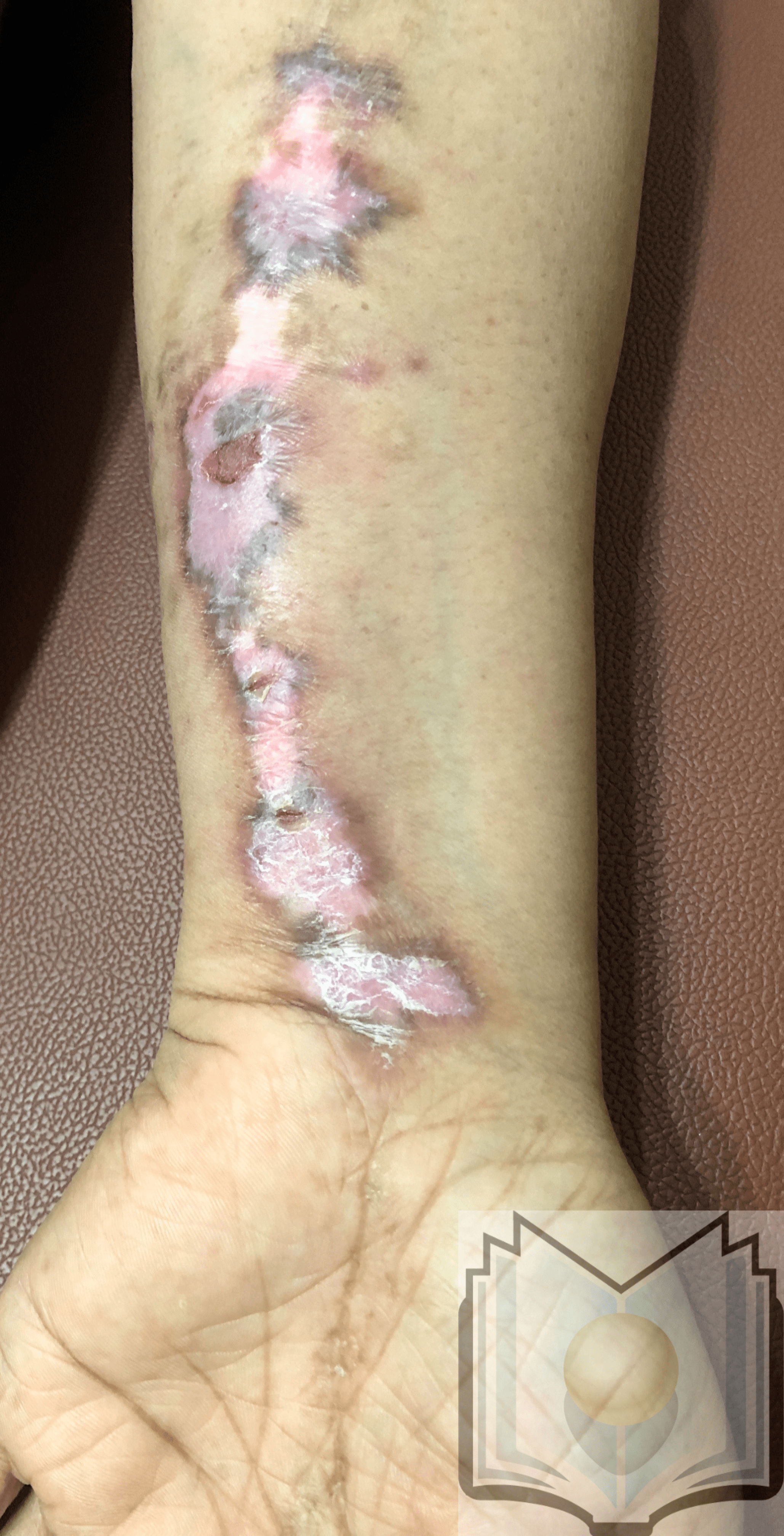
Linear Discoid Lupus Erythematosus Contributed by Dr. Shyam Verma, MBBS, DVD, FRCP, FAAD, Vadodara, India
References
Udompanich S, Chanprapaph K, Suchonwanit P. Hair and Scalp Changes in Cutaneous and Systemic Lupus Erythematosus. American journal of clinical dermatology. 2018 Oct:19(5):679-694. doi: 10.1007/s40257-018-0363-8. Epub [PubMed PMID: 29948959]
Dammacco R, Procaccio P, Racanelli V, Vacca A, Dammacco F. Ocular Involvement in Systemic Lupus Erythematosus: The Experience of Two Tertiary Referral Centers. Ocular immunology and inflammation. 2018:26(8):1154-1165. doi: 10.1080/09273948.2018.1501495. Epub 2018 Aug 10 [PubMed PMID: 30096011]
Abadías-Granado I, Sánchez-Bernal J, Felipo-Berlanga F, Ara-Martín M. Coexistence of Tumid Lupus Erythematosus and Discoid Lupus Erythematosus. Actas dermo-sifiliograficas. 2019 Apr:110(3):253-255. doi: 10.1016/j.ad.2018.02.023. Epub 2018 Jul 30 [PubMed PMID: 30072022]
Sontheimer RD. The lexicon of cutaneous lupus erythematosus--a review and personal perspective on the nomenclature and classification of the cutaneous manifestations of lupus erythematosus. Lupus. 1997:6(2):84-95 [PubMed PMID: 9061656]
Level 3 (low-level) evidenceSalah E. Clinical and dermoscopic spectrum of discoid lupus erythematosus: novel observations from lips and oral mucosa. International journal of dermatology. 2018 Jul:57(7):830-836. doi: 10.1111/ijd.14015. Epub 2018 Apr 27 [PubMed PMID: 29700807]
Drenkard C, Parker S, Aspey LD, Gordon C, Helmick CG, Bao G, Lim SS. Racial Disparities in the Incidence of Primary Chronic Cutaneous Lupus Erythematosus in the Southeastern US: The Georgia Lupus Registry. Arthritis care & research. 2019 Jan:71(1):95-103. doi: 10.1002/acr.23578. Epub [PubMed PMID: 29669194]
Gaüzère L, Gerber A, Renou F, Ferrandiz D, Bagny K, Osdoit S, Yvin JL, Raffray L. [Epidemiology of systemic lupus erythematosus in Reunion Island, Indian Ocean: A case-series in adult patients from a University Hospital]. La Revue de medecine interne. 2019 Apr:40(4):214-219. doi: 10.1016/j.revmed.2018.07.004. Epub 2018 Jul 21 [PubMed PMID: 30041816]
Level 3 (low-level) evidenceOh EH, Kim EJ, Ro YS, Ko JY. Ten-year retrospective clinicohistological study of cutaneous lupus erythematosus in Korea. The Journal of dermatology. 2018 Apr:45(4):436-443. doi: 10.1111/1346-8138.14233. Epub 2018 Feb 9 [PubMed PMID: 29423919]
Level 2 (mid-level) evidenceKahn JS, Deverapalli SC, Rosmarin DM. JAK-STAT signaling pathway inhibition: a role for treatment of discoid lupus erythematosus and dermatomyositis. International journal of dermatology. 2018 Aug:57(8):1007-1014. doi: 10.1111/ijd.14064. Epub 2018 Jun 5 [PubMed PMID: 29873082]
Two A, So JK, Paravar T. Discoid Lupus and Human Immunodeficiency Virus: A Retrospective Chart Review to Determine the Prevalence and Progression of Co-occurrence of these Conditions at a Single Academic Center. Indian journal of dermatology. 2017 Mar-Apr:62(2):226. doi: 10.4103/0019-5154.201750. Epub [PubMed PMID: 28400654]
Level 2 (mid-level) evidencePatel P, Werth V. Cutaneous lupus erythematosus: a review. Dermatologic clinics. 2002 Jul:20(3):373-85, v [PubMed PMID: 12170873]
Level 3 (low-level) evidenceMutasim DF, Adams BB. Immunofluorescence in dermatology. Journal of the American Academy of Dermatology. 2001 Dec:45(6):803-22; quiz 822-4 [PubMed PMID: 11712024]
Dahl MV. Usefulness of direct immunofluorescence in patients with lupus erythematosus. Archives of dermatology. 1983 Dec:119(12):1010-7 [PubMed PMID: 6360051]
Harrist TJ, Mihm MC Jr. The specificity and clinical usefulness of the lupus band test. Arthritis and rheumatism. 1980 Apr:23(4):479-90 [PubMed PMID: 6989372]
George R, Kurian S, Jacob M, Thomas K. Diagnostic evaluation of the lupus band test in discoid and systemic lupus erythematosus. International journal of dermatology. 1995 Mar:34(3):170-3 [PubMed PMID: 7751090]
Level 2 (mid-level) evidenceChong BF, Song J, Olsen NJ. Determining risk factors for developing systemic lupus erythematosus in patients with discoid lupus erythematosus. The British journal of dermatology. 2012 Jan:166(1):29-35. doi: 10.1111/j.1365-2133.2011.10610.x. Epub 2011 Dec 5 [PubMed PMID: 21910708]
Inamadar AC. Perforation of paper with pen: Simple technique to explain the carpet tack sign in discoid lupus erythematosus. Journal of the American Academy of Dermatology. 2019 Dec:81(6):e159-e160. doi: 10.1016/j.jaad.2019.03.039. Epub 2019 Mar 22 [PubMed PMID: 30905795]
Rowell NR. Laboratory abnormalities in the diagnosis and management of lupus erythematosus. The British journal of dermatology. 1971 Mar:84(3):210-6 [PubMed PMID: 4102091]
Zaalberg A, Moradi Tuchayi S, Ameri AH, Ngo KH, Cunningham TJ, Eliane JP, Livneh M, Horn TD, Rosman IS, Musiek A, Anadkat MJ, Demehri S. Chronic Inflammation Promotes Skin Carcinogenesis in Cancer-Prone Discoid Lupus Erythematosus. The Journal of investigative dermatology. 2019 Jan:139(1):62-70. doi: 10.1016/j.jid.2018.06.185. Epub 2018 Jul 17 [PubMed PMID: 30030152]
Farley-Loftus R, Elmariah SB, Ralston J, Kamino H, Franks AG Jr. Hypertrophic discoid lupus erythematosus. Dermatology online journal. 2010 Nov 15:16(11):1 [PubMed PMID: 21163152]
Level 3 (low-level) evidenceFlynn A, Gilhooley E, O'Shea F, Wynne B. The use of SLICC and ACR criteria to correctly label patients with cutaneous lupus and systemic lupus erythematosus. Clinical rheumatology. 2018 Mar:37(3):817-818. doi: 10.1007/s10067-018-3999-0. Epub 2018 Feb 1 [PubMed PMID: 29392510]
Xie HH, Shen H, Zhang L, Cui MY, Xia LP, Lu J. Elevated Serum Interleukin-34 Level in Patients with Systemic Lupus Erythematosus Is Associated with Disease Activity. Scientific reports. 2018 Feb 22:8(1):3462. doi: 10.1038/s41598-018-21859-z. Epub 2018 Feb 22 [PubMed PMID: 29472590]
Kretz CC, Norpo M, Abeler-Dörner L, Linke B, Haust M, Edler L, Krammer PH, Kuhn A. Anti-annexin 1 antibodies: a new diagnostic marker in the serum of patients with discoid lupus erythematosus. Experimental dermatology. 2010 Oct:19(10):919-21. doi: 10.1111/j.1600-0625.2010.01145.x. Epub [PubMed PMID: 20701627]
Level 3 (low-level) evidenceCallen JP. Discoid lupus erythematosus--variants and clinical associations. Clinics in dermatology. 1985 Jul-Sep:3(3):49-57 [PubMed PMID: 3880023]
Muangchan C, van Vollenhoven RF, Bernatsky SR, Smith CD, Hudson M, Inanç M, Rothfield NF, Nash PT, Furie RA, Senécal JL, Chandran V, Burgos-Vargas R, Ramsey-Goldman R, Pope JE. Treatment Algorithms in Systemic Lupus Erythematosus. Arthritis care & research. 2015 Sep:67(9):1237-1245. doi: 10.1002/acr.22589. Epub [PubMed PMID: 25777803]
Powers DB. Systemic lupus erythematosus and discoid lupus erythematosus. Oral and maxillofacial surgery clinics of North America. 2008 Nov:20(4):651-62. doi: 10.1016/j.coms.2008.07.001. Epub [PubMed PMID: 18940631]
Francès C, Barète S, Piette JC. [Dermatologic manifestations in lupus erythematosus]. La Revue de medecine interne. 2008 Sep:29(9):701-9. doi: 10.1016/j.revmed.2008.04.021. Epub 2008 Jun 17 [PubMed PMID: 18562046]
Bjornberg A, Hellgren L. Topical treatment of chronic discoid lupus erythematosus with betamethason-17-valerate and fluocinolone acetonie-a double blind study. Indian journal of dermatology. 1966 Oct:12(1):17-8 [PubMed PMID: 5341750]
Level 1 (high-level) evidenceJessop S, Whitelaw DA, Delamere FM. Drugs for discoid lupus erythematosus. The Cochrane database of systematic reviews. 2009 Oct 7:(4):CD002954. doi: 10.1002/14651858.CD002954.pub2. Epub 2009 Oct 7 [PubMed PMID: 19821298]
Level 1 (high-level) evidenceTzellos TG, Kouvelas D. Topical tacrolimus and pimecrolimus in the treatment of cutaneous lupus erythematosus: an evidence-based evaluation. European journal of clinical pharmacology. 2008 Apr:64(4):337-41 [PubMed PMID: 18157526]
Level 1 (high-level) evidenceSárdy M, Ruzicka T, Kuhn A. Topical calcineurin inhibitors in cutaneous lupus erythematosus. Archives of dermatological research. 2009 Jan:301(1):93-8. doi: 10.1007/s00403-008-0894-6. Epub 2008 Sep 17 [PubMed PMID: 18797893]
Level 3 (low-level) evidenceMarmor MF, Kellner U, Lai TY, Melles RB, Mieler WF, American Academy of Ophthalmology. Recommendations on Screening for Chloroquine and Hydroxychloroquine Retinopathy (2016 Revision). Ophthalmology. 2016 Jun:123(6):1386-94. doi: 10.1016/j.ophtha.2016.01.058. Epub 2016 Mar 16 [PubMed PMID: 26992838]
Cavazzana I, Sala R, Bazzani C, Ceribelli A, Zane C, Cattaneo R, Tincani A, Calzavara-Pinton PG, Franceschini F. Treatment of lupus skin involvement with quinacrine and hydroxychloroquine. Lupus. 2009 Jul:18(8):735-9. doi: 10.1177/0961203308101714. Epub [PubMed PMID: 19502270]
Level 2 (mid-level) evidenceSeiger E, Roland S, Goldman S. Cutaneous lupus treated with topical tretinoin: a case report. Cutis. 1991 May:47(5):351-5 [PubMed PMID: 2070657]
Level 3 (low-level) evidenceKuhn A, Ruland V, Bonsmann G. Cutaneous lupus erythematosus: update of therapeutic options part II. Journal of the American Academy of Dermatology. 2011 Dec:65(6):e195-213. doi: 10.1016/j.jaad.2010.06.017. Epub 2010 Aug 30 [PubMed PMID: 20800319]
Wenzel J, Brähler S, Bauer R, Bieber T, Tüting T. Efficacy and safety of methotrexate in recalcitrant cutaneous lupus erythematosus: results of a retrospective study in 43 patients. The British journal of dermatology. 2005 Jul:153(1):157-62 [PubMed PMID: 16029342]
Level 2 (mid-level) evidenceOrtiz Z, Shea B, Suarez-Almazor ME, Moher D, Wells GA, Tugwell P. The efficacy of folic acid and folinic acid in reducing methotrexate gastrointestinal toxicity in rheumatoid arthritis. A metaanalysis of randomized controlled trials. The Journal of rheumatology. 1998 Jan:25(1):36-43 [PubMed PMID: 9458200]
Level 1 (high-level) evidencePelle MT, Werth VP. Thalidomide in cutaneous lupus erythematosus. American journal of clinical dermatology. 2003:4(6):379-87 [PubMed PMID: 12762830]
Chasset F, Tounsi T, Cesbron E, Barbaud A, Francès C, Arnaud L. Efficacy and tolerance profile of thalidomide in cutaneous lupus erythematosus: A systematic review and meta-analysis. Journal of the American Academy of Dermatology. 2018 Feb:78(2):342-350.e4. doi: 10.1016/j.jaad.2017.09.059. Epub 2017 Oct 5 [PubMed PMID: 28989111]
Level 1 (high-level) evidenceGénéreau T, Chosidow O, Danel C, Chérin P, Herson S. High-dose intravenous immunoglobulin in cutaneous lupus erythematosus. Archives of dermatology. 1999 Sep:135(9):1124-5 [PubMed PMID: 10490126]
Level 3 (low-level) evidenceChen P, Broadbent E, Coomarasamy C, Jarrett P. Illness perception in association with psychological functioning in patients with discoid lupus erythematosus. The British journal of dermatology. 2015 Sep:173(3):824-6. doi: 10.1111/bjd.13709. Epub 2015 Jul 20 [PubMed PMID: 25639859]
Level 3 (low-level) evidenceKuhn A, Sonntag M, Richter-Hintz D, Oslislo C, Megahed M, Ruzicka T, Lehmann P. Phototesting in lupus erythematosus: a 15-year experience. Journal of the American Academy of Dermatology. 2001 Jul:45(1):86-95 [PubMed PMID: 11423840]
Wozniacka A, Carter A, McCauliffe DP. Antimalarials in cutaneous lupus erythematosus: mechanisms of therapeutic benefit. Lupus. 2002:11(2):71-81 [PubMed PMID: 11958581]
Cusack C, Danby C, Fallon JC, Ho WL, Murray B, Brady J, O'Kelly P, Ambrose N, Kearns G, Murphy GM. Photoprotective behaviour and sunscreen use: impact on vitamin D levels in cutaneous lupus erythematosus. Photodermatology, photoimmunology & photomedicine. 2008 Oct:24(5):260-7. doi: 10.1111/j.1600-0781.2008.00373.x. Epub [PubMed PMID: 18811868]
Jessop S, Whitelaw DA, Grainge MJ, Jayasekera P. Drugs for discoid lupus erythematosus. The Cochrane database of systematic reviews. 2017 May 5:5(5):CD002954. doi: 10.1002/14651858.CD002954.pub3. Epub 2017 May 5 [PubMed PMID: 28476075]
Level 1 (high-level) evidenceDrucker AM, Su J, Mussani F, Siddha SK, Gladman DD, Urowitz MB. Prognostic implications of active discoid lupus erythematosus and malar rash at the time of diagnosis of systemic lupus erythematosus: Results from a prospective cohort study. Lupus. 2016 Apr:25(4):376-81. doi: 10.1177/0961203315610645. Epub 2015 Oct 8 [PubMed PMID: 26453664]
Kuhn A, Landmann A, Wenzel J. Advances in the treatment of cutaneous lupus erythematosus. Lupus. 2016 Jul:25(8):830-7. doi: 10.1177/0961203316641771. Epub [PubMed PMID: 27252259]
Level 3 (low-level) evidenceMeller S, Homey B, Ruzicka T. Socioeconomic factors in lupus erythematosus. Autoimmunity reviews. 2005 Apr:4(4):242-6 [PubMed PMID: 15893719]
Pincus D, Saccar CV Lead poisoning. American family physician. 1979 Jun; [PubMed PMID: 110123]