 Tools
Tools
 Lung Decortication
Introduction
Approximately 1 million patients are hospitalized in the United States each year with pneumonia. Of those hospitalized for pneumonia, 20% to 40% will develop a parapneumonic effusion, and 5% to 10% of these parapneumonic effusions will progress to an empyema (approximately 32,000 patients per year in the United States). Approximately 15% of these patients with empyema die, and 30% require an operation on their chest to clear the infection.[1][2]
Lung decortication is a well-established procedure that Delorme first performed in 1895 to treat empyema.[3] This procedure is primarily indicated in chronic empyema thoracis (pyogenic or tuberculous), hemothorax, pleural thickening, and other similar conditions. Decortication involves excision the restrictive layer of the thick fibrinous peel overlying the lung, chest wall, and diaphragm. The decortication extends over the visceral pleura covering the lung surfaces, including the fissures, along the parietal pleura of the chest wall and diaphragm.
Notably, the mediastinal surface of the lung is generally spared, except where adhesions can be safely released without risk to vital mediastinal structures. This thick fibrinous peel results from the ingrowth of the fibroblasts during the advanced stages of empyema.[4] Apart from its proven utility in advanced stages of empyema, favorable outcomes have been shown by Shin et al when decortication was adopted as the first-line treatment for empyema.[5] The primary objectives of this surgical procedure are to restore lung expansion, remove the source of infection, and prevent deformity due to fibrothorax.
Anatomy and Physiology
Register For Free And Read The Full Article
 Search engine and full access to all medical articles
- 10 free questions in your specialty
- Free CME/CE Activities
- Free daily question in your email
- Save favorite articles to your dashboard
- Emails offering discounts
Learn more about a Subscription to StatPearls Point-of-Care
Anatomy and Physiology
Lung decortication is a surgical procedure performed to remove a fibrous peel or thickened pleural cortex that restricts lung expansion, most commonly resulting from chronic empyema, hemothorax, or fibrothorax. The pleural space lies between the visceral pleura, which tightly invests the lungs, and the parietal pleura, which lines the chest wall, diaphragm, and mediastinum. Under normal conditions, this space contains a thin film of lubricating pleural fluid, produced primarily by the parietal pleura and absorbed via lymphatic stomata. This balance maintains a minimal volume of fluid, allowing for frictionless lung expansion and contraction. Thoracic empyema refers to the accumulation of purulent fluid within the pleural space, typically resulting from pneumonia, lung abscess, trauma, or postoperative complications. Infection disrupts the normal physiology of pleural fluid turnover, leading to exudation, bacterial proliferation, and impaired lymphatic drainage.[6][7]
As empyema progresses, fibrin deposition becomes organized into a dense fibrous layer encasing the lung, leading to the organized or chronic stage of the disease. This fibrous cortex causes lung entrapment, preventing complete expansion and resulting in a restrictive ventilatory defect and ventilation–perfusion mismatch. Normal lung expansion depends on negative intrapleural pressure and the compliance of the pleural membranes; when the fibrous peel compromises these, respiratory mechanics deteriorate, and gas exchange is impaired. During decortication, the surgeon carefully dissects and removes this inelastic fibrous layer from the visceral pleura, restoring lung compliance and the capacity for full reexpansion. This intervention reestablishes normal pleural mechanics, decreases restrictive physiology, improves oxygenation, and eliminates residual pleural space, ultimately restoring the anatomic and physiologic conditions necessary for efficient pulmonary ventilation and gas exchange.
Indications
Lung decortication is a surgical procedure performed to remove a fibrous pleural peel or thickened pleural cortex that restricts lung expansion, most commonly in the setting of chronic empyema, fibrothorax, or hemothorax (see Image. Empyema Seen During Video-Assisted Thoracoscopic Surgery and Image. Thickened Pleura Seen During Video-Assisted Thoracoscopic Surgery). The pleural space lies between the visceral pleura, which tightly invests the lungs, and the parietal pleura, which lines the chest wall, diaphragm, and mediastinum. Under normal conditions, this space contains a thin film of lubricating pleural fluid, produced primarily by the parietal pleura and absorbed via lymphatic stomata. This balance maintains a minimal fluid volume, allowing frictionless lung expansion and contraction. Thoracic empyema is the accumulation of purulent fluid within the pleural space, typically resulting from pneumonia, lung abscess, trauma, or postoperative complications. Infection disrupts the normal physiology of pleural fluid turnover, leading to exudation, bacterial proliferation, and impaired lymphatic drainage.[6][7]
Lung decortication is primarily indicated in cases of pleural empyema, which can be of pyogenic or tubercular origin. The most common etiologic agents include Streptococcus mitis, Streptococcus pneumoniae, Staphylococcus aureus, and Pseudomonas aeruginosa.[8] Other indications include chronic hemothorax, pleural thickening secondary to inflammatory conditions such as rheumatoid arthritis, and pleural tumors like malignant mesothelioma.[5][9] A 70% or less vital capacity can be an objective indicator for surgical decortication.[10]
Empyema typically progresses through 3 stages:
- Exudative stage
- A sterile pleural effusion with increased vascular permeability and inflammatory exudate occurs within the first 1 to 3 days.
- Fibrinopurulent stage
- Bacterial invasion with pus formation, fibrin deposition, and septation occurs within the pleural cavity over 4 to 14 days.
- Organizing stage
- Fibroblast proliferation and collagen deposition form a thick pleural peel encasing the lung, limiting expansion after 2 to 3 weeks of untreated or inadequately treated infection.[11]
The physiologic consequences of chronic empyema include respiratory impairment due to decreased lung compliance and restricted ventilation, chest wall and diaphragmatic dysfunction leading to reduced tidal volume and ventilation–perfusion mismatch, and systemic effects such as fever, sepsis, and catabolic stress that worsen the overall condition. Normal lung expansion depends on negative intrapleural pressure and pleural compliance; when these are disrupted by fibrosis, gas exchange deteriorates. During decortication, the surgeon meticulously removes the fibrous cortex from the visceral pleura, restoring lung expansion, improving oxygenation, and reestablishing normal pleural mechanics.
The optimal timing for surgery varies by etiology: space deloculation (1–2 weeks) for inadequately drained multiloculated empyema or early clotted hemothorax; early decortication (4–12 weeks) for organizing hemothorax, unresolved effusion, or empyema; and late decortication (>3 months) for posttraumatic fibrothorax, chronic empyema, idiopathic fibrothorax, or pleural tuberculosis.[12] Ultimately, lung decortication restores the anatomic and physiologic integrity of the pleural space, improving pulmonary compliance, ventilation, and overall respiratory function.
Contraindications
Contraindications to performing a decortication procedure include the following:
- Absolute
- Underlying severely diseased lung
- Although decortication is performed for a collapsed or trapped lung, there are occasions when it may not reexpand; this includes a severely damaged and diseased lung. These patients fail to show improvement in the postoperative period, particularly regarding symptom resolution. After a detailed evaluation, these patients might be offered a pneumonectomy at the same sitting or later during follow-up.[3]
- Bronchial stenosis
- This is also an absolute contraindication to performing a decortication procedure. These patients also require resection of the stenosed segment, bronchial anastomosis, and decortication.[3]
- Pleural malignancy
- Underlying severely diseased lung
- Relatively absolute
- Uncontrolled invasive infection
- Significant operative risk
- Patients who have hemodynamic instability, coagulation disorders, multiorgan failure, and poor general status will not withstand the morbidity of major surgery. Therefore, surgical decortication is generally contraindicated in these patients.
- Presence of intrapulmonary abscess or extensive parenchymal necrosis
- Relative
- Minimal symptoms
- Little evidence of physiologic impairment
Equipment
The following equipment is required to perform an open lung decortication:
- Skin preparation (using either 10% povidone-iodine or 2% chlorhexidine gluconate and 70% isopropyl alcohol solution)
- Personal protection (gown, mask, goggles, sterile gloves)
- Scalpel
- Electrocautery and bipolar forceps
- Rib spreader (Finochietto rib retractor)
- Bone instruments (if rib resection is required): Periosteal elevator, rib raspatory, bone cutter, and bone nibbler
- Lung grasping forceps (Duval lung grasping forceps)
- Sponge holding forceps
- Hemostats (curved or right angle)
- Sutures
- Intercostal drains
- Dressing
Equipment for thoracoscopic decortication includes:
- Basic operating room setup
- Standard thoracic surgery instrument set
- Double-lumen endotracheal tube (for single-lung ventilation)
- Video thoracoscopy tower with high-definition monitor, light source, and camera system
- CO2 insufflation system (optional, for space creation and visualization)
- Thoracoscopic access and visualization
- Trocar and cannula sets (5 mm and 10 to 12 mm ports)
- 30° or 0° thoracoscope (rigid endoscope, preferably high-definition)
- Endoscopic light cables
- Dissection and Decortication Instruments
- Endoscopic graspers and forceps (blunt and toothed)
- Endoscopic scissors (curved and straight)
- Endoscopic dissectors (Maryland, peanut dissectors)
- Suction-irrigation system with endoscopic suction tips
- Electrocautery hook, spatula, or Maryland dissector with cautery capability
- Energy devices (optional, eg, harmonic scalpel, LigaSure, or bipolar cautery)
- Drainage and dressings
- Intercostal drains
- Sterile gauze, tape
Personnel
Trained thoracic surgeons must perform decortication. The operating team comprises an anesthesiologist, surgical assistants, technical assistants, and nursing staff. An experienced pulmonologist and radiologist must be engaged in caring for these patients, as they play a vital role in preoperative and postoperative management and decision-making. Patients undergoing decortication for chronic empyema might also require intensive monitoring in the intensive care unit during the initial postoperative period.
Preparation
Preoperative Evaluation and Patient Preparation
Proper preoperative patient selection and surgical planning are essential for optimal outcomes following lung decortication. A thorough diagnostic assessment must include a chest radiograph and contrast-enhanced computed tomography of the thorax to evaluate the thickness of the pleural peel, degree of lung entrapment, condition of the lung parenchyma, and any mediastinal shift or associated abnormalities. In some centers, bronchoscopy is performed preoperatively to assess airway patency, rule out endobronchial obstruction, and aid in surgical planning. Standard preoperative laboratory workups are mandatory, including a complete blood count, coagulation profile, renal and hepatic function tests, and screening for infectious diseases. Because decortication involves stripping the fibrous rind from the visceral pleura and chest wall—procedures that can cause significant intraoperative bleeding—adequate supplies of cross-matched blood and blood products must be arranged to ensure hemodynamic stability throughout the operation.
Preprocedure Positioning
The patient is positioned in the lateral decubitus position with the diseased side up to optimize exposure of the operative hemithorax. A folded towel or roll is placed beneath the dependent side to elevate and stabilize the thorax. The dependent leg is flexed to approximately 90 degrees, and a pillow is positioned between the legs to prevent pressure injury. All bony prominences and pressure points are carefully padded to avoid discomfort. For left-sided decortication, an esophageal dilator, bougie, or wide-bore nasogastric tube may be inserted to help identify the esophagus intraoperatively and minimize the risk of esophageal injury during dissection. Once positioned, the chest wall is prepared and draped aseptically using a standard skin-prep solution, ensuring a sterile operative field before incision.
Technique or Treatment
Decortication is classically performed through a sixth or seventh interspace posterolateral thoracotomy because incision at this lower level offers better diaphragm exposure, where adhesions are often denser than those encountered elsewhere. Excision of the sixth rib is not always necessary, but it may improve exposure, especially when the contractile process of the thickened pleura has narrowed the intercostal spaces. The location of the thoracotomy should be tailored to imaging findings to optimize drainage of retained fluid and access to the remaining chest cavity.
Posterolateral Thoracotomy
A posterolateral thoracotomy involves the following steps:
- Skin incision
- The skin incision swings downwards, beginning at a level midway between the spinous process and the tip of the scapula. The anterior limit is the midaxillary or anterior axillary line. This incision extends around 2 inches below the tip of the scapula. The incision is deepened using the electrocautery. The latissimus dorsi and the serratus anterior muscles are divided using the electrocautery. The tip of the scapula is grasped using an Allis forceps, and the ribs are counted in the subscapular space.
- Entry into the thoracic cavity
- This is established via the fifth or the sixth interspace. Using electrocautery, the intercostal muscles must be divided at the upper border of the lower rib so that the neurovascular bundle is spared. A rib resection might be required if there is excessive crowding of the ribs.
- Entry into the extrapleural space
- The extrapleural space is entered after dividing the intercostal muscles. The parietal pleura is separated for several centimeters on each side of the incision to insert a rib spreader. If a space is present, it is opened and its contents are thoroughly evacuated. Care is taken not to enter the empyema cavity directly. The mediastinum is generally not involved in the inflammatory process. Therefore, care must be taken to avoid injury to the mediastinal structures. Similarly, the apex of the lung must be freed carefully. Injury to the subclavian vessels may occur during the apical dissection and can cause hemorrhage. Care must also be taken to avoid injury to the esophagus (left-side decortication) or vena cava (right-side decortication) during medial dissection and the diaphragm during the inferior dissection. When there is no free space, the peel must first be freed from the parietal pleura, starting over the mediastinal surface anteriorly toward the pericardial reflection of the parietal pleura, where it is usually free from adhesions.
- Exision of the fibrous peel
- As the next step, the peel is elevated from the visceral pleura. This is done by incising it with a scalpel until the visceral pleura, which is thin and pliable, is reached. The edges of the peel are then grasped with forceps and separated from the visceral pleura by gentle, blunt dissection with either a “pusher” or a gauze-covered finger. The initial incision in the peel may be vertical or horizontal, or several incisions may be made to start the decortication. Gentle reexpansion of the lung by the anesthetist often facilitates separation of the peel, which must be removed over the entire lung surface, including the interlobar fissures. The rind or the pleural peel must be removed from the lung parenchyma, including the fissures.[3]
- Lung inflation
- After removing the thick peel, the anesthesiologist is asked to inflate the lung to locate the air leaks. All the major air leaks must be formally closed with sutures. Adequate hemostasis must be ensured. Diathermy or bipolar forceps may be handy for achieving hemostasis.
- Drain insertion
- The intercostal drain is inserted in the thoracic interspace. Some surgeons insert 2 drains, 1 in the base (posterior) and 1 in the apex (anterior). These tubes remain in place until signs of lung expansion (both clinical and radiological) appear.
- Closure
- Subsequently, a layered chest wall closure is done.
Video-Assisted Thoracoscopic Surgery
Video-assisted thoracoscopic surgery (VATS) is particularly useful in managing empyemas seen in their early stages, where the cavity can be deloculated under direct vision; the lung can be reexpanded; and chest drains can be placed to drain the chest cavity fully. Specifics of VATS are as follows:
- VATS decortication is usually performed via an anterior approach. Three ports can be inserted at the surgeon's discretion. Some surgeons also favor a uniportal technique. A 30-degree camera is used for visualization during the procedure.[13] The preoperative computed tomogram is used as a guide to enter the uninvolved area of the thoracic cavity.
- The cautery hook and suction cannula are effective instruments for dissection.
- The limits of the dissection are the same as in open surgery.
- The camera port can be switched to perform adhesiolysis at different portions of the pleural cavity.
- The chest tube can be inserted in the port sites.
The efficacy of VATS for pleural toiletting in the early stages of empyema has already been proven. Compared with VATS, mortality, major morbidity, prolonged length of stay, and discharge to other than home were higher with thoracotomy.[14] A meta-analysis by Pan et al has shown similar outcomes for VATS decortication compared to thoracotomy and decortication. However, the relapse rate shows no significant difference.[15]
Robotic Lung Decortication
The increased adoption of robotic surgical platforms for use in complex thoracic procedures has led to the successful application of the robot for pleural decortication, even in cases of organized empyema. Although peer-reviewed data do not yet support it, using the robot, its wristed instruments, and precise camera control allows for more effective decortication than can be achieved with traditional thoracoscopy. The successful application of minimally invasive techniques for decortication relies on the ability to clear enough space for appropriate port placement using bedside VATS techniques.
For robotic decortication, 3 to 4 8-mm robotic ports are placed in the inferior interspace (approximately the eighth interspace, or as the pathology dictates). Once the robot is docked, complete adhesiolysis can be accomplished using electrocautery. Monopolar shears are an effective instrument for identifying the proper dissection plane for decortication. The wristed robotic suction irrigator can then be used to keep a bloodless field and to act as a blunt dissector to develop the plane between the visceral pleura and peel. Once decorticated, the rind is removed from the thoracic cavity after the robot is undocked. Chest tubes are placed in the typical fashion.
Postoperative Care
Postoperative care includes adequate analgesia, antibiotic therapy, hydration, and nutritional support. Sick patients often require mechanical ventilation, so intensive monitoring must be ensured during the initial postoperative period. Adequate care of the chest tubes must also be ensured. Apart from serial chest radiographs, periodic arterial blood gas analysis might be required in these patients.
Complications
The common complications of lung decortication include:
- Hemorrhage
- Blood loss from the raw lung surfaces can result in a significant hemorrhage. A postoperative blood profile should be performed to determine the need for a blood transfusion.
- Persistent air leak and bronchopleural fistula
- Minor air leaks can occur during the decortication process. However, these leaks resolve spontaneously after a few days. Large leaks must be closed with formal suturing to avoid the development of a bronchopleural fistula.
- Persistent lung collapse
- Collapse and nonexpansion of the lung parenchyma are frequently observed in the postoperative period following decortication. Incentive spirometry and chest physiotherapy play crucial roles in the re-expansion of the underlying parenchyma. However, some patients may not show adequate lung expansion due to diseased or destroyed lungs.
- Injury to vital structures
- Decortication must be performed carefully by experienced surgeons. If the limits of peel removal are not followed, injury to vital structures, including subclavian vessels, diaphragm, esophagus, and pericardium, is common.
- Retained infective focus and sepsis
- During decortication, the pus must be thoroughly removed, and pleural toileting performed. Retained pus is a nidus of infection and may lead to sepsis in the postoperative period.
- Severe postoperative pain
- Any thoracotomy, especially those with rib resection, may lead to significant pain in the postoperative period. Adequate postoperative analgesia is a must and may require a combination of intravenous and epidural analgesia.
- Chest wall deformity and scoliosis
Clinical Significance
Lung decortication holds substantial clinical significance as it directly restores pulmonary function in patients with trapped lung caused by chronic empyema, hemothorax, fibrothorax, or pleural thickening. The procedure relieves mechanical restriction by removing the dense, fibrous pleural rind that encases the lung, allowing for full reexpansion of the underlying parenchyma. This re-expansion improves lung compliance, tidal volume, and ventilation–perfusion matching, thereby reducing dyspnea and enhancing oxygenation. Decortication also helps reestablish negative intrathoracic pressure, facilitating normal respiratory mechanics and preventing residual pleural space that could harbor infection or fluid accumulation. From a broader clinical standpoint, decortication transforms a non-functional, trapped lung into a functioning one, often leading to significant symptomatic relief and measurable improvements in spirometric parameters, such as forced vital capacity and forced expiratory volume in 1 second.
The optimal result after decortication is highly dependent on the underlying lung condition. The duration of the fibrothorax does not necessarily predict postoperative outcome. In patients with extensive parenchymal lung disease, improvements in lung volumes may be limited or, in some cases, vital capacity may even decrease further. Therefore, judicious patient selection is essential—those with substantial pleural fibrosis but relatively well-preserved lung parenchyma, whose quality of life is significantly limited by exertional dyspnea, are most likely to benefit from surgery.[16] When performed in appropriately selected patients, lung decortication restores pulmonary physiology, prevents chronic infection, reduces the risk of bronchopleural fistula, and averts long-term restrictive lung sequelae, ultimately improving functional capacity, quality of life, and respiratory health.
Enhancing Healthcare Team Outcomes
Effective care for patients undergoing lung decortication requires a coordinated, multidisciplinary approach emphasizing clinical skill, strategic planning, and seamless communication among healthcare professionals. Clinicians must possess strong diagnostic and procedural skills to identify candidates who most benefit from surgery—those with significant pleural fibrosis but relatively preserved lung parenchyma. Radiologists contribute by providing detailed interpretation of chest radiographs and computed tomography scans, guiding operative planning, and confirming the extent of pleural thickening. Surgeons, anesthesiologists, and intensivists collaborate to ensure preoperative optimization, intraoperative precision, and vigilant postoperative monitoring. Nurses are critical in perioperative care, pain management, respiratory assessment, and early mobilization, which are key to preventing complications such as atelectasis or pneumonia.
Interprofessional communication and care coordination are essential for improving outcomes and patient safety. Pharmacists are crucial in selecting and managing antibiotics, anticoagulants, and analgesics. At the same time, respiratory therapists provide postoperative pulmonary hygiene and ventilatory support. Case managers and rehabilitation specialists coordinate discharge planning and follow-up care to facilitate a smooth recovery. Regular interdisciplinary case discussions, standardized care pathways, and shared documentation systems enhance team performance, reduce errors, and ensure patient-centered continuity of care. This collaborative framework improves functional recovery and quality of life and reinforces a culture of safety and accountability throughout the surgical continuum.
Media
(Click Image to Enlarge)
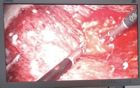
Empyema Seen During Video-Assisted Thoracoscopic Surgery. This is an intraoperative image of an empyema as seen during video-assisted thoracoscopic surgery (VATS).
Contributed by A Kumar, MD, FACC
(Click Image to Enlarge)
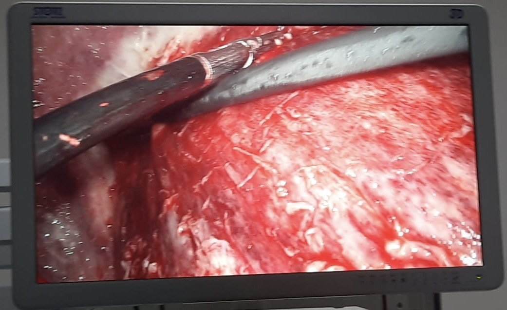
Thickened Pleura Seen During Video-Assisted Thoracoscopic Surgery. This is an intraoperative image of thickened pleura with underlying trapped lung as seen during video-assisted thoracoscopic surgery (VATS).
Contributed by A Kumar, MD, FACC
References
Addala DN, Bedawi EO, Rahman NM. Parapneumonic Effusion and Empyema. Clinics in chest medicine. 2021 Dec:42(4):637-647. doi: 10.1016/j.ccm.2021.08.001. Epub [PubMed PMID: 34774171]
Merchant N, Liu C. Thoracic empyema: aetiology, diagnosis, treatment, and prevention. Current opinion in pulmonary medicine. 2024 May 1:30(3):204-209. doi: 10.1097/MCP.0000000000001054. Epub 2024 Feb 7 [PubMed PMID: 38323933]
Level 3 (low-level) evidenceLYNN RB, WELLINGTON JL. DECORTICATION OF THE LUNG. Canadian Medical Association journal. 1963 Dec 21:89(25):1260-5 [PubMed PMID: 14098889]
Yu H. Management of pleural effusion, empyema, and lung abscess. Seminars in interventional radiology. 2011 Mar:28(1):75-86. doi: 10.1055/s-0031-1273942. Epub [PubMed PMID: 22379278]
Shin JA, Chang YS, Kim TH, Haam SJ, Kim HJ, Ahn CM, Byun MK. Surgical decortication as the first-line treatment for pleural empyema. The Journal of thoracic and cardiovascular surgery. 2013 Apr:145(4):933-939.e1. doi: 10.1016/j.jtcvs.2012.07.035. Epub 2012 Aug 25 [PubMed PMID: 22929218]
Level 2 (mid-level) evidencePiazzolla M, Tondo P. Causes, management and treatment of empyema. Minerva surgery. 2024 Apr:79(2):210-218. doi: 10.23736/S2724-5691.23.09992-6. Epub 2024 Jan 24 [PubMed PMID: 38264874]
Yalcin NG, Choong CK, Eizenberg N. Anatomy and pathophysiology of the pleura and pleural space. Thoracic surgery clinics. 2013 Feb:23(1):1-10, v. doi: 10.1016/j.thorsurg.2012.10.008. Epub [PubMed PMID: 23206712]
Doub JB, Putnam N. Clinical Ramifications of Bacterial Aggregation in Pleural Fluid. Infectious disease reports. 2024 Jul 18:16(4):608-614. doi: 10.3390/idr16040046. Epub 2024 Jul 18 [PubMed PMID: 39051246]
Miyazaki T, Yamasaki N, Tsuchiya T, Matsumoto K, Kamohara R, Hatachi G, Nagayasu T. Is Pleurectomy/Decortication Superior to Extrapleural Pneumonectomy for Patients with Malignant Pleural Mesothelioma? A Single-Institutional Experience. Annals of thoracic and cardiovascular surgery : official journal of the Association of Thoracic and Cardiovascular Surgeons of Asia. 2018 Apr 20:24(2):81-88. doi: 10.5761/atcs.oa.17-00192. Epub 2018 Jan 23 [PubMed PMID: 29367501]
Lopez-Majano V, Joshi RC. Indications for decortication. Respiration; international review of thoracic diseases. 1970:27(6):565-81 [PubMed PMID: 5490008]
Jung Y, Yi E, Lee S, Chung JH. Clinical outcome of urgent thoracoscopic surgery on complicated parapneumonic infection with short-term preoperative antibiotic usage. Journal of thoracic disease. 2025 Jan 24:17(1):357-368. doi: 10.21037/jtd-24-1331. Epub 2025 Jan 22 [PubMed PMID: 39975742]
Level 2 (mid-level) evidenceLin CM, Chen YL, Cheng YF, Cheng CY, Huang CL, Hung WH, Wang BY. Optimal timing for video assisted thoracic surgery decortication for improved survival in chronic empyema. Scientific reports. 2024 Oct 19:14(1):24548. doi: 10.1038/s41598-024-75569-w. Epub 2024 Oct 19 [PubMed PMID: 39427034]
Reichert M, Pösentrup B, Hecker A, Schneck E, Pons-Kühnemann J, Augustin F, Padberg W, Öfner D, Bodner J. Thoracotomy versus video-assisted thoracoscopic surgery (VATS) in stage III empyema-an analysis of 217 consecutive patients. Surgical endoscopy. 2018 Jun:32(6):2664-2675. doi: 10.1007/s00464-017-5961-7. Epub 2017 Dec 7 [PubMed PMID: 29218675]
Shen KR. Current state of the art for the surgical management of empyema thoracis. The Journal of thoracic and cardiovascular surgery. 2019 Mar:157(3):1300-1301. doi: 10.1016/j.jtcvs.2018.11.006. Epub 2018 Nov 10 [PubMed PMID: 30503730]
Pan H, He J, Shen J, Jiang L, Liang W, He J. A meta-analysis of video-assisted thoracoscopic decortication versus open thoracotomy decortication for patients with empyema. Journal of thoracic disease. 2017 Jul:9(7):2006-2014. doi: 10.21037/jtd.2017.06.109. Epub [PubMed PMID: 28840000]
Level 1 (high-level) evidenceAbraham SV, Chikkahonnaiah P. Change in Pulmonary Function Following Decortication for Chronic Pleural Empyema. Turkish thoracic journal. 2020 Jan:21(1):27-31. doi: 10.5152/TurkThoracJ.2019.180146. Epub 2020 Jan 1 [PubMed PMID: 32163360]