 Tools
Tools
 Lumbar Plexus Block
Introduction
The concept of a regional anesthesia technique that provides neural blockade of the entire lumbar plexus, a lumbar plexus block, dates back nearly 50 years. The first description of such a block by Winnie et al in 1973 was an “inguinal perivascular technique,” alternatively referred to as a “3 in 1 technique”.[1] Winnie proposed that a large volume of local anesthetic injected in the femoral nerve sheath could spread proximally to produce blockade of the obturator, lateral femoral cutaneous, as well as femoral nerve (and presumably the other nerves of the lumbar plexus). Later work would show that this rarely succeeded and typically only blocked the femoral and lateral femoral cutaneous nerves. In 1976, Chayen et al described a “posterior lumbar plexus block” or “psoas compartment block,” which proved to be a more reliable realization of the goal of blocking the whole of the lumbar plexus with a single injection.[2] Touray et al were among those who demonstrated that whereas both approaches effectively block femoral and lateral femoral cutaneous nerves, only the posterior lumbar plexus approach can block the obturator nerve.[3] Thus, today the term “lumbar plexus block” is generally not used to refer to the inguinal-femoral nerve block and is considered synonymous with the posterior approach.
Since the initial description of the posterior lumbar plexus block, several variations of the technique have been described. Most of these differ from the original technique of Chayen et al only in minor details and relate to the distance of the needle insertion point from the midline or the lumbar level at which the block is performed.[4][5] Perhaps the most significant changes have been in how the block’s endpoint is defined. The original technique relied on a “loss of resistance,” but this transitioned to the more common use of a nerve stimulator technique (typically looking for motor stimulation of the femoral nerve with quadriceps twitch). More recently, nerve stimulation has given way to ultrasound-guided lumbar plexus block techniques, for which several approaches have been described. Thus far, the evidence is insufficient to support the superiority of any one ultrasound-guided technique.
Anatomy and Physiology
Register For Free And Read The Full Article
 Search engine and full access to all medical articles
- 10 free questions in your specialty
- Free CME/CE Activities
- Free daily question in your email
- Save favorite articles to your dashboard
- Emails offering discounts
Learn more about a Subscription to StatPearls Point-of-Care
Anatomy and Physiology
The lumbar plexus is formed within the body of the psoas muscle by the 4 spinal nerves of L1 to L4. In 60% of people, the lumbar plexus receives a contribution from the nerve root of T12 as well.[6] Anatomical variations in the formation of the lumbar plexus have been described with a prevalence of 20% to 40%.[7][8] Most common variants are related to the presence of an accessory obturator nerve or an isolated obturator nerve surrounded by a muscular fold, different nerve root derivation of the femoral cutaneous nerve, or a bifurcation of the femoral nerve within the psoas muscle. Whether these anatomical variations have clinical implications for lumbar plexus block is unknown.[8]
The psoas major muscle is composed of 2 portions: an anterior portion that arises from the anterolateral surface of the intervertebral discs and vertebral bodies, and a posterior portion that originates from the anterior aspect of the transverse processes. These 2 portions of the psoas are separated by a fascia, creating in effect a “compartment” between them. In this fascial plane, the lumbar plexus is formed from its component nerve roots alongside the lumbar ascending vein and branches of the lumbar artery. This plane extends medially, opening into a wedge-shaped space at the medial surface of the psoas muscle, the lumbar paravertebral space.[9] The component nerve roots of the lumbar plexus each exit the neural canal through their respective intervertebral foramen, turn in a steep caudal direction across the lumbar paravertebral space, and access the psoas muscle 1 vertebral level below. Once formed in this fascial plane between the psoas muscle bodies, the component nerves of the lumbar plexus take their paths, each diving in a separate direction into the body of the psoas muscle from which they each exist separately.
The main branches of the lumbar plexus include:
- Iliohypogastric nerve: Formed by T12 and L1, it travels across the psoas muscle and arises at the upper lateral border of this muscle between the anterior surface of the quadratus lumborum and the posterior surface of the kidney.
- Ilioinguinal nerve: Formed by L1, the ilioinguinal nerve arises from the lateral border of the psoas muscle just below the iliohypogastric nerve. It follows a downward trajectory on the anterior surface of the quadratus lumborum and the upper part of the iliacus.
- Genitofemoral nerve: Formed mainly by L1 to L2, the genitofemoral nerve arises from the anterior surface of the psoas muscle along the medial border. It follows a downward trajectory on the psoas muscle within the fascia iliaca and crosses posterior to the ureter and peritoneum. The genitofemoral nerve provides innervation to the cremaster muscle, skin of the femoral triangle, scrotum, pubis, and labium major.
- Lateral femoral cutaneous nerve: Formed by the posterior division of L2 and L3 lumbar nerves. Arises beneath the lateral margin of the psoas muscle and follows downward laterally deep to the fascia over the iliacus muscle and finally crosses under the inguinal ligament medial to the anterior superior iliac spine. The lateral femoral cutaneous nerve provides sensory innervation to the skin of the anterolateral aspect of the thigh, and some of its terminal branches contribute to the patellar plexus.
- Femoral nerve: Formed by the posterior division of L2, L3, and L4, within the psoas muscle, and exits from the lateral border of the psoas major in the fossa iliaca, roughly 4 cm above the inguinal ligament. From here, the femoral nerve enters the femoral triangle, laterally to the femoral sheath, and after a short distance, divides into its terminal branches. The femoral nerve gives motor innervation to the quadriceps muscle and sensory innervation to the knee through the peripatellar plexus and the medial aspect of the leg through the saphenous nerve.
- Obturator nerve: Formed by the anterior division of L2, L3, L4, within the psoas muscle and follows a downward trajectory within the muscle body to arise on the medial side of the psoas at the level of the pelvic brim. Upon exiting the psoas, the nerve continues in a postero-lateral direction towards the sacroiliac joint. The obturator nerve provides motor innervation to the adductor muscles in the medial aspect of the thigh and sensory innervation to the hip joint and knee joint.
As the lumbar plexus is formed from the component nerve roots, it acquires a triangular shape, wider at its caudal portion, while fanning out medial-laterally, with the femoral nerve situated in the middle, the obturator nerve to the medial, and the lateral cutaneous nerve to the lateral. Magnetic resonance imaging studies have demonstrated that the spread of local anesthetic in a psoas block is usually within the psoas muscle, most often along the internal fascial plane, with cephalad spread to the lumbar nerve roots and around the lumbar branches.[10] The terminology is confusing because the term “psoas sheath” block has been used to mean blocking the lumbar plexus within the psoas, whereas the term “psoas compartment” block has been used to mean injection external and posterior to the psoas between the psoas and quadratus muscles rather than the compartment between the muscle bodies of the psoas. It is believed, based on dye injection studies, that 20% to 40% of the time, a traditional lumbar plexus block is actually in this space rather than in the psoas.
Several studies have specifically described the distance from the skin to the lumbar plexus. In general, that distance ranges from 9 to 10 cm and is slightly deeper in males than in females.[6] The distance between the anterior border of the transverse process and the lumbar plexus ranges between 1.5 and 2.0 cm, with a median value of 1.8 cm in both sexes. Caution is urged regarding the depth of needle insertion, as insertion more than 2.0 to 3.0 cm beyond the transverse process may increase the risk of retroperitoneal or even intraperitoneal injury. Similarly, a needle insertion deeper than 10 to 12 cm may increase the risk of injury.[11]
Indications
The lumbar plexus block is most commonly used to provide perioperative analgesia but may also be used as a surgical anesthetic, particularly when combined with a sciatic nerve block. The lumbar plexus block is typically used to provide analgesia following injuries or surgeries of the hip or thigh (eg, acetabular fractures, femoral neck or mid-shaft fractures, hip replacement, hip arthroscopy, and knee replacement) but has also been used for chronic pain conditions (eg, herpes zoster). Notably, the lumbar plexus block is unlikely by itself to produce complete anesthesia for hip replacement surgery due to the innervation of the posteromedial hip capsule deriving from branches of the sacral plexus and sciatic nerve.[5]
Contraindications
The contraindications for lumbar plexus block are similar to those of other regional anesthesia techniques, including:
- Patient refusal
- Allergy to local anesthetics
- Local infection
- Systemic anticoagulation (INR >1.5 or inadequate time since cessation of anticoagulant)
The following are relative contraindications or instances where clinicians might consider an alternative technique:
- Presence of an intrathecal pump or a spinal cord stimulator
- Major lumbar spine deformity
- Prior major spine surgery, implanted hardware, fusion
- Preexisting neurologic deficit
Equipment
Neurostimulation Technique
The following equipment is typically needed when neurstimulation techniques are used:
- Insulated stimulating needle (10 cm length; caution: 15 cm needle will only rarely be needed)
- Peripheral nerve block stimulator plus surface electrode
- One 23- to 25-gauge needle for skin infiltration
- One 8 to 10 cm 25-gauge spinal needle
- 20 mL of local anesthetic
- 1 pack of gauze 4-inch x 4-inch
- Chlorhexidine gluconate solution for skin asepsis
- Sterile gloves
- Marking pen
Ultrasound-Guided Technique
The following equipment is typically needed when ultrasound guidance is used for needle placement:
- Low frequency (5 to 10 Hz) curved transducer
- One 23- to 25-gauge needle for skin infiltration
- 20 mL of local anesthetic
- 1 pack of gauze 4-inch x 4-inch
- Chlorhexidine gluconate solution for skin asepsis
- Sterile gloves
- Marking pen
Personnel
The lumbar plexus block is a deep block, considered an advanced regional anesthesia technique that should only be performed by trained physicians. Having a nurse trained in regional anesthesia, both to assist with performing the block and to help administer sedation to the patient, may be helpful.
Preparation
A standard preoperative assessment should be conducted for any anesthesia procedure, including medical history, physical exam, airway assessment, and review of test results. Of special note are an allergic history, a history of neuropathy or demyelinating disease, coagulopathy, and a history of chronic pain. Current patient prescriptions, including analgesics and anticoagulants, should be noted. The patient should be informed about the benefits and risks of the lumbar plexus block, as well as what the procedure entails, as part of his informed consent. As with all nerve blocks, this procedure should be performed in a proper setting that has available monitoring, oxygen, suction, and resuscitation equipment.
Technique or Treatment
Several variants of the landmark technique are used in conjunction with neurostimulation to identify the femoral nerve within the psoas by evoking a motor response in the quadriceps muscle. While these techniques differ somewhat in the needle insertion point (lumbar level and distance from the midline), the following method is the most common.
Standard American Society of Anesthesiologists monitoring is applied, and the patient is positioned in lateral decubitus on the side opposite the surgery, with the side of the block upward. The ipsilateral knee flexed at 90 degrees, and the hip flexed at 30 degrees. The needle insertion point is marked 4 to 5 cm lateral to the midline at the level of the iliac crest along the intercristal line. The block area is then sterilely prepped and draped with sterile towels. A safety time-out is performed with the participation of the patient, nurse, and physician to confirm the patient's identity, the procedure, and the laterality. As the lumbar plexus block may be uncomfortable for the patient, mild sedation is often given using small doses of intravenous midazolam and/or fentanyl (see Image. Lumbar Plexus Block Technique). The most common technique for lumbar plexus block comprises the following steps:
- Local anesthesia using lidocaine 1% is injected using the 23- to 25-gauge needle at the marked site of needle introduction along the same trajectory planned for the nerve block needle.
- The 25-gauge spinal needle is introduced at this site, perpendicular to the skin, attempting to locate the transverse process. If the transverse process is not found, the needle is withdrawn to a subcutaneous depth and reinserted with a slight caudad or cephalad angulation. This fanning exploration is repeated until the transverse process is located. Its depth and direction are noted, or it can be left in place with the nerve block needle to be inserted alongside it.
- The neurostimulator is set to 1.5 mA with a 0.1-millisecond pulse width at 2 Hz and is attached to both the block needle and an electrocardiogram (ECG) pad on the skin. The block needle is inserted alongside, or in place of, the spinal finder needle, and at the depth determined by the finder needle, the transverse process is contacted.
- The block needle is now withdrawn and redirected either caudally or cephalad to a depth 2 cm greater than the depth of contact with the transverse process. This may need to be repeated several times until a twitch response of the quadriceps muscle is elicited.
- Precise needle positioning is guided by reducing the neurostimulator current until a motor response is still visible at 0.5 mA.
- Inject the local anesthetic in fractionated aliquots, aspirating between each to be sure the needle is not intravascular.
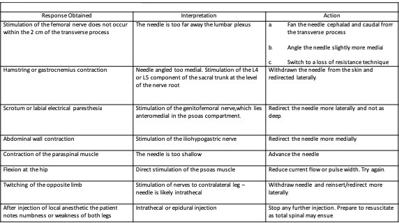
Although the technique described above appears straightforward, challenges may arise and require troubleshooting to ensure successful execution and patient safety.
Additional considerations apply when placing a continuous lumbar plexus catheter. Needle positioning follows the same principles as a single-shot lumbar plexus block; however, a small risk of intravascular or neuraxial catheter misplacement persists despite correct needle placement. Catheters require confirmation of negative aspiration for blood or cerebrospinal fluid, and local anesthetic administration should occur in slow, fractionated doses. Final catheter placement should extend 4 to 5 cm beyond the depth reached by the needle. A continuous infusion of ropivacaine 0.2% at 8 to 10 mL/hr provides effective analgesia. Infusion rates may be reduced when motor weakness limits participation in physical therapy and increased when analgesia remains inadequate. Development of foot drop does not indicate excessive blockade and should not be attributed to the procedure; a surgical cause, eg, hematoma, bone fragment, or dislocation leading to sciatic nerve compression, requires urgent evaluation. Ultrasound-guided techniques fall outside the scope of this activity.
Complications
Complications related to the lumbar plexus block are infrequent. Although there are authors who report a high incidence of local anesthetic spread to the epidural space, our experience at the University of Pittsburgh is quite the opposite.[5][12] With the epidural spread, one would expect contralateral spread, bilateral weakness, hypotension, and difficulty with micturition. Indeed, this is, in our experience, exceedingly rare. More medial needle insertion and a more cephalic approach (L3) may increase the likelihood of this complication.[5]
Intrathecal injection and spinal anesthesia as a complication of lumbar plexus block are also rare. Its prevalence is unknown, as the vast majority of information comes from case reports and a few observational studies.[13][14][15] Auroy et al reported 5 cases of major complications after lumbar plexus block in a sample of 394 patients.[16] This report is greatly at odds with the experience of the University of Pittsburgh, where the acute pain service has performed many thousands of lumbar plexus blocks over the last 2 decades with few complications altogether, no case of direct intrathecal injection, and only 1 incident of intrathecal catheter placement.
Renal injury, eg, a subcapsular hematoma, is a rare complication of lumbar plexus block and has been associated with more cephalad injection sites, including L3. Use of the intercristal line helps guide needle placement toward the L4 or L5 transverse process, reducing this risk. Avoid advancing the needle more than 2 cm beyond the transverse process to further minimize the likelihood of renal injury. Local anesthetic systemic toxicity remains a potential complication of any nerve block, most commonly resulting from intravascular injection, although excessive dosing may also contribute. Management requires immediate administration of intravenous intralipid along with supportive care.
Retroperitoneal or psoas hematoma and other vascular injuries constitute major but rare complications. Bleeding within a noncompressible space, eg, the psoas compartment, warrants heightened concern, particularly when direct observation and compression are limited. Patients receiving anticoagulation therapy or those with coagulopathy may not represent appropriate candidates for this procedure. Deep plexus blocks should adhere to the 2018 American Society of Regional Anesthesia (ASRA) guidelines for neuraxial techniques in patients receiving anticoagulation or antiplatelet therapy.[17]
Peripheral nerve injury remains a common patient concern, yet occurs infrequently. Long-term injury has an incidence of 2 to 4 cases per 10,000 peripheral nerve blocks overall, with reported rates as low as 0.1% for lumbar plexus block.[18][19][18] Neurostimulation producing a muscle twitch at currents below 0.2 mA correlates with a higher risk of histological nerve injury.[20] In such cases, slight needle withdrawal should be performed until a motor response is elicited by 0.5 mA stimulation.
Clinical Significance
Lumbar plexus block represents an advanced regional anesthesia technique with well-established efficacy over many years. Safety concerns, particularly in experienced centers where the technique remains standard practice, appear overstated. Clinical experience supports its use as part of multimodal analgesia for lower limb surgery, where it serves as a reliable and effective modality for postoperative pain control. A systematic review and meta-analysis demonstrated reductions in opioid consumption, improved patient comfort, and enhanced postoperative ambulation and participation in physical therapy.[21] This technique also contributes significantly to enhanced recovery pathways for hip surgery.
Comprehensive knowledge of lumbar plexus anatomy, procedural variations, indications, and contraindications remains essential for anesthesiologists and acute pain physicians. Familiarity with alternative regional anesthesia techniques for hip surgery, including the pericapsular nerve group, quadratus lumborum types 1 and 3, and fascia iliaca blocks, supports optimal selection of techniques.[22][23][24][25][24] Pediatric patients with cerebral palsy undergoing hip reconstruction represent another population in which this technique may be applied. Comparative data demonstrate that lumbar plexus catheters provide pain control comparable to lumbar epidural catheters in this group.[26]
Table. Lumbar Plexus Block Comparison
| Lumbar Plexus Block Comparisons | Reported Lumbar Plexus Block Pros | Reported Lumbar Plexus Block Cons |
| Quadratus lumborum type 3 [23][24] | Noninferiority trial with lumbar plexus block as standard |
Quadriceps weakness Less time efficient |
| Pericapsular nerve group [22] |
Lower postoperative opioid consumption Higher incidence of sensory block at 6H and 12H Longer duration of analgesia |
Higher incidence of motor block of knee extension but not hip adduction at 6H and 12H, which did not impact physiotherapy participation at 24H Lower satisfaction scores |
| Fascia iliaca [25] |
Faster block effect onset Lower intraoperative and postoperative opioid requirements |
Longer ultrasound imaging time Longer length of stay |
Enhancing Healthcare Team Outcomes
Lumbar plexus block is an advanced regional anesthesia technique that provides effective perioperative analgesia for hip and thigh procedures by targeting nerve roots L1 to L4 within the psoas muscle. The posterior psoas compartment approach offers more reliable blockade of the obturator nerve compared with anterior techniques. Anatomical variability and deep needle placement increase procedural complexity and risk, including intrathecal injection, vascular injury, retroperitoneal hematoma, and local anesthetic systemic toxicity. Accurate identification of landmarks, appropriate use of neurostimulation or ultrasound guidance, and adherence to safety thresholds for needle depth and stimulation current are essential for optimizing outcomes. While commonly used for postoperative analgesia, a lumbar plexus block alone may not provide complete anesthesia for hip surgery due to contributions from the sacral plexus.
Interprofessional collaboration strengthens patient safety and outcomes through coordinated care. Physicians and advanced practitioners perform patient selection, procedural planning, and block placement, while primary care clinicians contribute to preoperative optimization and risk assessment. Nurses support monitoring, sedation, and early recognition of complications, and pharmacists guide safe dosing and management of local anesthetic toxicity. Physical therapists facilitate early mobilization, and all team members communicate findings, reinforce shared decision-making, and ensure timely follow-up to reduce complications and improve recovery.
Media
(Click Image to Enlarge)
Lumbar Plexus Block Technique
Created and Contributed by J Polania Gutierrez, MD
References
Winnie AP, Ramamurthy S, Durrani Z. The inguinal paravascular technic of lumbar plexus anesthesia: the "3-in-1 block". Anesthesia and analgesia. 1973 Nov-Dec:52(6):989-96 [PubMed PMID: 4796576]
Chayen D, Nathan H, Chayen M. The psoas compartment block. Anesthesiology. 1976 Jul:45(1):95-9 [PubMed PMID: 937760]
Touray ST, de Leeuw MA, Zuurmond WW, Perez RS. Psoas compartment block for lower extremity surgery: a meta-analysis. British journal of anaesthesia. 2008 Dec:101(6):750-60. doi: 10.1093/bja/aen298. Epub 2008 Oct 22 [PubMed PMID: 18945717]
Level 1 (high-level) evidenceAwad IT, Duggan EM. Posterior lumbar plexus block: anatomy, approaches, and techniques. Regional anesthesia and pain medicine. 2005 Mar-Apr:30(2):143-9 [PubMed PMID: 15765457]
de Leeuw MA, Zuurmond WW, Perez RS. The psoas compartment block for hip surgery: the past, present, and future. Anesthesiology research and practice. 2011:2011():159541. doi: 10.1155/2011/159541. Epub 2011 May 22 [PubMed PMID: 21716721]
Farny J, Drolet P, Girard M. Anatomy of the posterior approach to the lumbar plexus block. Canadian journal of anaesthesia = Journal canadien d'anesthesie. 1994 Jun:41(6):480-5 [PubMed PMID: 8069987]
Sim IW, Webb T. Anatomy and anaesthesia of the lumbar somatic plexus. Anaesthesia and intensive care. 2004 Apr:32(2):178-87 [PubMed PMID: 15957714]
Anloague PA, Huijbregts P. Anatomical variations of the lumbar plexus: a descriptive anatomy study with proposed clinical implications. The Journal of manual & manipulative therapy. 2009:17(4):e107-14 [PubMed PMID: 20140146]
Gadsden J, Latmore M, Levine DM, Robinson A. High Opening Injection Pressure Is Associated With Needle-Nerve and Needle-Fascia Contact During Femoral Nerve Block. Regional anesthesia and pain medicine. 2016 Jan-Feb:41(1):50-5. doi: 10.1097/AAP.0000000000000346. Epub [PubMed PMID: 26650431]
Mannion S, Barrett J, Kelly D, Murphy DB, Shorten GD. A description of the spread of injectate after psoas compartment block using magnetic resonance imaging. Regional anesthesia and pain medicine. 2005 Nov-Dec:30(6):567-71 [PubMed PMID: 16326342]
Di Benedetto P, Pinto G, Arcioni R, De Blasi RA, Sorrentino L, Rossifragola I, Baciarello M, Capotondi C. Anatomy and imaging of lumbar plexus. Minerva anestesiologica. 2005 Sep:71(9):549-54 [PubMed PMID: 16166916]
Capdevila X, Coimbra C, Choquet O. Approaches to the lumbar plexus: success, risks, and outcome. Regional anesthesia and pain medicine. 2005 Mar-Apr:30(2):150-62 [PubMed PMID: 15765458]
Pousman RM, Mansoor Z, Sciard D. Total spinal anesthetic after continuous posterior lumbar plexus block. Anesthesiology. 2003 May:98(5):1281-2 [PubMed PMID: 12717153]
Level 3 (low-level) evidenceDuarte LT, Saraiva RA. [Total spinal block after posterior lumbar plexus blockade: case report.]. Revista brasileira de anestesiologia. 2006 Oct:56(5):518-23 [PubMed PMID: 19468598]
Level 3 (low-level) evidenceDogan Z, Bakan M, Idin K, Esen A, Uslu FB, Ozturk E. Total spinal block after lumbar plexus block: a case report. Brazilian journal of anesthesiology (Elsevier). 2014 Mar-Apr:64(2):121-3. doi: 10.1016/j.bjane.2013.03.002. Epub 2013 Oct 11 [PubMed PMID: 24794455]
Level 3 (low-level) evidenceAuroy Y, Benhamou D, Bargues L, Ecoffey C, Falissard B, Mercier FJ, Bouaziz H, Samii K. Major complications of regional anesthesia in France: The SOS Regional Anesthesia Hotline Service. Anesthesiology. 2002 Nov:97(5):1274-80 [PubMed PMID: 12411815]
Narouze S, Benzon HT, Provenzano D, Buvanendran A, De Andres J, Deer T, Rauck R, Huntoon MA. Interventional Spine and Pain Procedures in Patients on Antiplatelet and Anticoagulant Medications (Second Edition): Guidelines From the American Society of Regional Anesthesia and Pain Medicine, the European Society of Regional Anaesthesia and Pain Therapy, the American Academy of Pain Medicine, the International Neuromodulation Society, the North American Neuromodulation Society, and the World Institute of Pain. Regional anesthesia and pain medicine. 2018 Apr:43(3):225-262. doi: 10.1097/AAP.0000000000000700. Epub [PubMed PMID: 29278603]
Neal JM, Barrington MJ, Brull R, Hadzic A, Hebl JR, Horlocker TT, Huntoon MA, Kopp SL, Rathmell JP, Watson JC. The Second ASRA Practice Advisory on Neurologic Complications Associated With Regional Anesthesia and Pain Medicine: Executive Summary 2015. Regional anesthesia and pain medicine. 2015 Sep-Oct:40(5):401-30. doi: 10.1097/AAP.0000000000000286. Epub [PubMed PMID: 26288034]
Brull R, McCartney CJ, Chan VW, El-Beheiry H. Neurological complications after regional anesthesia: contemporary estimates of risk. Anesthesia and analgesia. 2007 Apr:104(4):965-74 [PubMed PMID: 17377115]
Sondekoppam RV, Tsui BC. Factors Associated With Risk of Neurologic Complications After Peripheral Nerve Blocks: A Systematic Review. Anesthesia and analgesia. 2017 Feb:124(2):645-660. doi: 10.1213/ANE.0000000000001804. Epub [PubMed PMID: 28067709]
Level 1 (high-level) evidenceAlMutiri WA, AlMajed E, Alneghaimshi MM, AlAwadh A, AlSarhan R, AlShebel MN, AlMatrody RAM, Hadaddi R, AlTamimi R, Bin Salamah R, AlZelfawi LA, AlBatati SK, AlHarthi A, AlMazroa G, AlHossan AM. Efficacy of Continuous Lumbar Plexus Blockade in Managing Post-Operative Pain after Hip or Femur Orthopedic Surgeries: A Systematic Review and Meta-Analysis. Journal of clinical medicine. 2024 May 29:13(11):. doi: 10.3390/jcm13113194. Epub 2024 May 29 [PubMed PMID: 38892904]
Level 1 (high-level) evidenceLee TY, Chung CJ, Park SY. Comparing the Pericapsular Nerve Group Block and the Lumbar Plexus Block for Hip Fracture Surgery: A Single-Center Randomized Double-Blinded Study. Journal of clinical medicine. 2023 Dec 25:13(1):. doi: 10.3390/jcm13010122. Epub 2023 Dec 25 [PubMed PMID: 38202129]
Level 1 (high-level) evidenceKelly T, Wolla CD, Wolf BJ, Hay E, Babb S, Wilson SH. Comparison of lateral quadratus lumborum and lumbar plexus blocks for postoperative analgesia following total hip arthroplasty: a randomized clinical trial. Regional anesthesia and pain medicine. 2022 Sep:47(9):541-546. doi: 10.1136/rapm-2022-103598. Epub 2022 Jun 10 [PubMed PMID: 35688515]
Level 1 (high-level) evidencePolania Gutierrez JJ, Ben-David B, Rest C, Grajales MT, Khetarpal SK. Quadratus lumborum block type 3 versus lumbar plexus block in hip replacement surgery: a randomized, prospective, non-inferiority study. Regional anesthesia and pain medicine. 2021 Feb:46(2):111-117. doi: 10.1136/rapm-2020-101915. Epub 2020 Nov 11 [PubMed PMID: 33177220]
Level 1 (high-level) evidenceWu J, Mou H, Luo X. Comparison between lumbar plexus block and fascia iliaca block in hip surgery: A systematic review and meta-analysis. Medicine. 2025 Sep 5:104(36):e43744. doi: 10.1097/MD.0000000000043744. Epub [PubMed PMID: 40922263]
Level 1 (high-level) evidenceTrionfo A, Zimmerman R, Gillock K, Budziszewski R, Hasan A. Lumbar Plexus Nerve Blocks for Perioperative Pain Management in Cerebral Palsy Patients Undergoing Hip Reconstruction: More Effective Than General Anesthesia and Epidurals. Journal of pediatric orthopedics. 2023 Jan 1:43(1):e54-e59. doi: 10.1097/BPO.0000000000002285. Epub 2022 Oct 26 [PubMed PMID: 36509456]