 Tools
Tools
 Histology, Lipofuscin
Introduction
Lipofuscin is a pigmented, heterogeneous byproduct of failed intracellular catabolism, conventionally found within lysosomes or the cytosol of aging postmitotic cells. Although lipofuscin is present in virtually any cell type, phenotypically proliferative cells often dilute its concentration to insignificant levels.[1] Hannover first discovered lipofuscin in 1842, but its progressive accumulation with age was not recognized until the end of the nineteenth century.[2][3] While this historical description remains accurate, the literature from the past few decades has shifted focus toward the newly recognized role of lipofuscin as a tissue photosensitizer and a potentiator of intracellular dyshomeostasis. Because of its extensively cross-linked tertiary structure and nondegradable nature, lipofuscin is hypothesized to play a critical role in inhibiting proteasome function, mitophagy, autophagy, and lysosomal stability, and in promoting the propagation of reactive oxygen species.[1][4][5]
Lipofuscin is frequently used interchangeably with "ceroid" or "ceroid lipofuscin," as both ceroid and lipofuscin are autofluorescent intracellular accumulations of similar composition.[6] However, this equivalence may be misleading, as the etymological distinction between lipofuscin and ceroid was traditionally intended to label the former as associated with normal aging and the latter with pathological conditions.[7][8] This distinction has become muddied as traditional lipofuscin has continued to gain attention for its risk-modifying effects on many diseases.[4]
Structure
Register For Free And Read The Full Article
 Search engine and full access to all medical articles
- 10 free questions in your specialty
- Free CME/CE Activities
- Free daily question in your email
- Save favorite articles to your dashboard
- Emails offering discounts
Learn more about a Subscription to StatPearls Point-of-Care
Structure
Lipofuscin’s Latin namesake translates to “dark lipid,” although this description only partially characterizes its variable composition.[3] Lipofuscin is a heterogeneous amalgam composed primarily of oxidized proteins (30% to 70%) and lipids, including triglycerides, free fatty acids, cholesterol, and lipoproteins (20% to 50%). Carbohydrates make a small contribution that may increase proportionally with age (4% to 7%). This idiosyncratic composition varies among tissue types, although many lipofuscin researchers agree that mitochondrial components contribute substantially to the protein fraction.[3][9][10] Jung et al suggested that adenosine triphosphate (ATP) synthase subunit residues account for up to 50% of the protein content in certain lipofuscin aggregates associated with congenital ceroid lipofuscinoses.[6]
Metals such as iron, copper, zinc, aluminum, manganese, and calcium constitute only 2% of lipofuscin.[11] However, despite their small proportion within lipofuscin, iron is believed to be the primary contributor to free radical generation through Fenton reactions.[12][13]
Fluorophores represent another important component of lipofuscin structure. The 400 to 700 nm fluorescence emission spectrum of lipofuscin reflects substantial tissue-to-tissue variability.[1][4] Cell-specific breakdown products generate a broad range of photosensitive substances, among which the fluorophore A2E is of particular interest because of its proposed association with wet macular degeneration. Numerous phototoxic effects have been described.[1][14][15] Ultimately, these components aggregate in response to a maladaptive intracellular environment characterized by lipid peroxidation, inhibition of proteasome function and autophagy, and erratic polymerization of granule constituents.[4][13] This process results in the formation of nondegradable, insoluble lipofuscin accumulations within lysosomes and the cytosol. Repeated cycles of ineffective lysosomal autophagy and mitophagy, compounded by uncontrolled reactive oxygen species, lead to a positive feedback loop of lipofuscin buildup.[13]
The continuous net positive gain of intracellular lipofuscin can have a dramatic effect on lysosomal structure. As demonstrated by Sitte and collaborators, progressive formation within fibroblast lysosomes, along with spillover into the cytoplasm, can exert toxic effects and hasten apoptosis.[16] Eventually, cellular macrostructure may become compromised due to shear lipofuscin volume. Research shows that lipofuscin may constitute up to 75% of the total cytoplasmic volume in the motor neurons of humans older than 100 years.[17] G0-phase cells, such as myocytes and neurons, will display the largest accumulations.[18]
Function
Outside of congenital lysosomal storage disease diagnostics, lipofuscin has little traditional histological or clinical value beyond indicating oxidative dyshomeostasis and aging cellular machinery. For over a century, the primary histological utility of lipofuscin has been the identification of cellular senescence, which inversely correlates with longevity.[2][4]
As previously discussed, recent discoveries have led to a greater understanding of lipofuscin’s function as a potentiator, rather than bystander, of apoptosis and intracellular dysregulation. While the exact details of lipofuscin’s effect on the cell are still hypothetical, there is agreement on several areas of research.[19] The most widely accepted theory of lipofuscin formation is the “mitochondrial-lysosomal axis theory of postmitotic cellular aging” proposed by Brunk and Terman: lipofuscin forms as a result of chronic lysosomal uptake of iron-rich mitochondrial breakdown products that propagate reactive oxygen species.[20][21] The subsequent lipid peroxidation breakdown products cause extensive cross-linking that prevents degradation and reduces lysosomal stability. The resulting sequelae include the release of toxic lipofuscin and excess iron into the cytosol, thereby restarting the cycle as another lysosome attempts to degrade the nondegradable material.[12] Recent experimental studies further support this model by demonstrating that authentic lipofuscin aggregates increase mitochondrial reactive oxygen species production, impair lysosomal integrity, and reduce cathepsin activity and degradative capacity. These findings provide experimental evidence linking lipofuscin accumulation to measurable mitochondrial and lysosomal dysfunction.[22]
Additional mechanisms of collateral damage have also been reported. Cross-linked lipofuscin proteins, such as those modified by 4-hydroxynonenal, are poor proteasomal substrates and consequently reduce proteasome efficacy.[23] Furthermore, exposed hydrophobic residues promote the attachment of unfolded, oxidized protein aggregates to 20S and 26S proteasomes, thereby inhibiting further degradation.[24][25] Autophagy and mitophagy gradually become impaired, allowing for a cytosolic accumulation of lipofuscin.[4][26] Because pro-apoptotic proteins such as c-jun, bax, and p27 are not efficiently degraded, lipofuscin correlates with increased rates of apoptosis.[4][27] Hohn et al showed that artificial lipofuscin with increased iron stores also increased caspase-3 activity.[12] Evidence indicates that lipofuscin directly contributes to impaired mitophagy by decreasing PINK1 expression on senescent mitochondria; PINK1 is an essential protein for marking malfunctioning mitochondria for destruction. Lipofuscinogenesis also demonstrates inhibition of mitochondrial fission.[26][28]
Tissue Preparation
Lipofuscin can accumulate in nearly any tissue; therefore, tissue preparation methods vary significantly. Extraction for analysis is the preferred method in postmitotic cells, in which lipofuscin is most abundant. Traditionally, organic solvent extraction or density gradient ultracentrifugation were used to isolate lipofuscin due to its high lipid content.[4][29] However, modern methods for lipofuscin analysis and quantification rely on its intrinsic autofluorescence properties.[30]
Histochemistry and Cytochemistry
Because of its highly variable and polymeric nature, no antibodies specific to lipofuscin exist. Jung et al pointed out that combinations of nonoverlapping antibodies and probes can be used to localize lipofuscin's fluorescence with other cellular structures of interest.[30] Many classical methods for identifying lipofuscin involved lipid and carbohydrate histochemical staining, including but not limited to Sudan Black, Fontana-Masson, Schmorl, Ziehl-Nielson, copper sulfate, picric acid, Nile Blue, osmium tetroxide (OsO4), eosin, hematoxylin, and ferric ferricyanide. Arguably, Sudan Black has been the most commonly used stain.[2][3][6][31]
Microscopy, Light
Both light and electron microscopy are viable methods for identifying lipofuscin, with or without histochemical or immunocytochemical staining; however, fluorescence microscopy remains the gold standard for quantitative identification.[30] Lipofuscin can be detected under ultraviolet light (330 to 380 nm) as well as across the visible light spectrum (380 to 700 nm).[1][8] The maximum fluorescence emission occurs at approximately 578 nm with excitation at 364 nm.[32]
The spatial arrangement of lipofuscin granules varies, although distribution patterns may be more predictable depending on the cell type. While lipofuscin may appear free-floating in the cytoplasm with light microscopy, electron microscopy clearly shows detailed lysosomal membranes surrounding the granular aggregations (see Image. Lipofuscin in Muscle).[6]
In diagnostic histopathology, lipofuscin can be difficult to distinguish from other endogenous pigments, such as melanin and hemosiderin, due to its intrinsic autofluorescence and yellow-brown appearance. Hemosiderin is typically coarse and golden-brown and stains positively with Prussian blue because of its iron content. Melanin stains with Fontana-Masson and is bleached by oxidizing agents, features that are not characteristic of lipofuscin.[33] Sudan Black B and autofluorescence-quenching techniques may further aid in differentiating lipofuscin during fluorescence-based analyses.[34]
Clinical Significance
The clinical significance of lipofuscin has not yet been fully elucidated, despite its ubiquity in aging and numerous diseases. As discussed earlier, its hypothesized role in cellular degeneration through oxidative sequelae is likely widespread across many common pathologies; however, lipofuscin accumulation may represent a legitimate risk factor in certain diseases.
Congenital neuronal ceroid lipofuscinoses provide the strongest evidence supporting a direct pathological role of lipofuscin. These early-onset lysosomal storage disorders cause marked accumulation of lipofuscin that is visible on biopsy. In neuronal ceroid lipofuscinoses such as Batten disease and Niemann-Pick C disease, inevitable and progressive accumulation leads to cell dysfunction and death at a young age.[4][6]
Elderly patients often present with hyperpigmented macules and patches on the dorsum of their hands and other sun-exposed areas of the skin. These benign melanin- and lipofuscin-rich areas are colloquially known as "age spots" or "liver spots" and have no clinical significance.[3] However, another skin-related disease in which lipofuscin abundance clearly indicates its role in pathology is apocrine chromhidrosis, a rare condition in which the patient’s sweat appears red. Identification of extensive lipofuscin granules in apocrine glands helps differentiate the etiologic process from toxin-induced pseudochromhidrosis and extrinsic pigment–induced eccrine chromhidrosis.[35]
The role of lipofuscin in most other pathologies remains poorly defined. The correlation between age-related macular degeneration and lipofuscin accumulation has been hypothesized but remains highly controversial. Most agree that lipofuscin accumulation inhibits phagocytosis by retinal pigment epithelial cells and that its fluorophores sensitize lysosomes to visible light, leading to cellular instability.[1][6][36] However, some studies suggest that lipofuscin concentration does not correlate with the rate of macular degeneration.[37]
Lipofuscin accumulation has been observed, though not directly implicated, in numerous other common diseases encountered in clinical practice. A prominent area of investigation focuses on its presence and potential role in advancing major age-related neurodegenerative diseases, including Alzheimer disease and Parkinson disease. Recent investigations of human postmortem brain tissue have quantified lipofuscin distribution, revealing higher lipofuscin number density and area fraction in late-stage Alzheimer disease cases compared with age-matched controls.[38] These findings demonstrate that lipofuscin burden and spatial distribution differ in Alzheimer pathology and support a mechanistic overlap between lipofuscin accumulation and lysosomal dysfunction in neurodegeneration.[38] In Parkinson disease–related research, studies in ATP13A2-deficient mouse models demonstrate concurrent accumulation of lipofuscin storage material and α-synuclein pathology alongside lysosomal dysfunction, implicating shared intracellular clearance deficits in Parkinson-like neurodegeneration.[39] Lipofuscin accumulation has also been noted in several other diseases, including atherosclerosis, Niemann-Pick disease, brown bowel syndrome, pigmented cysts, dermal hyperpigmentation, and numerous neoplastic processes, such as pancreatic tumors, non-choroidal melanomas, and mammary gland carcinomas.[4][6]
Although these pathologies may have unique relationships with lipofuscin, the clinical significance of identifying lipofuscin in a biopsy specimen is generally limited to indicating cellular senescence or oxidative stress. Because lipofuscin can occur in any cell type of older patients, an incidental biopsy finding does not warrant further workup unless a specific indication is present.
Media
(Click Image to Enlarge)
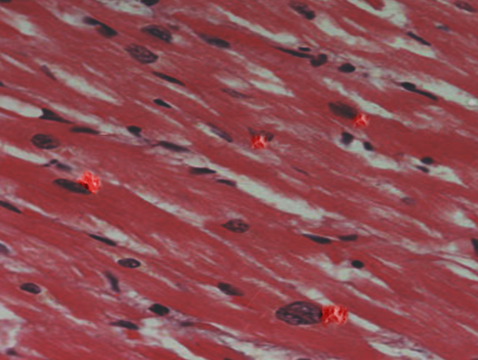
Lipofuscin in Muscle.
Contributed by S Bhimji, MD
References
Brunk UT, Terman A. Lipofuscin: mechanisms of age-related accumulation and influence on cell function. Free radical biology & medicine. 2002 Sep 1:33(5):611-9 [PubMed PMID: 12208347]
Level 3 (low-level) evidenceSalmonowicz H, Passos JF. Detecting senescence: a new method for an old pigment. Aging cell. 2017 Jun:16(3):432-434. doi: 10.1111/acel.12580. Epub 2017 Feb 9 [PubMed PMID: 28185406]
Skoczyńska A, Budzisz E, Trznadel-Grodzka E, Rotsztejn H. Melanin and lipofuscin as hallmarks of skin aging. Postepy dermatologii i alergologii. 2017 Apr:34(2):97-103. doi: 10.5114/ada.2017.67070. Epub 2017 Apr 13 [PubMed PMID: 28507486]
Moreno-García A, Kun A, Calero O, Medina M, Calero M. An Overview of the Role of Lipofuscin in Age-Related Neurodegeneration. Frontiers in neuroscience. 2018:12():464. doi: 10.3389/fnins.2018.00464. Epub 2018 Jul 5 [PubMed PMID: 30026686]
Level 3 (low-level) evidenceTerman A, Brunk UT. Aging as a catabolic malfunction. The international journal of biochemistry & cell biology. 2004 Dec:36(12):2365-75 [PubMed PMID: 15325578]
Level 3 (low-level) evidenceJung T, Bader N, Grune T. Lipofuscin: formation, distribution, and metabolic consequences. Annals of the New York Academy of Sciences. 2007 Nov:1119():97-111 [PubMed PMID: 18056959]
Level 3 (low-level) evidenceSeehafer SS, Pearce DA. You say lipofuscin, we say ceroid: defining autofluorescent storage material. Neurobiology of aging. 2006 Apr:27(4):576-88 [PubMed PMID: 16455164]
Level 3 (low-level) evidenceTohma H, Hepworth AR, Shavlakadze T, Grounds MD, Arthur PG. Quantification of ceroid and lipofuscin in skeletal muscle. The journal of histochemistry and cytochemistry : official journal of the Histochemistry Society. 2011 Aug:59(8):769-79. doi: 10.1369/0022155411412185. Epub [PubMed PMID: 21804079]
Level 3 (low-level) evidenceDouble KL, Dedov VN, Fedorow H, Kettle E, Halliday GM, Garner B, Brunk UT. The comparative biology of neuromelanin and lipofuscin in the human brain. Cellular and molecular life sciences : CMLS. 2008 Jun:65(11):1669-82. doi: 10.1007/s00018-008-7581-9. Epub [PubMed PMID: 18278576]
Level 2 (mid-level) evidenceHöhn A, Grune T. Lipofuscin: formation, effects and role of macroautophagy. Redox biology. 2013 Jan 19:1(1):140-4. doi: 10.1016/j.redox.2013.01.006. Epub 2013 Jan 19 [PubMed PMID: 24024146]
Level 3 (low-level) evidenceJolly RD, Douglas BV, Davey PM, Roiri JE. Lipofuscin in bovine muscle and brain: a model for studying age pigment. Gerontology. 1995:41 Suppl 2():283-95 [PubMed PMID: 8821339]
Level 3 (low-level) evidenceHöhn A, Jung T, Grimm S, Grune T. Lipofuscin-bound iron is a major intracellular source of oxidants: role in senescent cells. Free radical biology & medicine. 2010 Apr 15:48(8):1100-8. doi: 10.1016/j.freeradbiomed.2010.01.030. Epub 2010 Jan 29 [PubMed PMID: 20116426]
Brunk UT, Terman A. The mitochondrial-lysosomal axis theory of aging: accumulation of damaged mitochondria as a result of imperfect autophagocytosis. European journal of biochemistry. 2002 Apr:269(8):1996-2002 [PubMed PMID: 11985575]
Level 3 (low-level) evidenceWinkler BS, Boulton ME, Gottsch JD, Sternberg P. Oxidative damage and age-related macular degeneration. Molecular vision. 1999 Nov 3:5():32 [PubMed PMID: 10562656]
Tonolli PN, Chiarelli-Neto O, Santacruz-Perez C, Junqueira HC, Watanabe IS, Ravagnani FG, Martins WK, Baptista MS. Lipofuscin Generated by UVA Turns Keratinocytes Photosensitive to Visible Light. The Journal of investigative dermatology. 2017 Nov:137(11):2447-2450. doi: 10.1016/j.jid.2017.06.018. Epub 2017 Jul 12 [PubMed PMID: 28711386]
Sitte N, Merker K, Grune T, von Zglinicki T. Lipofuscin accumulation in proliferating fibroblasts in vitro: an indicator of oxidative stress. Experimental gerontology. 2001 Mar:36(3):475-86 [PubMed PMID: 11250119]
Yin D. Biochemical basis of lipofuscin, ceroid, and age pigment-like fluorophores. Free radical biology & medicine. 1996:21(6):871-88 [PubMed PMID: 8902532]
Level 3 (low-level) evidenceNowotny K, Jung T, Grune T, Höhn A. Accumulation of modified proteins and aggregate formation in aging. Experimental gerontology. 2014 Sep:57():122-31. doi: 10.1016/j.exger.2014.05.016. Epub 2014 May 28 [PubMed PMID: 24877899]
Level 3 (low-level) evidenceKorovila I, Hugo M, Castro JP, Weber D, Höhn A, Grune T, Jung T. Proteostasis, oxidative stress and aging. Redox biology. 2017 Oct:13():550-567. doi: 10.1016/j.redox.2017.07.008. Epub 2017 Jul 12 [PubMed PMID: 28763764]
Terman A, Gustafsson B, Brunk UT. The lysosomal-mitochondrial axis theory of postmitotic aging and cell death. Chemico-biological interactions. 2006 Oct 27:163(1-2):29-37 [PubMed PMID: 16737690]
Level 3 (low-level) evidenceTerman A, Kurz T, Navratil M, Arriaga EA, Brunk UT. Mitochondrial turnover and aging of long-lived postmitotic cells: the mitochondrial-lysosomal axis theory of aging. Antioxidants & redox signaling. 2010 Apr:12(4):503-35. doi: 10.1089/ars.2009.2598. Epub [PubMed PMID: 19650712]
Level 3 (low-level) evidenceBaldensperger T, Jung T, Heinze T, Schwerdtle T, Höhn A, Grune T. The age pigment lipofuscin causes oxidative stress, lysosomal dysfunction, and pyroptotic cell death. Free radical biology & medicine. 2024 Nov 20:225():871-880. doi: 10.1016/j.freeradbiomed.2024.10.311. Epub 2024 Oct 31 [PubMed PMID: 39486751]
Shringarpure R, Grune T, Sitte N, Davies KJ. 4-Hydroxynonenal-modified amyloid-beta peptide inhibits the proteasome: possible importance in Alzheimer's disease. Cellular and molecular life sciences : CMLS. 2000 Nov:57(12):1802-9 [PubMed PMID: 11130184]
Reinheckel T, Ullrich O, Sitte N, Grune T. Differential impairment of 20S and 26S proteasome activities in human hematopoietic K562 cells during oxidative stress. Archives of biochemistry and biophysics. 2000 May 1:377(1):65-8 [PubMed PMID: 10775442]
Höhn A, Jung T, Grimm S, Catalgol B, Weber D, Grune T. Lipofuscin inhibits the proteasome by binding to surface motifs. Free radical biology & medicine. 2011 Mar 1:50(5):585-91. doi: 10.1016/j.freeradbiomed.2010.12.011. Epub 2010 Dec 16 [PubMed PMID: 21167934]
König J, Ott C, Hugo M, Jung T, Bulteau AL, Grune T, Höhn A. Mitochondrial contribution to lipofuscin formation. Redox biology. 2017 Apr:11():673-681. doi: 10.1016/j.redox.2017.01.017. Epub 2017 Jan 25 [PubMed PMID: 28160744]
Powell SR, Wang P, Divald A, Teichberg S, Haridas V, McCloskey TW, Davies KJ, Katzeff H. Aggregates of oxidized proteins (lipofuscin) induce apoptosis through proteasome inhibition and dysregulation of proapoptotic proteins. Free radical biology & medicine. 2005 Apr 15:38(8):1093-101 [PubMed PMID: 15780767]
Level 3 (low-level) evidenceNgo JK, Davies KJ. Importance of the lon protease in mitochondrial maintenance and the significance of declining lon in aging. Annals of the New York Academy of Sciences. 2007 Nov:1119():78-87 [PubMed PMID: 18056957]
Level 3 (low-level) evidenceSiakotos AN. Procedures for the isolation of brain lipopigments: ceroid and lipofuscin. Methods in enzymology. 1974:31():478-85 [PubMed PMID: 4419292]
Level 3 (low-level) evidenceJung T, Höhn A, Grune T. Lipofuscin: detection and quantification by microscopic techniques. Methods in molecular biology (Clifton, N.J.). 2010:594():173-93. doi: 10.1007/978-1-60761-411-1_13. Epub [PubMed PMID: 20072918]
Level 3 (low-level) evidenceTerman A, Brunk UT. Lipofuscin. The international journal of biochemistry & cell biology. 2004 Aug:36(8):1400-4 [PubMed PMID: 15147719]
Warburton S, Davis WE, Southwick K, Xin H, Woolley AT, Burton GF, Thulin CD. Proteomic and phototoxic characterization of melanolipofuscin: correlation to disease and model for its origin. Molecular vision. 2007 Mar 1:13():318-29 [PubMed PMID: 17392682]
Hughes J, Russell L, Khoshnoodi P, Smith K, Burkemper N, Hurley MY, Heinecke G. Fontana-Masson Staining in Aneurysmal and Hemosiderotic Dermatofibromas: A Study of 79 Cases. Journal of cutaneous pathology. 2026 Apr:53(4):383-387. doi: 10.1111/cup.70052. Epub 2026 Jan 4 [PubMed PMID: 41486081]
Level 3 (low-level) evidenceHe D, Li T, Yang X, Xu Y, Sun H. Sudan Black B treatment for reducing autofluorescence in human glioma tissue and improving fluorescent signals of bacterial LPS staining. Journal of biophotonics. 2023 May:16(5):e202200357. doi: 10.1002/jbio.202200357. Epub 2023 Jan 18 [PubMed PMID: 36633394]
Blalock TW, Crowson AN, Danford B. A case of generalized red sweating. Dermatology online journal. 2014 Dec 14:21(3):. pii: 13030/qt73k8k695. Epub 2014 Dec 14 [PubMed PMID: 25780968]
Level 3 (low-level) evidenceSatoh M, Yamasaki Y, Nagake Y, Kasahara J, Hashimoto M, Nakanishi N, Makino H. Oxidative stress is reduced by the long-term use of vitamin E-coated dialysis filters. Kidney international. 2001 May:59(5):1943-50 [PubMed PMID: 11318967]
Smith RT. New Understanding of Age-Related Macular Degeneration Through Quantitative Autofluorescence. JAMA ophthalmology. 2016 Jul 1:134(7):824-6. doi: 10.1001/jamaophthalmol.2016.1466. Epub [PubMed PMID: 27254789]
Level 3 (low-level) evidenceAbdelhakeem AA, Chang S, Novoseltseva A, Hyman M, Mckee AC, Bigio IJ, Boas DA, Huber BR, Wang H. Quantitative analysis of lipofuscin in neurodegenerative diseases using serial sectioning two-photon microscopy and fluorescence lifetime imaging microscopy. Neurophotonics. 2025 Jul:12(3):035007. doi: 10.1117/1.NPh.12.3.035007. Epub 2025 Aug 13 [PubMed PMID: 40809089]
Schultheis PJ, Fleming SM, Clippinger AK, Lewis J, Tsunemi T, Giasson B, Dickson DW, Mazzulli JR, Bardgett ME, Haik KL, Ekhator O, Chava AK, Howard J, Gannon M, Hoffman E, Chen Y, Prasad V, Linn SC, Tamargo RJ, Westbroek W, Sidransky E, Krainc D, Shull GE. Atp13a2-deficient mice exhibit neuronal ceroid lipofuscinosis, limited α-synuclein accumulation and age-dependent sensorimotor deficits. Human molecular genetics. 2013 May 15:22(10):2067-82. doi: 10.1093/hmg/ddt057. Epub 2013 Feb 7 [PubMed PMID: 23393156]