 Tools
Tools
 Lichen Nitidus
Introduction
Lichen nitidus is a rare chronic inflammatory condition first described in 1907 by Pinkus. This uncommon cutaneous eruption has clearly defined clinical and histopathological characteristics, although its etiology and pathogenesis remain unknown.[1] Lichen nitidus most commonly presents in children and young adults and has no sex or racial predilection.[2][3] Lichen nitidus presents as multiple, discrete, shiny, flat-topped, pale to skin-colored papules measuring 1 to 2 mm in diameter. These lesions commonly present on the extremities, abdomen, chest, and penile shaft (see Image. Lichen Nitidus).[2][3] The condition is usually asymptomatic, so treatment is generally reserved for symptomatic or cosmetically disturbing lesions.[2]
Etiology
Register For Free And Read The Full Article
 Search engine and full access to all medical articles
- 10 free questions in your specialty
- Free CME/CE Activities
- Free daily question in your email
- Save favorite articles to your dashboard
- Emails offering discounts
Learn more about a Subscription to StatPearls Point-of-Care
Etiology
Lichen nitidus is a rare inflammatory dermatosis with an incompletely understood etiology. Genetic predisposition has been proposed because familial cases have been reported in the literature.[3] Although the close clinical association between lichen nitidus and lichen planus has led to speculation that they may represent related disorders, results from immunohistochemical studies supported lichen nitidus as a distinct entity rather than a variant of lichen planus.[1] Lichen nitidus has also been reported in association with systemic conditions, including Crohn disease, Niemann-Pick disease, amenorrhea, Down syndrome, advanced HIV infection, and after tattoo placement.[3][4][5] Rare cases have additionally been described after interferon-alfa and ribavirin therapy for hepatitis C.[1][5] Generalized purpuric lichen nitidus is an exceptionally uncommon variant and has been reported following nivolumab therapy, suggesting a possible immune-mediated mechanism related to immune checkpoint inhibition.[6][7]
Epidemiology
Lichen nitidus is an uncommon inflammatory dermatosis that most frequently affects children and young adults, although cases have been reported across all age groups, including older adults.[2][3] The condition appears to occur with similar frequency in both sexes and does not demonstrate a consistent racial or ethnic predilection.[4]
Pathophysiology
Lichen nitidus is a chronic inflammatory condition with distinct clinical and histopathological features but unclear pathophysiology.[1][2] Although the pathophysiology is unclear, lichen nitidus may result from an underlying immune mechanism or may be associated with immune alterations.[1][3] Reports have described lichen nitidus associated with other cutaneous diseases, including lichen planus, erythema nodosum, vitiligo, and lichen spinulosus.[1] The suggested pathophysiology is that an allergen may activate antigen-presenting cells (Langerhans cells), triggering a cell-mediated response and leading to lymphocyte accumulation that forms the discrete inflammatory papules seen in lichen nitidus, which contain many Langerhans cells.[8] Additionally, in theory, viruses, mycobacteria, treponemal species, streptococci, and other active infectious agents could activate a cell-mediated response, leading to the accumulation of lymphocytes and the formation of the inflammatory papules seen in lichen nitidus.[1]
Histopathology
Lichen nitidus has distinct histopathological characteristics. The hallmark of lichen nitidus is a well-circumscribed granulomatous infiltrate of lymphocytes, macrophages, Langerhans giant cells, and multinucleated epithelioid histiocytes in the papillary dermis bordered by extensions of epidermal ridges. This sharply circumscribed inflammatory infiltrate generally spans 4 to 5 dermal papillae within the dermis, giving a ball-in-clutch configuration.[1][3][4][9][10][11] Early lichen nitidus lesions have more lymphocytes, whereas later lesions are more granulomatous and may include occasional giant cells.[3] The epidermis is thinned and parakeratotic with hydropic degeneration of basal keratinocytes and cytoid bodies.[3][10] Generalized purpuric lichen nitidus has the same histopathological features as lichen nitidus, with subepidermal hemorrhages.[6]
Reports have suggested that lichen nitidus is a variant of lichen planus, but both cutaneous conditions have distinct histopathological features. Lichen planus has more uniform lymphocytes within the infiltrate, which are mostly T helper cells (CD4+). Both conditions have inflammatory changes: a ball-in-claw configuration involving the stratum spinosum, a thinned epidermis, and hyperkeratotic papules. However, parakeratotic papules are present in lichen nitidus, whereas orthokeratotic papules occur in lichen planus.[3]
History and Physical
Lichen nitidus usually presents as asymptomatic, shiny, flat-topped, fleshy pink-to-dark-brown papules. Lesions typically measure 1 to 2 mm in diameter. In patients with darker skin, lesions may appear lighter.[11] Facial hypopigmentation has been increasingly recognized as an atypical manifestation of lichen nitidus in patients with darker skin, particularly among pediatric populations.[12] Lesions commonly present on the neck, trunk, extremities, abdomen, and penile shaft. Involvement of the mucous membranes, palms, soles, and nails is rare, but cases have been reported.[2][3][4] If lichen nitidus is symptomatic, patients primarily report mild sporadic pruritus.[3] Koebner phenomenon is a hallmark of lichen nitidus, as seen in lichen planus, but is not present in all cases.[3][4]
Rarely, lichen nitidus presents only on the palms and soles and usually occurs alongside lesions in other locations;[13] however, one case report described isolated palmar lichen nitidus.[14] Perforating lichen nitidus represents an exceptionally rare variant, with approximately 11 cases reported worldwide, and has been described in association with atopic dermatitis. Notably, findings from a reported case demonstrated progression of perforating lichen nitidus lesions despite effective dupilumab therapy for underlying atopic dermatitis, suggesting a distinct, potentially unique pathophysiologic mechanism.[15] In addition, a rare presentation of generalized follicular spinous lichen nitidus accompanied by perifollicular granuloma formation has been reported, further expanding the clinical spectrum of this condition and highlighting its potential to create diagnostic uncertainty.[16]
If lesions are present in the oral mucosa, they appear as flat, grayish papules.[3][17] Nail involvement is rare but may present with longitudinal furrowing and ridging of the nail plate due to inflammation of the nail matrix. Nail involvement usually presents with the generalized variant of lichen nitidus, and patients are more likely to have palmar involvement. If the nail fold is involved, nonspecific periungual inflammation or violaceous shiny papules can be seen.[2] Thickening, ridging, pitting, or detachment can also occur in affected nails.[17] Reported variants of lichen nitidus include the following:
Purpuric lichen nitidus (hemorrhagic) is rare and presents with red-brown papules due to underlying hemorrhage within the lesions. Cases have been reported in patients with features similar to pigmented purpuric dermatosis.[6] Purpuric generalized lichen nitidus is very uncommon. Histologically, subepidermal hemorrhages within lesions are noted. Degenerative changes occur within capillary vessels, which explains the extravasation of erythrocytes.[18]
Actinic lichen nitidus usually presents as pinpoint lichenoid papules in photodistributed areas. This variant commonly presents in both pediatric and adult patients with Fitzpatrick skin types IV to VI. Actinic lichen nitidus characteristically demonstrates annular plaques and can have similar histological features to lichen planus. This condition is thought to be a variant of polymorphous light eruption.[4][19] Lichen nitidus in a linear distribution can mimic lichen spinulosus.[1]
Evaluation
The diagnosis of lichen nitidus can be made clinically. Skin biopsy provides a definitive diagnosis and has distinct histopathological features, as mentioned above.
Dermoscopy
Dermoscopy can be a useful tool in the clinical diagnosis of lichen nitidus. Smooth, white, well-circumscribed 1- to 2-mm circular areas are seen with a reflected brown shadow. The well-circumscribed white circles can correlate with the epidermal acanthosis seen on histopathology. The reflected brownish shadow corresponds to the underlying epithelioid cells and lymphocytes in the dermal papilla. Dermoscopy can help differentiate lichen nitidus from other cutaneous conditions.[9] In patients with palmoplantar involvement, hyperkeratotic pits and well-defined depressions with thin scale have been noted on dermoscopic assessment.[13]
| Pause and Reflect |
A 12-year-old child presents with multiple asymptomatic, shiny, flesh-colored papules on the abdomen and upper extremities that have gradually increased over several months. The lesions are cosmetically concerning to the patient’s family, but the child has no systemic symptoms, and physical examination findings are otherwise unremarkable. The clinician suspects lichen nitidus based on the characteristic appearance of the lesions. How should the clinician counsel the patient and family regarding the diagnosis, prognosis, need for biopsy, and the most appropriate next step in management? |
Treatment / Management
Patients are usually asymptomatic, and lesions usually resolve within months to a year. Treatment is usually reserved for symptomatic or cosmetically disturbing lesions.[2][3] Reported treatment options include the following:(B3)
- Topical corticosteroids
- Systemic corticosteroids
- Topical calcineurin inhibitors (tacrolimus)
- Acitretin
- Narrow UVB rays
- Photochemotherapy with psoralen plus ultraviolet A (PUVA)
- H1 receptor antagonist
- Astemizole
- Dinitrochlorobenzene immunotherapy
- Itraconazole
- Isoniazid
- Cyclosporine
- Systemic isotretinoin [6][8][17][20] (B3)
In chronic or persistent forms of lichen nitidus, topical or systemic corticosteroids are options.[3] In patients with generalized lichen nitidus, resolution is less predictable.[8] PUVA is effective for generalized involvement of the upper and lower extremities.[3] Topical or systemic corticosteroids, astemizole, and dinitrochlorobenzene immunotherapy have also been reported as effective treatment options for generalized lichen nitidus.[8](B3)
Acitretin has been reported to have good results in patients with involvement of the palms and soles.[3] If nail involvement is present, intramatricial corticosteroid injections are a treatment option.[2] Narrowband UVB has a lower carcinogenic risk than broadband UVB or PUVA. The mechanism of treatment is unknown, but it is thought to impair cellular immunity by depleting epidermal Langerhans cells.[8](B3)
Cyclosporine was also reported in rare case reports as beneficial for generalized purpuric lichen nitidus.[6][21] Results from a case report described successful treatment with systemic isotretinoin. Retinoids are thought to suppress inflammation by reducing the migration of neutrophils and eosinophils to the dermis and to have antiproliferative effects. The reported dosage was isotretinoin 40 mg daily, and researchers noted regression after 4 months. This case involved a patient who had cosmetic concerns and diffuse pruritus.[20] The papules seen in lichen nitidus can lead to postinflammatory hyperpigmentation, which usually resolves within a few months.[17](B3)
Differential Diagnosis
Differential diagnoses include but are not limited to lichen planus, keratosis pilaris, follicular eczema, lichen sclerosus et atrophicus, lichen spinulosus, and pigmented purpuric dermatosis.[1][4][6][9]
Prognosis
Lichen nitidus is a self-limiting dermatosis that is usually asymptomatic and does not require treatment. Lesions usually resolve within months to 1 year.[3] If lichen nitidus becomes symptomatic or is cosmetically disturbing, the treatment options described above may be used.[2]
Complications
Lichen nitidus is usually asymptomatic. Occasionally, lesions can be mildly pruritic or affect the nails, palms, soles, and mucosa. Treatment is usually unnecessary, and lesions typically resolve within months to 1 year.[3]
Consultations
A dermatologist consultation is an option for further evaluation and treatment of patients with lichen nitidus.
Deterrence and Patient Education
Patients require education about the benign nature of lichen nitidus and the likelihood of spontaneous resolution of lesions. If patients become symptomatic or have cosmetic concerns, they should also understand the available treatment options.
Enhancing Healthcare Team Outcomes
Lichen nitidus is a rare, benign inflammatory dermatosis characterized by multiple, discrete, flat-topped papules most commonly involving the trunk, extremities, abdomen, and genital region. The condition primarily affects children and young adults and is usually asymptomatic, although some patients experience pruritus or cosmetic distress. The exact pathogenesis remains unclear but is thought to involve a cell-mediated immune response. Diagnosis is often clinical and may be supported by dermoscopy or skin biopsy demonstrating the characteristic ball-in-clutch histopathologic appearance. Most presentations resolve spontaneously within months to 1 year, and treatment is generally reserved for symptomatic, generalized, or cosmetically concerning disease. Therapeutic options include topical corticosteroids, calcineurin inhibitors, phototherapy, and systemic therapies in refractory cases.
Interprofessional collaboration improves diagnostic accuracy, reduces unnecessary interventions, and supports patient-centered treatment of lichen nitidus. Primary care clinicians, dermatologists, and advanced practice clinicians coordinate evaluation, differential diagnosis, treatment selection, and follow-up care. Nurses reinforce patient education about the benign, self-limited nature of the condition, monitor adherence to treatment, and identify adverse effects or disease progression. Pharmacists assist with medication counseling, dosing verification, and monitoring for complications associated with topical or systemic therapies. Timely dermatology referral, shared decision-making, and coordinated communication among healthcare professionals help optimize symptom control, minimize treatment-related risks, and improve quality of care while addressing cosmetic and psychosocial concerns.
Media
(Click Image to Enlarge)
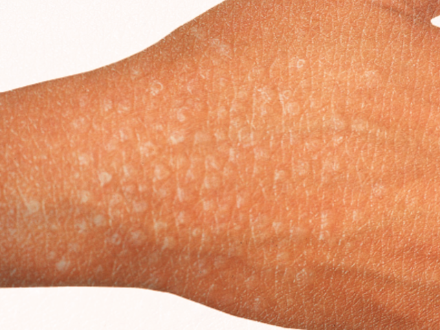
Lichen Nitidus. This photo depicts multiple discrete, glistening, skin-colored, flat-topped papules characteristic of lichen nitidus.
Contributed by S Bhimji, MD
References
Cho EB, Kim HY, Park EJ, Kwon IH, Kim KH, Kim KJ. Three cases of lichen nitidus associated with various cutaneous diseases. Annals of dermatology. 2014 Aug:26(4):505-9. doi: 10.5021/ad.2014.26.4.505. Epub 2014 Jul 31 [PubMed PMID: 25143682]
Level 3 (low-level) evidenceKataria V, Singal A, Arora VK. Lichen Nitidus Associated with Onychodystrophy and Response to Therapy: Report of Two Cases. Skin appendage disorders. 2019 Apr:5(3):158-161. doi: 10.1159/000493534. Epub 2018 Oct 10 [PubMed PMID: 31049338]
Level 3 (low-level) evidenceSynakiewicz J, Polańska A, Bowszyc-Dmochowska M, Żaba RW, Adamski Z, Reich A, Dańczak-Pazdrowska A. Generalized lichen nitidus: a case report and review of the literature. Postepy dermatologii i alergologii. 2016 Dec:33(6):488-490. doi: 10.5114/ada.2016.63890. Epub 2016 Dec 2 [PubMed PMID: 28035230]
Level 3 (low-level) evidencePayette MJ, Weston G, Humphrey S, Yu J, Holland KE. Lichen planus and other lichenoid dermatoses: Kids are not just little people. Clinics in dermatology. 2015 Nov-Dec:33(6):631-43. doi: 10.1016/j.clindermatol.2015.09.006. Epub 2015 Sep 12 [PubMed PMID: 26686015]
Botelho LF, Magalhães JP, Ogawa MM, Enokihara MM, Cestari Sda C. Generalized Lichen nitidus associated with Down's syndrome: case report. Anais brasileiros de dermatologia. 2012 May-Jun:87(3):466-8 [PubMed PMID: 22714765]
Level 3 (low-level) evidenceRallis E, Verros C, Moussatou V, Sambaziotis D, Papadakis P. Generalized purpuric lichen nitidus. Report of a case and review of the literature. Dermatology online journal. 2007 May 1:13(2):5 [PubMed PMID: 17498424]
Level 3 (low-level) evidenceCho M, Nonomura Y, Kaku Y, Dainichi T, Otsuka A, Kabashima K. Generalized Lichen Nitidus Following Anti-PD-1 Antibody Treatment. JAMA dermatology. 2018 Mar 1:154(3):367-369. doi: 10.1001/jamadermatol.2017.5670. Epub [PubMed PMID: 29344609]
Do MO, Kim MJ, Kim SH, Myung KB, Choi YW. Generalized lichen nitidus successfully treated with narrow-band UVB phototherapy: two cases report. Journal of Korean medical science. 2007 Feb:22(1):163-6 [PubMed PMID: 17297274]
Level 3 (low-level) evidenceMalakar S, Save S, Mehta P. Brown Shadow in Lichen Nitidus: A Dermoscopic Marker! Indian dermatology online journal. 2018 Nov-Dec:9(6):479-480. doi: 10.4103/idoj.IDOJ_338_17. Epub [PubMed PMID: 30505802]
Shockman S, Lountzis N. Lichen nitidus. Cutis. 2013 Dec:92(6):288, 297-8 [PubMed PMID: 24416748]
Palaniappan V, Karthikeyan K. Lichen nitidus. Clinical and experimental dermatology. 2025 Jun 25:50(7):1305-1314. doi: 10.1093/ced/llaf048. Epub [PubMed PMID: 39882979]
Polcari IC, Mancini AJ, Stein SL. Facial hypopigmentation in skin of color: An atypical presentation of lichen nitidus. Pediatric dermatology. 2021 Nov:38 Suppl 2():110-112. doi: 10.1111/pde.14692. Epub 2021 Jul 16 [PubMed PMID: 34272759]
Qian G, Wang H, Wu J, Meng Z, Xiao C. Different dermoscopic patterns of palmoplantar and nonpalmoplantar lichen nitidus. Journal of the American Academy of Dermatology. 2015 Sep:73(3):e101-3. doi: 10.1016/j.jaad.2015.06.024. Epub [PubMed PMID: 26282806]
Level 2 (mid-level) evidenceDurusu İN, Güler D, Gürel G, Yalçın GŞ. A very rare localization of a rare disease: palmar lichen nitidus. Anais brasileiros de dermatologia. 2022 Jan-Feb:97(1):96-98. doi: 10.1016/j.abd.2021.03.009. Epub 2021 Nov 24 [PubMed PMID: 34836740]
Gustafson D, Wolf JE, Lester L, Sander H. Perforating lichen nitidus in the setting of atopic dermatitis. Journal of cutaneous pathology. 2024 Feb:51(2):108-113. doi: 10.1111/cup.14545. Epub 2023 Oct 12 [PubMed PMID: 37828706]
Taneja N, Mehta N, Arava S, Gupta V. An unusual variant of lichen nitidus: Generalized follicular spinous with perifollicular granulomas. Journal of cutaneous pathology. 2020 Sep:47(9):834-839. doi: 10.1111/cup.13712. Epub 2020 Jul 2 [PubMed PMID: 32285461]
Chu J, Lam JM. Lichen nitidus. CMAJ : Canadian Medical Association journal = journal de l'Association medicale canadienne. 2014 Dec 9:186(18):E688. doi: 10.1503/cmaj.140434. Epub 2014 Oct 27 [PubMed PMID: 25349001]
Level 3 (low-level) evidenceYáñez S, Val-Bernal JF. Purpuric generalized lichen nitidus: an unusual eruption simulating pigmented purpuric dermatosis. Dermatology (Basel, Switzerland). 2004:208(2):167-70 [PubMed PMID: 15057011]
Level 3 (low-level) evidenceBlalock TW, Kannan S, Davis LS. Actinic lichen nitidus. Dermatology reports. 2010 Aug 31:2(2):e10. doi: 10.4081/dr.2010.e10. Epub 2010 Jul 13 [PubMed PMID: 25386247]
Level 3 (low-level) evidenceTopal IO, Gokdemir G, Sahin IM. Generalized lichen nitidus: successful treatment with systemic isotretinoin. Indian journal of dermatology, venereology and leprology. 2013 Jul-Aug:79(4):554. doi: 10.4103/0378-6323.113108. Epub [PubMed PMID: 23760340]
Level 3 (low-level) evidenceKumar L, Gupta LK, Balai M, Chouhan N. Generalized Lichen Nitidus Successfully Treated with Cyclosporine. Indian dermatology online journal. 2026 Jan 1:17(1):105-106. doi: 10.4103/idoj.idoj_881_24. Epub 2025 Sep 9 [PubMed PMID: 40924681]