 Tools
Tools
 Leishmaniasis
Introduction
Leishmaniasis is a disease caused by protozoan parasites of the genus Leishmania and is most commonly transmitted through the bites of infected sand flies. Leishmaniasis has historically been widespread in tropical climates across multiple continents, including the Americas, East Africa, the Middle East, Central Asia, and India. Leishmaniasis is broadly classified into 2 main forms: cutaneous and visceral.
Leishmaniasis is an ancient disease, with a history extending from prehistoric origins in the Americas and Africa to the present day. Results from archeological and molecular studies showed that cutaneous and mucocutaneous leishmaniasis existed as early as 2500 BC in Egyptian and Christian Nubian mummies. During the Middle Kingdom, Egyptian trade and military expeditions to Nubia (modern Sudan) are thought to have introduced leishmaniasis into Egypt, because samples from earlier periods did not contain detectable DNA.
Furthermore, some sources identify Sudan as the original focus of visceral leishmaniasis.[1] Additionally, the Ebers Papyrus, a medical manuscript dating to 1500 BC, described a condition thought to be cutaneous leishmaniasis, then termed the Nile pimple. Based on this ancient understanding of the disease, practitioners attempted an early form of vaccination in the Middle East and Central Asia. The procedure involved collecting exudate from active lesions and inoculating the buttocks of children.[2]
Over the next 1000 years, cutaneous sores consistent with leishmaniasis were described, including a condition in the region now known as northern Afghanistan, termed the Balkh sore, attributed to infection with Leishmania tropica. In Asia and the Middle East, the disease continued to be documented under names such as the Aleppo boil, Jericho boil, and Baghdad boil. Many of these names remain in use today.[2] The infectious nature of leishmaniasis was first recognized when a Scottish clinician observed parasites in a Delhi boil. However, similar findings reported by the Russian clinician Piotr Fokich Borovsky led to the identification of the organisms in Asian sores as protozoa, a finding published in 1898. In 1900, a British pathologist observed ovoid bodies, proposed that they represented degenerated trypanosomes, and termed the illness dum-dum fever.
Around the same time, Irish clinician Charles Donovan published a paper on similar ovoid bodies from the spleens of patients in India. An investigation into kala-azar and the splenic ovoid bodies described by the pathologist Leishman and clinician Donovan concluded that these ovoid bodies were not degenerated trypanosomes but represented a new protozoan species later termed Leishmania donovani. Over the next decades, additional species were identified, including Leishmania tropica and Leishmania aethiopica. Classification of the disease into New World and Old World leishmaniasis was subsequently based on these organisms and their geographic distribution.[2]
Etiology
Register For Free And Read The Full Article
 Search engine and full access to all medical articles
- 10 free questions in your specialty
- Free CME/CE Activities
- Free daily question in your email
- Save favorite articles to your dashboard
- Emails offering discounts
Learn more about a Subscription to StatPearls Point-of-Care
Etiology
Leishmaniasis is a disease caused by a protozoan parasite in the order Trypanosomatida, family Trypanosomatidae, and genus Leishmania. Transmission occurs most commonly through the bites of female Phlebotomus sand flies in the Eastern Hemisphere (Old World) and female Lutzomyia sand flies in the Western Hemisphere (New World). More than 20 Leishmania species are pathogenic to humans. Species commonly associated with visceral leishmaniasis include Leishmania donovani and L infantum, whereas species associated with cutaneous leishmaniasis include L major, L mexicana, L tropica, and L aethiopica. L donovani is found throughout Central Africa, South Asia, the Middle East, India, and Bangladesh. L major is endemic to North Africa, the Middle East, Central Asia, and northwestern India and China, while L mexicana is highly endemic in Central America and Mexico and is also endemic in Texas, United States.[2][3][4]
The 2 developmental stages are the amastigote and the promastigote, with the former infecting lysosomal vacuoles in phagocytic cells. The promastigote is an extracellular form that attaches to the insect vector’s microvilli. Adult sand flies are very small, measuring approximately one-third the size of a small mosquito and less than 3.5 mm in length.
Because sand flies are susceptible to dehydration, they thrive in moist climates, which helps explain the geographic distribution of the disease. Sand flies are nocturnal and rest during the day in burrows, under rocks, or in other shelters. At rest, their wings form a characteristic V shape over the back, and a distinct thoracic hump angles the head downward (see Image. Phlebotomus papatasi Sand Fly After a Blood Meal). Both male and female sand flies obtain carbohydrates from plant juices, whereas only females require a blood meal and can transmit leishmaniasis. During the blood meal, an infected female sand fly transmits the protozoan parasites to the human host.[5]
Epidemiology
Leishmaniasis is endemic in tropical and subtropical regions of at least 89 countries across Africa, Asia, the Middle East, the Mediterranean, and Central and South America.[6] The World Health Organization estimates that more than 1 billion people live in areas endemic for leishmaniasis and are at risk of infection, with an estimated 30,000 new cases of visceral leishmaniasis and more than 1 million new cases of cutaneous leishmaniasis occurring annually.[WHO. Leishmaniasis] Clinical manifestations correlate strongly with the infecting Leishmania species and its geographic distribution.
Because L donovani is the most common cause of visceral leishmaniasis and occurs throughout Africa, the Middle East, and Asia, most cases of visceral leishmaniasis arise in the Eastern Hemisphere, with Brazil representing a notable exception. More than 90% of global cases of visceral leishmaniasis occur in Brazil, Ethiopia, India, Somalia, South Sudan, and Sudan, whereas more than 90% of cutaneous leishmaniasis cases occur in 11 countries, predominantly Afghanistan, Algeria, Brazil, Colombia, Iran, and Syria. Risk factors for leishmaniasis include deforestation, urbanization, and climate change, which alter sand fly habitats and increase human-vector contact.[7] Additional risk factors include malnutrition, poor housing, and a lack of bed nets or window screens.[8]
Human immunodeficiency virus infection, organ transplant, and the use of immunosuppressive medications also increase the risk of leishmaniasis.[9] Additionally, conflict, famine, and population migration into highly endemic areas may increase the occurrence of leishmaniasis.[10] Seasonal peaks in visceral and cutaneous leishmaniasis occur during spring and are likely associated with temperature, humidity, and vector behavior.[11]
Pathophysiology
Leishmania parasites are transmitted by sand fly bites, with the genera Phlebotomus and Lutzomyia serving as the most common vectors in the Eastern and Western Hemispheres, respectively.[2] Only female sand flies require blood meals and can transmit leishmaniasis. During these meals, Leishmania parasites may be acquired from an infected host or transmitted to a susceptible host.
When a previously uninfected sand fly feeds on an infected host, the insect uses its saw-like mouthparts to penetrate the skin and create a small wound in which blood from injured capillaries pools. The infectious promastigote enters the sand fly foregut and replicates (see Image. Life Cycle of Leishmania Parasites).[6][12][13] Subsequent feeding on dogs, rodents, marsupials, or humans transmits the parasites to a new host.[2]
Once the protozoa enter the host, they localize within phagolysosomes. The type of infected phagocytic cell influences whether cutaneous or visceral leishmaniasis develops. In cutaneous leishmaniasis, the parasites infect resident macrophages within the skin, and once an infected macrophage becomes filled with amastigotes, the cell ruptures, releasing the organisms and promoting infection of adjacent macrophages. Visceral leishmaniasis differs because amastigotes disseminate hematogenously to mononuclear cells in the liver, spleen, bone marrow, and intestinal lymph nodes.[2]
Histopathology
Histopathologic examination of tissue affected by leishmaniasis reveals a dense, chronic inflammatory infiltrate containing numerous infected macrophages. The hallmark of the disease is the presence of numerous small, ovoid or round amastigotes within these macrophages. Amastigotes are small, round or ovoid structures measuring 2 to 4 µm. Each amastigote contains a distinct nucleus and a tiny rod-shaped structure called a kinetoplast. The organisms frequently cluster around the periphery of the macrophage vacuole, producing a pattern resembling a carousel or marquee (see Image. Leishmania donovani Amastigotes in a Bone Marrow Histiocyte).
History and Physical
Clinical manifestations of leishmaniasis are traditionally classified into 3 main syndromes: cutaneous, mucocutaneous, and visceral.[11][14]
Cutaneous Leishmaniasis
Cutaneous disease is the most common manifestation of leishmaniasis and is further subdivided into localized cutaneous and diffuse cutaneous disease. According to the World Health Organization, localized cutaneous leishmaniasis has an incubation period from 2 weeks to 6 months.[WHO. Leishmaniasis] Cutaneous lesions develop at the site of the sand fly bite, manifesting as an asymptomatic papule, multiple papules, or nodules. The lesions enlarge and progress to well-circumscribed ulcers with a raised violaceous border and epidermal breakdown (see Image. Ulcerative Lesion of Cutaneous Leishmaniasis). These lesions often heal spontaneously within 2 to 5 years, depending on the species, and leave a depressed scar.[6][14]
Diffuse cutaneous leishmaniasis also begins as a painless nodule but may progress to involve the entire cutaneous surface. The disease has a predilection for the face, ears, and extensor surfaces such as the knees and elbows. Invasion of the nasopharyngeal and oral mucosa may occur in up to one-third of individuals. Leishmania species most commonly associated with diffuse cutaneous leishmaniasis are L aethiopica in the Eastern Hemisphere and L mexicana in the Western Hemisphere. Skin lesions may further progress to diffuse hypopigmented plaques and patches.
Mucocutaneous Leishmaniasis
The incubation period for mucosal disease ranges from several months to a few years after the initial cutaneous sores have healed.[WHO. Leishmaniasis] Mucosal disease occurs when the parasite spreads to the mucous membranes of the nose and throat, most commonly through hematogenous or lymphatic spread. Mucosal manifestations usually develop within 2 years but may be delayed for decades. L braziliensis accounts for the majority of mucocutaneous disease, although other species can be implicated. The oral and nasal mucosa are preferentially affected, although ulcerative involvement may extend to the vocal cords and tracheal cartilage. Mucosal disease may be severe and life-threatening.[14]
Visceral Leishmaniasis
Visceral leishmaniasis, first described in India during the 1800s, is also known as kala-azar.[2][11][14] The incubation period is generally 2 to 8 months but may range from 10 days to more than 2 years.[WHO. Leishmaniasis] Clinical findings associated with visceral involvement commonly include fever, splenomegaly, hypergammaglobulinemia, and pancytopenia. These manifestations often result from direct infection of the liver, spleen, bone marrow, or other viscera. The most common causative species are L donovani, L infantum, and L chagasi.[11][14] Occasionally, cutaneous involvement may develop after treatment of visceral leishmaniasis and present as a papular rash on the face and upper extremities. Post–kala-azar dermal leishmaniasis is the term for this manifestation, and the lesions are nondisfiguring and self-limited.[14]
Evaluation
Cutaneous Leishmaniasis
Diagnosis of cutaneous leishmaniasis most commonly relies on parasite identification through histopathologic examination of fixed tissue, culture, or molecular analysis using polymerase chain reaction (PCR). Tissue obtained from the margin of a cutaneous ulcer provides the highest diagnostic yield. Giemsa staining is typically used, and CD1a immunostaining can highlight Leishmania amastigotes.[15] Results from a study comparing scraping smears with fine-needle aspiration cytology showed that needle aspiration improved amastigote detection and patient comfort.[16]
A simplified collection technique, known as the press-imprint smear or touch preparation method, can also be used. Results from a study comparing this technique with histopathologic examination for cutaneous disease showed significantly greater sensitivity with this technique. The technique uses a 3-mm punch biopsy, similar to the specimen obtained for formalin-fixed histopathologic examination. With the press imprint smear method, the tissue fragment is positioned longitudinally between 2 glass slides, with the epidermis perpendicular to the slides, and compressed.
Pressure applied to the center of the slides spreads tissue fluid and material across both surfaces. After the slides are separated, the tissue is removed, and the slides are allowed to air-dry. The slides are then fixed with methanol, stained with Giemsa, and examined microscopically using an oil-immersion objective.[17] Histopathologic examination has sensitivity values ranging from approximately 40% to 70% and specificity values approaching 100%.[18][19]
Culture may also be performed and is most useful for skin lesions that have been present for 3 to 12 months.[20] Novy-MacNeal-Nicolle (NNN) medium is typically used and consists of a solid blood agar slant overlaid with liquid NNN medium. Monophasic liquid media, such as Schneider insect medium, Roswell Park Memorial Institute 1640 medium, or medium 199 supplemented with fetal bovine serum, are typically used to amplify parasite numbers. Culture provides specificity values approaching 100%, but sensitivity values are relatively low at 50% to 75%. The limited sensitivity primarily results from a low parasite burden in some specimens.[21]
Molecular testing with PCR yields higher sensitivity values than either histopathologic examination or culture. Results from one systematic review and meta-analysis found a pooled sensitivity of 95% and a pooled specificity of 91% for PCR. Results from the review found no difference in diagnostic accuracy when PCR was performed on aspirates, skin biopsy specimens, or smears, including press-imprint and touch-preparation smears, suggesting that PCR analysis of a simple smear may provide the least invasive and most effective diagnostic strategy.[22]
Serologic tests, including enzyme-linked immunosorbent assays, Western blot testing, and direct agglutination tests, are rarely used because the limited humoral response yields low sensitivity. Intradermal skin testing has demonstrated good sensitivity and specificity, with a delayed-type hypersensitivity reaction measuring 5 mm considered a positive result. However, skin testing cannot distinguish a previous infection from a current one.
Visceral Leishmaniasis
Diagnosis of visceral leishmaniasis similarly relies on parasite identification through histopathologic examination of fixed tissue, culture, or PCR. Histopathologic sampling requires biopsy or needle aspiration of an affected organ. Bone marrow is the preferred diagnostic specimen because splenic aspiration carries a rare but potentially fatal risk of hemorrhage.[23]
Alternatively, splenic or lymph node biopsy may be performed. Giemsa staining and CD1a immunostaining of aspirated material should be performed in addition to inoculation of biopsy material into culture medium. Histopathologic examination yields specificity values of approximately 100%, but sensitivity values may be as low as 50%. Diagnostic accuracy varies by tissue type, with splenic aspirates yielding the highest sensitivity and bone marrow aspirates the lowest.[24]
Culture should be performed using NNN medium or another parasitic growth medium. Culture sensitivity varies by sampling site and exceeds 93% for splenic aspirates. Sensitivity values range from 53% to 86% for bone marrow aspirates and from 52% to 58% for lymph node aspirates.
PCR testing also achieves high sensitivity in the diagnosis of visceral leishmaniasis. The highest sensitivity values are observed with splenic and bone marrow aspirates, whereas peripheral blood yields slightly lower values. Results from one systematic review found a pooled sensitivity of 93% and a pooled specificity of 95% for PCR. The pooled sensitivity and specificity values were 93% and 96% for peripheral blood, and 95% and 93% for bone marrow, respectively.[25]
Treatment / Management
Cutaneous Leishmaniasis
Limited cutaneous leishmaniasis often resolves clinically without treatment. Clinical observation alone is a reasonable approach for immunocompetent individuals with lesions that are healing spontaneously. Local therapy is reasonable for individuals who desire treatment, have lesions that are not healing spontaneously, or have lesions that remain unhealed after 4 to 6 weeks of observation.
Systemic treatment should be initiated for individuals with complicated cutaneous leishmaniasis, individuals who are immunocompromised, and individuals at risk of mucocutaneous leishmaniasis. Local therapy includes pentavalent antimonials (eg, sodium stibogluconate and meglumine antimoniate) and paromomycin (eg, paromomycin ointment or cream). Pentavalent antimonials are injected intralesionally every 7 days for up to 3 weeks. Topical paromomycin is applied for 20 days.
Systemic therapy may include oral fluconazole or ketoconazole, oral miltefosine, or parenteral treatment with one of the pentavalent antimonials, amphotericin, or pentamidine isethionate. Fluconazole is administered daily for 6 weeks, whereas ketoconazole and miltefosine are administered daily for 28 days. Pentavalent antimonials are administered for 10 to 20 days, amphotericin for 25 to 30 days, and pentamidine isethionate weekly for 3 doses.
Mucocutaneous Leishmaniasis
Mucocutaneous leishmaniasis requires prompt treatment because the disease is expected to progress and cause tissue destruction if left untreated. Treatment options include pentavalent antimonials, amphotericin, miltefosine, and pentamidine. Sodium stibogluconate is administered intravenously or intramuscularly daily for 28 days, whereas meglumine antimoniate is administered intravenously or intramuscularly daily for 30 days. Miltefosine is administered for 28 days, and pentamidine isethionate is administered intravenously or intramuscularly for at least 15 doses and until the lesions heal.
Visceral Leishmaniasis
Visceral leishmaniasis requires treatment. For immunocompetent individuals in East Africa, the preferred regimen combines intravenous or intramuscular sodium stibogluconate daily with paromomycin daily for 17 days. Alternative regimens include intravenous liposomal amphotericin B administered daily for 10 days or oral miltefosine plus intramuscular paromomycin daily for 14 days. In South Asia, intravenous liposomal amphotericin B is administered on days 1 to 5, 14, and 21. Oral miltefosine administered daily for 28 days is an alternative. In Europe and the Americas, intravenous liposomal amphotericin B administered daily for 7 days is the preferred treatment.
For people living with HIV infection in East Africa, the preferred regimen consists of intravenous liposomal amphotericin B administered on days 1, 3, 5, 7, 9, and 11 plus oral miltefosine for 28 days. In South Asia, liposomal amphotericin B is administered on days 1, 3, 5, 7, 9, and 11, whereas miltefosine is administered daily for 14 days. In Europe and the Americas, liposomal amphotericin B is administered to achieve a total dose of 40 mg/kg.
Differential Diagnosis
Cutaneous Leishmaniasis
- Leprosy
- Cutaneous tuberculosis
- Nontuberculous mycobacterial infections
- Syphilis
- Deep fungal infections
- Basal cell carcinoma or squamous cell carcinoma
- Granuloma annulare
Mucocutaneous Leishmaniasis
- Syphilis
- Yaws
- Rhinoscleroma
- Granulomatosis with polyangiitis
- Oral squamous cell carcinoma
Visceral Leishmaniasis
- Malaria
- Typhoid fever
- Brucellosis
- Miliary tuberculosis
- Leukemia
- Lymphoma
- Schistosomiasis
Pertinent Studies and Ongoing Trials
As of July 2026, the following clinical trials were ongoing:
Cutaneous Leishmaniasis
- NCT07652021 is a single-center randomized controlled trial comparing healing outcomes between intralesional meglumine antimoniate alone and intralesional meglumine antimoniate combined with fractional carbon dioxide laser therapy in adults with cutaneous leishmaniasis.
- NCT07463040 is a phase 3, multicenter, multiarm, multistage randomized controlled trial comparing intramuscular sodium stibogluconate with oral miltefosine, oral miltefosine plus intramuscular paromomycin, intravenous liposomal amphotericin B, and intravenous pentamidine isethionate for the treatment of cutaneous leishmaniasis.
- NCT04268524 is a phase 3, multicenter, randomized open-label noninferiority trial evaluating oral miltefosine, thermotherapy, oral miltefosine plus thermotherapy, and intralesional meglumine antimoniate for the treatment of cutaneous leishmaniasis caused by L tropica.
- NCT06997159 is a phase 2, multicenter, randomized observer-blinded trial evaluating the efficacy, safety, and pharmacokinetics of 2 LXE408 regimens compared with miltefosine in participants with localized cutaneous leishmaniasis.
- NCT07149753 is a phase 1, single-center trial evaluating the safety and dosing of topical miltefosine gel in individuals with uncomplicated cutaneous leishmaniasis.
- NCT06798402 is a phase 2, single-center, randomized open-label trial comparing the efficacy of intralesional ciprofloxacin with intralesional sodium stibogluconate for the treatment of cutaneous leishmaniasis.
- NCT06797544 is a phase 2, single-center, single-group, open-label trial evaluating the efficacy of intralesional levofloxacin for the treatment of cutaneous leishmaniasis.
- NCT06798415 is a phase 2, single-center, single-group, open-label trial evaluating the efficacy of intralesional metronidazole for the treatment of cutaneous leishmaniasis.
- NCT06822478 is a phase 3, single-center, randomized single-blind trial evaluating the safety and efficacy of topical arnica tincture for the treatment of cutaneous leishmaniasis.
Mucocutaneous Leishmaniasis
- NCT06550609 is a phase 2, single-center, open-label, proof-of-concept trial evaluating inhaled pentamidine in combination with oral miltefosine for the treatment of mucosal leishmaniasis caused by L braziliensis.
Prognosis
Limited cutaneous leishmaniasis often resolves spontaneously within 3 to 18 months, depending on the host's innate immune response. If treatment is initiated, the prognosis is also excellent, particularly when treatment begins early. Conversely, mucocutaneous and visceral leishmaniasis can be life-threatening and, without treatment, may progress with high mortality rates.
With prompt treatment, the prognosis for mucocutaneous and visceral leishmaniasis is generally favorable, with cure rates exceeding 75% and 95%, respectively. HIV infection is one of the strongest predictors of a poor prognosis, with a 5-fold increase in mortality compared with individuals without human immunodeficiency virus infection. Other factors associated with increased mortality include tuberculosis, extremes of age, severe malnutrition, and prolonged illness.[26]
Complications
Complications of leishmaniasis range from severe skin disfigurement to life-threatening organ failure. Cutaneous leishmaniasis can result in severe scarring, secondary bacterial infections, and leishmaniasis recidivans, a condition in which the initial skin lesions appear to heal but recur years later along the edge of the scar. Mucocutaneous leishmaniasis can result in tissue disfigurement, laryngeal erosion with breathing difficulties, and difficulty speaking, eating, and swallowing. Visceral leishmaniasis can result in life-threatening infections, pancytopenia, immunodeficiency, weight loss and malnutrition, hemorrhage, and post–kala-azar dermal leishmaniasis, a condition in which Leishmania parasites persist in the skin after treatment, causing disfiguring rashes, nodules, and lesions.
Deterrence and Patient Education
Initial preventive measures focus on avoiding sand fly bites. Awareness of endemic areas, nocturnal sand fly activity, and animal exposure in leishmaniasis-endemic areas is important. Because sand flies are very small, bed nets require a mesh size approximately 3 times smaller than standard. Treating bed nets with permethrin may further decrease the risk of infection. Results from studies showed that vaccinating dogs and using insecticidal dog collars decreased the disease burden.[27]
Enhancing Healthcare Team Outcomes
Leishmaniasis is a global disease, and efforts to provide education, diagnostic tools, and treatment in endemic areas are needed. Coordination through the World Health Organization and findings published by countries worldwide have provided important insights into treatment, drug resistance, and transmission. Individual treatments have been evaluated worldwide.
The efficacy of amphotericin in the treatment of visceral leishmaniasis is well established.[28] Results from studies of populations in India demonstrated that miltefosine, the only oral agent approved for the treatment of visceral leishmaniasis, had efficacy similar to that of amphotericin.[29] The pentavalent antimonial compound sodium stibogluconate is another commonly used agent with favorable treatment results.[30] For individuals living with HIV, results from studies showed that antiretroviral treatment significantly decreased the risk of relapse after visceral leishmaniasis.[31]
Media
(Click Image to Enlarge)
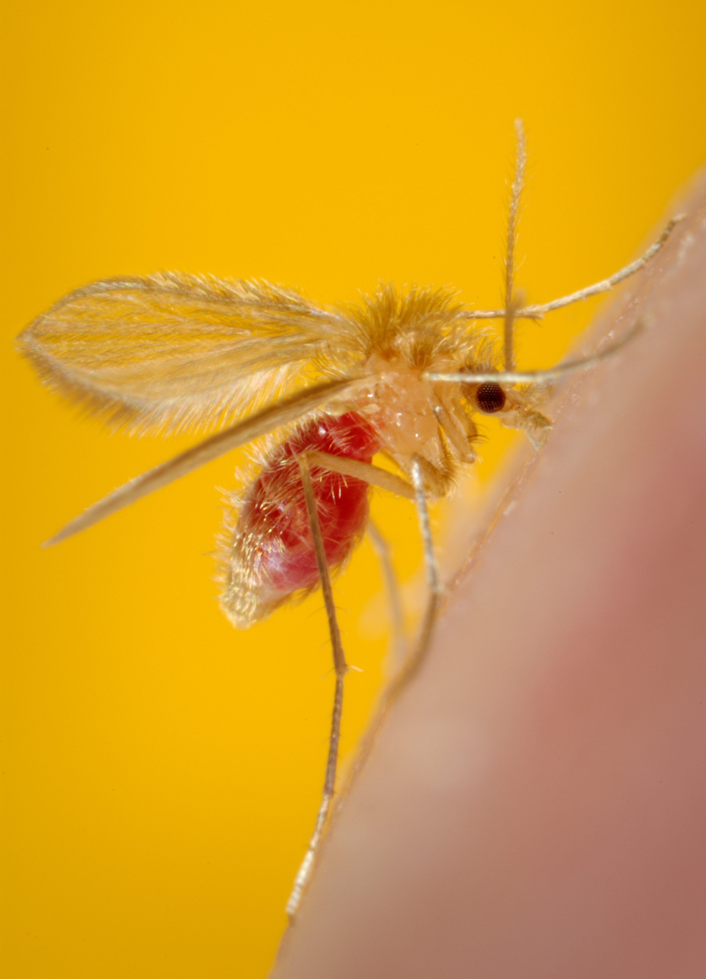
Female Phlebotomus papatasi Sand Fly After a Blood Meal. This sand fly had just completed its blood meal, as evidenced by its distended, transparent abdomen. Sand flies like this P papatasi are responsible for the spread of the vector-borne, parasitic disease leishmaniasis, which is caused by the obligate intracellular protozoa of the genus Leishmania.
Frank Hadley Collins, Public Health Image Library, Public Domain, Centers for Disease Control and Prevention
(Click Image to Enlarge)
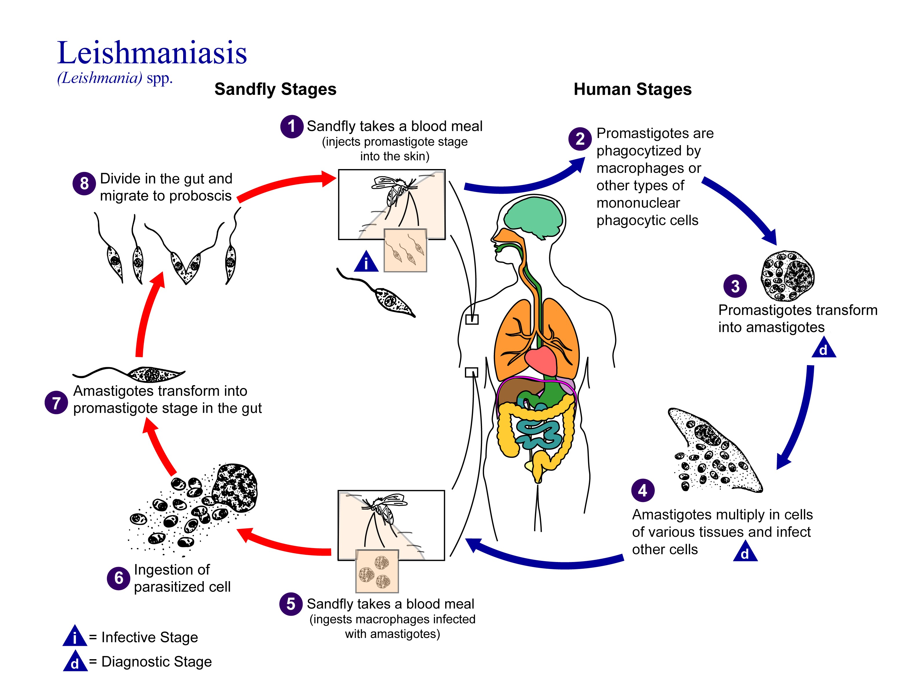
Life Cycle of Leishmania Parasites. The transmission of Leishmania parasites between sand flies and humans starts when promastigotes are injected during a blood meal, transform into intracellular amastigotes within human phagocytic cells, and are subsequently ingested by another sand fly, where they develop into promastigotes and migrate to the proboscis.
Alexander J da Silva, Public Health Image Library, Public Domain, Centers for Disease Control and Prevention
(Click Image to Enlarge)
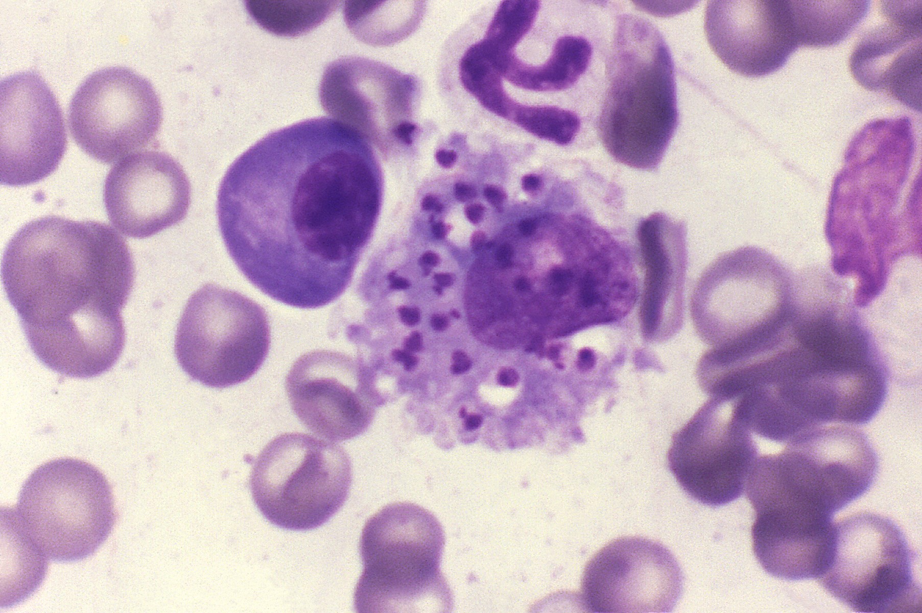
Leishmania donovani Amastigotes in a Bone Marrow Histiocyte. Giemsa-stained canine bone marrow smear demonstrates numerous intracellular Leishmania donovani amastigotes within a histiocyte. The small organisms contain characteristic nuclei and kinetoplasts.
Public Health Image Library, Public Domain, Centers for Disease Control and Prevention
(Click Image to Enlarge)
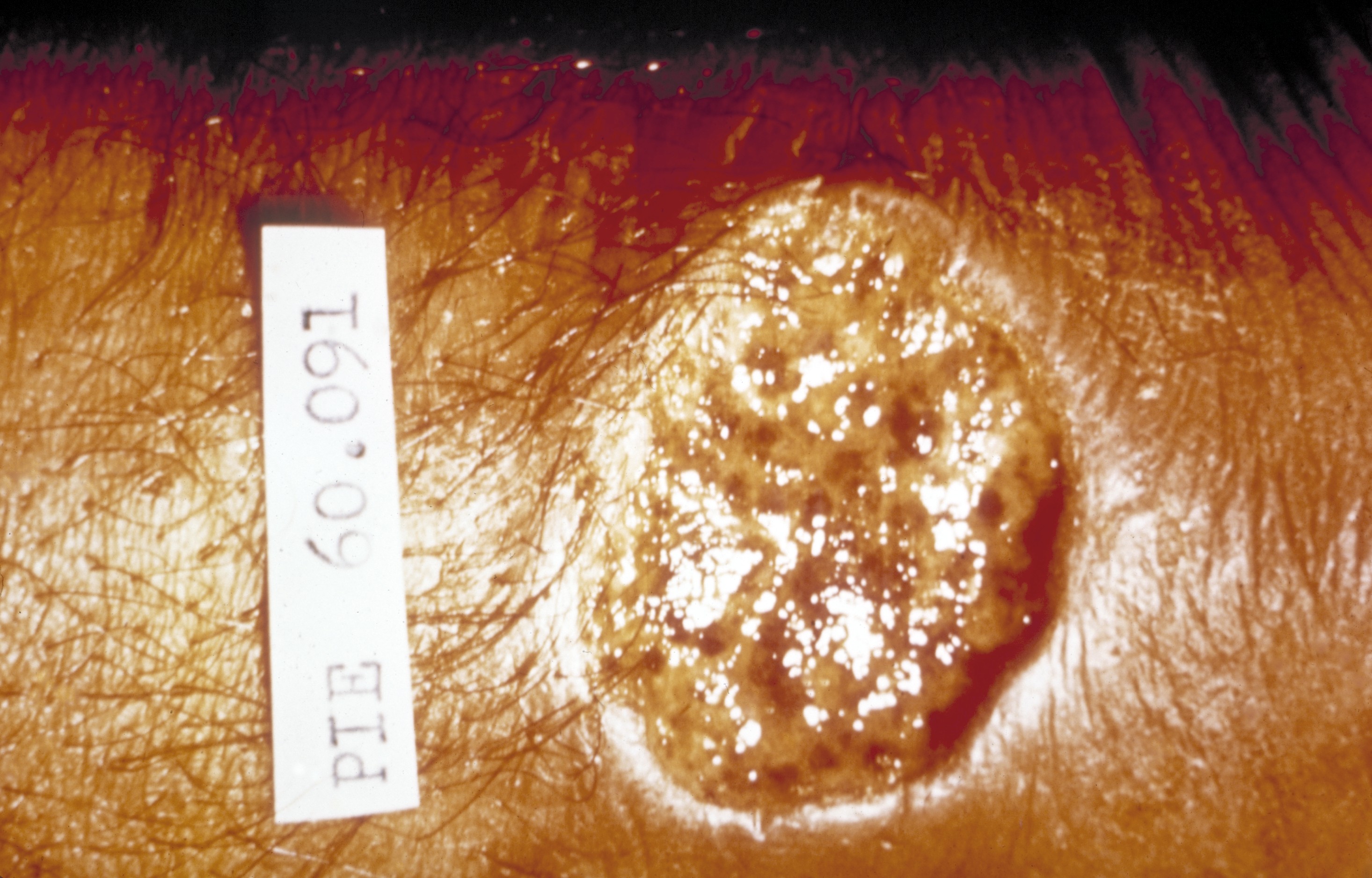
Ulcerative Lesion of Cutaneous Leishmaniasis. A large, well-circumscribed cutaneous ulcer with a raised border and moist, irregular base is characteristic of ulcerative cutaneous leishmaniasis.
Public Health Image Library, Public Domain, Centers for Disease Control and Prevention
References
Zink AR, Spigelman M, Schraut B, Greenblatt CL, Nerlich AG, Donoghue HD. Leishmaniasis in ancient Egypt and Upper nubia. Emerging infectious diseases. 2006 Oct:12(10):1616-7 [PubMed PMID: 17176592]
Level 3 (low-level) evidenceSteverding D. The history of leishmaniasis. Parasites & vectors. 2017 Feb 15:10(1):82. doi: 10.1186/s13071-017-2028-5. Epub 2017 Feb 15 [PubMed PMID: 28202044]
Sosa-Bibiano EI, Sánchez-Martínez LA, López-Ávila KB, Chablé-Santos JB, Torres-Castro JR, Fernández-Figueroa EA, Rangel-Escareño C, Loría-Cervera EN. Leishmania (Leishmania) mexicana Infection in Wild Rodents from an Emergent Focus of Cutaneous Leishmaniasis in Yucatan, Mexico. Journal of tropical medicine. 2022:2022():8392005. doi: 10.1155/2022/8392005. Epub 2022 May 31 [PubMed PMID: 35686208]
Hopke K, Meyers A, Auckland L, Hamer S, Florin D, Diesel A, Patterson A. Leishmania mexicana in a central Texas cat: clinical presentation, molecular identification, sandfly vector collection and novel management. JFMS open reports. 2021 Jan-Jun:7(1):2055116921999595. doi: 10.1177/2055116921999595. Epub 2021 Mar 22 [PubMed PMID: 33815814]
Claborn DM. The biology and control of leishmaniasis vectors. Journal of global infectious diseases. 2010 May:2(2):127-34. doi: 10.4103/0974-777X.62866. Epub [PubMed PMID: 20606968]
Torres-Guerrero E, Quintanilla-Cedillo MR, Ruiz-Esmenjaud J, Arenas R. Leishmaniasis: a review. F1000Research. 2017:6():750. doi: 10.12688/f1000research.11120.1. Epub 2017 May 26 [PubMed PMID: 28649370]
Carrer DC, Papera F, Ríos DN. Cutaneous leishmaniasis: epidemiology, treatment access and translational challenges of topical therapies. Frontiers in microbiology. 2025:16():1588311. doi: 10.3389/fmicb.2025.1588311. Epub 2026 Jan 12 [PubMed PMID: 41602766]
Heidari A, Dashtaki NM, Mizbani S, Rejali M, Maracy MR. Residential environment, human behavior and socio-economic status in transmission of cutaneous leishmaniasis in central Iran. Scientific reports. 2025 Mar 1:15(1):7271. doi: 10.1038/s41598-025-91999-6. Epub 2025 Mar 1 [PubMed PMID: 40025204]
Georgiadou SP, Makaritsis KP, Dalekos GN. Leishmaniasis revisited: Current aspects on epidemiology, diagnosis and treatment. Journal of translational internal medicine. 2015 Apr-Jun:3(2):43-50 [PubMed PMID: 27847886]
Kato H. Epidemiology of Leishmaniasis: Risk factors for its pathology and infection. Parasitology international. 2025 Apr:105():102999. doi: 10.1016/j.parint.2024.102999. Epub 2024 Nov 26 [PubMed PMID: 39592080]
Tofighi Naeem A, Mahmoudi S, Saboui F, Hajjaran H, Pourakbari B, Mohebali M, Zarkesh MR, Mamishi S. Clinical Features and Laboratory Findings of Visceral Leishmaniasis in Children Referred To Children Medical Center Hospital, Tehran, Iran during 2004-2011. Iranian journal of parasitology. 2014 Mar:9(1):1-5 [PubMed PMID: 25642253]
Handman E, Bullen DV. Interaction of Leishmania with the host macrophage. Trends in parasitology. 2002 Aug:18(8):332-4 [PubMed PMID: 12377273]
Level 3 (low-level) evidenceRogers ME, Chance ML, Bates PA. The role of promastigote secretory gel in the origin and transmission of the infective stage of Leishmania mexicana by the sandfly Lutzomyia longipalpis. Parasitology. 2002 May:124(Pt 5):495-507 [PubMed PMID: 12049412]
Level 3 (low-level) evidenceHandler MZ, Patel PA, Kapila R, Al-Qubati Y, Schwartz RA. Cutaneous and mucocutaneous leishmaniasis: Differential diagnosis, diagnosis, histopathology, and management. Journal of the American Academy of Dermatology. 2015 Dec:73(6):911-26; 927-8. doi: 10.1016/j.jaad.2014.09.014. Epub [PubMed PMID: 26568336]
Riyal H, Samaranayake N, Amarathunga P, Munidasa D, Karunaweera N. Evaluation of CD1a immunostaining in the diagnosis of cutaneous leishmaniasis caused by Leishmania donovani in Sri Lanka. Parasitology. 2024 Sep:151(10):1148-1153. doi: 10.1017/S0031182024000799. Epub 2024 Nov 11 [PubMed PMID: 39523649]
Hosseinzadeh M, Omidifar N, Lohrasb MH. Use of fine needle aspiration cytology in the diagnosis of cutaneous leishmaniasis: a comparison with the conventional scraping method. Tropical doctor. 2012 Apr:42(2):112-3. doi: 10.1258/td.2011.110420. Epub [PubMed PMID: 22431831]
Level 3 (low-level) evidenceSousa AQ, Pompeu MM, Frutuoso MS, Lima JW, Tinel JM, Pearson RD. Press imprint smear: a rapid, simple, and cheap method for the diagnosis of cutaneous leishmaniasis caused by Leishmania (Viannia) braziliensis. The American journal of tropical medicine and hygiene. 2014 Nov:91(5):905-7. doi: 10.4269/ajtmh.14-0160. Epub 2014 Aug 11 [PubMed PMID: 25114007]
Cardozo RS, García-Montero PP, Chicharro C, Tardío JC. Cutaneous leishmaniasis: A pathological study of 360 cases with special emphasis on the contribution of immunohistochemistry and polymerase chain reaction to diagnosis. Journal of cutaneous pathology. 2020 Nov:47(11):1018-1025. doi: 10.1111/cup.13785. Epub 2020 Sep 8 [PubMed PMID: 32578232]
Level 3 (low-level) evidenceManamperi NH, Chandu de Silva MV, Pathirana N, Abeyewickreme W, Karunaweera ND. Tissue Impression Smears as a Supplementary Diagnostic Method for Histopathology in Cutaneous Leishmaniasis in Sri Lanka. The American journal of tropical medicine and hygiene. 2018 Mar:98(3):759-762. doi: 10.4269/ajtmh.17-0748. Epub 2018 Jan 11 [PubMed PMID: 29345220]
Boggild AK, Ramos AP, Espinosa D, Valencia BM, Veland N, Miranda-Verastegui C, Arevalo J, Low DE, Llanos-Cuentas A. Clinical and demographic stratification of test performance: a pooled analysis of five laboratory diagnostic methods for American cutaneous leishmaniasis. The American journal of tropical medicine and hygiene. 2010 Aug:83(2):345-50. doi: 10.4269/ajtmh.2010.09-0414. Epub [PubMed PMID: 20682880]
Pourmohammadi B, Motazedian M, Hatam G, Kalantari M, Habibi P, Sarkari B. Comparison of three methods for diagnosis of cutaneous leishmaniasis. Iranian journal of parasitology. 2010 Dec:5(4):1-8 [PubMed PMID: 22347259]
Mesa LE, Manrique R, Muskus C, Robledo SM. Test accuracy of polymerase chain reaction methods against conventional diagnostic techniques for Cutaneous Leishmaniasis (CL) in patients with clinical or epidemiological suspicion of CL: Systematic review and meta-analysis. PLoS neglected tropical diseases. 2020 Jan:14(1):e0007981. doi: 10.1371/journal.pntd.0007981. Epub 2020 Jan 21 [PubMed PMID: 31961871]
Level 1 (high-level) evidenceSakkas H, Gartzonika C, Levidiotou S. Laboratory diagnosis of human visceral leishmaniasis. Journal of vector borne diseases. 2016 Mar:53(1):8-16 [PubMed PMID: 27004573]
Scarpini S, Dondi A, Totaro C, Biagi C, Melchionda F, Zama D, Pierantoni L, Gennari M, Campagna C, Prete A, Lanari M. Visceral Leishmaniasis: Epidemiology, Diagnosis, and Treatment Regimens in Different Geographical Areas with a Focus on Pediatrics. Microorganisms. 2022 Sep 21:10(10):. doi: 10.3390/microorganisms10101887. Epub 2022 Sep 21 [PubMed PMID: 36296164]
de Ruiter CM, van der Veer C, Leeflang MM, Deborggraeve S, Lucas C, Adams ER. Molecular tools for diagnosis of visceral leishmaniasis: systematic review and meta-analysis of diagnostic test accuracy. Journal of clinical microbiology. 2014 Sep:52(9):3147-55. doi: 10.1128/JCM.00372-14. Epub 2014 May 14 [PubMed PMID: 24829226]
Level 1 (high-level) evidenceAbongomera C, van Henten S, Vogt F, Buyze J, Verdonck K, van Griensven J. Prognostic factors for mortality among patients with visceral leishmaniasis in East Africa: Systematic review and meta-analysis. PLoS neglected tropical diseases. 2020 May:14(5):e0008319. doi: 10.1371/journal.pntd.0008319. Epub 2020 May 15 [PubMed PMID: 32413028]
Level 1 (high-level) evidencede Vries HJ, Reedijk SH, Schallig HD. Cutaneous leishmaniasis: recent developments in diagnosis and management. American journal of clinical dermatology. 2015 Apr:16(2):99-109. doi: 10.1007/s40257-015-0114-z. Epub [PubMed PMID: 25687688]
Level 3 (low-level) evidenceBerman JD, Badaro R, Thakur CP, Wasunna KM, Behbehani K, Davidson R, Kuzoe F, Pang L, Weerasuriya K, Bryceson AD. Efficacy and safety of liposomal amphotericin B (AmBisome) for visceral leishmaniasis in endemic developing countries. Bulletin of the World Health Organization. 1998:76(1):25-32 [PubMed PMID: 9615494]
Sundar S, Jha TK, Thakur CP, Engel J, Sindermann H, Fischer C, Junge K, Bryceson A, Berman J. Oral miltefosine for Indian visceral leishmaniasis. The New England journal of medicine. 2002 Nov 28:347(22):1739-46 [PubMed PMID: 12456849]
Level 3 (low-level) evidenceAbongomera C, Gatluak F, Buyze J, Ritmeijer K. A Comparison of the Effectiveness of Sodium Stibogluconate Monotherapy to Sodium Stibogluconate and Paromomycin Combination for the Treatment of Severe Post Kala Azar Dermal Leishmaniasis in South Sudan - A Retrospective Cohort Study. PloS one. 2016:11(9):e0163047. doi: 10.1371/journal.pone.0163047. Epub 2016 Sep 22 [PubMed PMID: 27658288]
Level 2 (mid-level) evidenceAbongomera C, Diro E, Vogt F, Tsoumanis A, Mekonnen Z, Admassu H, Colebunders R, Mohammed R, Ritmeijer K, van Griensven J. The Risk and Predictors of Visceral Leishmaniasis Relapse in Human Immunodeficiency Virus-Coinfected Patients in Ethiopia: A Retrospective Cohort Study. Clinical infectious diseases : an official publication of the Infectious Diseases Society of America. 2017 Oct 30:65(10):1703-1710. doi: 10.1093/cid/cix607. Epub [PubMed PMID: 29020196]
Level 2 (mid-level) evidence