 Tools
Tools
 Anatomy, Head and Neck: Larynx
Introduction
The larynx is a cartilaginous segment of the respiratory tract located in the anterior aspect of the neck. The primary function of the larynx in humans and other vertebrates is to protect the lower respiratory tract from aspiration of food into the trachea during breathing. The larynx also contains the vocal cords and functions as the voice box responsible for sound production, or phonation. From a phylogenetic perspective, the human larynx has achieved the highest level of evolutionary development, with the capacity to articulate speech, which is absent in invertebrates and fish. The larynx is approximately 4 to 5 cm in length and width, with a slightly shorter anteroposterior diameter. The structure is smaller in women than in men and larger in adults than in children because of growth during puberty. A large larynx correlates with a deeper voice.
The larynx is located at the level of the C3 to C7 vertebrae and is held in position by muscles and ligaments. The superior region of the larynx is the epiglottis, which is attached to the hyoid bone and connected to the inferior part of the pharynx. The inferior aspect of the larynx connects to the superior portion of the trachea.
Structure and Function
Register For Free And Read The Full Article
 Search engine and full access to all medical articles
- 10 free questions in your specialty
- Free CME/CE Activities
- Free daily question in your email
- Save favorite articles to your dashboard
- Emails offering discounts
Learn more about a Subscription to StatPearls Point-of-Care
Structure and Function
The larynx consists of a cartilaginous skeleton, ligaments and muscles that move and stabilize it, and a mucous membrane. The laryngeal skeleton consists of 9 cartilages: the thyroid cartilage, cricoid cartilage, epiglottis, arytenoid cartilages, corniculate cartilages, and cuneiform cartilages. The first 3 are unpaired cartilages, and the latter 3 are paired cartilages. The thyroid cartilage functions as a protective shield surrounding the anterior part of the larynx and extends verticallyfrom the superior to the inferior regions. This structure is the largest of the laryngeal cartilages and resembles a partially opened book, with the posterior surface facing anteriorly. The 2 laminae meet in the midline to form a protrusion called the laryngeal prominence, popularly known as the Adam’s apple. The cricoid cartilage is also known as the cricoid ring or signet ring because it is the only cartilage that completely encircles the trachea. This cartilage lies in the inferior part of the larynx at the level of the C6 vertebra and has 2 parts: the anterior part, also called the arch, and the posterior portion, which is much wider than the anterior portion and is referred to as the lamina.
The epiglottis is an elastic, leaf-shaped cartilaginous flap that covers the opening of the larynx. This structure is attached to the internal surface of the thyroid cartilage and projects over the pharynx, allowing the passage of air into the larynx, trachea, and lungs. As the hyoid bone rises, it draws the larynx upward during swallowing to allow food or liquid to enter the esophagus and to prevent these substances from entering the trachea.
The second group consists of 3 pairs of cartilages. The arytenoid cartilages are a pair of small, hard but flexible, pyramid-shaped cartilages that rest on the posterior portion of the cricoid cartilage. The base of each cartilage has 2 processes: the anterior angle forms the vocal process, and the lateral angle forms the muscular process. The corniculate cartilages, also known as the cartilages of Santorini, are small, elastic, cone-shaped cartilages that articulate with the apices of the arytenoid cartilages. Finally, the cuneiform cartilages, also known as the cartilages of Wrisberg, are 2 elongated, fibrous pieces of yellow cartilage located bilaterally within the aryepiglottic folds. These cartilages do not attach directly to other cartilages but help support the vocal folds and the lateral aspects of the epiglottis.
The laryngeal cartilages move through several joints between them. The cricothyroid joint connects the thyroid cartilage to the cricoid arch. The cricoarytenoid joints connect each arytenoid cartilage to the cricoid cartilage, and the arycorniculate joint connects the arytenoid cartilage to the corniculate cartilage.
Laryngeal Ligaments
Laryngeal ligaments are classified as extrinsic or intrinsic. Extrinsic ligaments attach the larynx to other structures, such as the hyoid bone or trachea, whereas intrinsic ligaments connect the laryngeal cartilages to one another. The intrinsic ligaments are the cricothyroid, cricocorniculate, thyroepiglottic, thyroarytenoid, and arytenoepiglottic ligaments. The cricothyroid ligament, or cricothyroid membrane, is pyramid-shaped, with its apex attached to the middle of the thyroid cartilage and its base attached to the superior border of the cricoid cartilage. The cricocorniculate ligaments are 2 fibrous bands linking the cricoid cartilage to the corniculate cartilages. The thyroepiglottic ligament connects the thyroid cartilage to the epiglottis. The thyroarytenoid ligaments extend from the external surfaces of the arytenoid cartilages to the middle part of the thyroid cartilage and are divided into the superior ligament, which lies adjacent to the superior vocal folds, and the inferior ligament, which lies along the inferior vocal folds. The arytenoepiglottic ligaments connect the arytenoid cartilages to the epiglottis.
The extrinsic ligaments are the thyrohyoid, hyoepiglottic, and cricotracheal ligaments. The thyrohyoid ligament, or membrane, connects the posterior surface of the body of the hyoid bone to the upper border of the thyroid cartilage. The hyoepiglottic ligament connects the epiglottis to the upper border of the hyoid bone. The cricotracheal ligament connects the cricoid cartilage to the first ring of the trachea.
Laryngeal Cavity
The internal space of the larynx extends from the laryngeal inlet to the lower border of the cricoid cartilage. The cavity is pyramid-shaped, with its superior base directed toward the tongue and its apex directed toward the trachea. This cavity has a base, an apex, and 3 walls: 1 posterior and 2 lateral.
The posterior part of the internal space of the larynx forms part of the anterior wall of the pharynx and has 2 vertical recesses referred to as the piriform sinuses. The shape of the lateral walls is determined by the laryngeal cartilages and consists of 3 parts: a superior portion corresponding to the thyroid cartilage, an inferior portion corresponding to the cricoid cartilage, and a middle portion called the cricothyroid space. The apex of the larynx forms an opening that connects with the trachea. The base of the larynx is oval-shaped and communicates with the pharynx.
The internal space of the larynx is wide in the superior and inferior parts but narrows in the middle, forming the glottis and dividing the cavity into 3 sections: the supraglottic, glottic, and infraglottic regions. The vocal folds, glottis, and laryngeal ventricles comprise the glottic space. The vocal folds consist of 4 folds of fibroelastic tissue, 2 superior and 2 inferior, that attach anteriorly to the thyroid cartilage and posteriorly to the arytenoid cartilage. The superior vocal folds are thin and ribbon-shaped and have no muscular components, whereas the inferior vocal folds are wider and contain a muscular fascicle along their entire length. The space between the superior vocal folds is larger than the space between the inferior vocal folds, and 4 vocal folds can be seen within the laryngeal cavity when viewed from above. The inferior vocal folds are the only pair capable of adduction; therefore, they are considered the true vocal folds, whereas the superior folds are referred to as the false vocal folds.
The glottis is the portion of the laryngeal cavity formed by the 4 vocal folds and the opening between them. The laryngeal ventricles, or sinuses of Morgagni, are fusiform fossae located between the inferior true vocal folds and the superior vestibular folds. The subglottic section is the space below the glottis and has an inverted bottleneck shape bounded superiorly by the vocal folds and inferiorly by the trachea. The supraglottic section forms an oval cavity that extends from the free edge of the epiglottis and the aryepiglottic folds to the arytenoid cartilages. The hyoepiglottic ligament is generally considered the roof of this cavity.
Embryology
The human larynx first appears within the primitive pharyngeal floor during the first 4 weeks of embryonic development as a longitudinal depression known as the laryngotracheal groove. This groove eventually contributes to the formation of the esophagotracheal septum around the fifth week of embryonic development, separating the anterior respiratory tract from the dorsally located esophagus. The connective tissue, smooth muscle, and cartilage that form the larynx arise from splanchnic mesenchyme located ventral to the foregut. The laryngeal cartilages develop from the third, fourth, and sixth pharyngeal arches. During this period, the primitive glottis assumes a T-shaped configuration because paired arytenoid swellings produced by mesenchymal proliferation reduce the laryngeal lumen to a small opening. The laryngeal epithelium proliferates rapidly and temporarily obliterates the laryngeal lumen, which recanalizes during the 10th week as the epithelium breaks down. The future vocal folds develop from a pair of lateral depressions associated with the anteroposterior folds of the mucous membrane.
Blood Supply and Lymphatics
The blood supply to the larynx is provided by the superior and inferior laryngeal arteries. The superior laryngeal artery is a branch of the superior thyroid artery that supplies the epiglottis, supraglottic region, and superior vocal folds. The inferior laryngeal artery arises from the inferior thyroid artery and supplies the subglottic region and inferior vocal folds. A small branch of the superior thyroid artery may also supply the posterior cricoarytenoid and arytenoid muscles.
The laryngeal veins accompany the arteries and are similarly named the superior and inferior laryngeal veins. These veins drain into the superior thyroid and inferior thyroid veins, which drain into the internal jugular and subclavian veins, respectively. Lymphatic drainage of the larynx is divided into supraglottic and infraglottic pathways. The supraglottic lymphatic network is dense and drains into the deep cervical lymph nodes. The subglottic lymphatics are less dense and drain into the lower deep cervical lymph nodes through the pretracheal, paratracheal, and prelaryngeal lymph nodes. The vocal folds within the glottic region lack lymphatic vessels.
Nerves
The larynx receives innervation from the superior laryngeal nerve and recurrent laryngeal nerve, whose terminal portion is the inferior laryngeal nerve. The superior laryngeal nerve is a branch of the vagus nerve (cranial nerve X) that arises from the inferior ganglion of the vagus nerve. The nerve also receives branches from the superior cervical ganglion of the sympathetic nervous system and descends between the carotid vessels and the pharynx, reaching the larynx just below the hyoid bone, where it divides into internal and external branches. The external laryngeal nerve supplies the cricothyroid muscle. The internal laryngeal nerve descends with the superior laryngeal artery toward the thyrohyoid membrane, passes through the membrane, and distributes branches over the epiglottis and mucous membrane surrounding the laryngeal inlet.
The recurrent laryngeal nerve provides motor innervation to all intrinsic muscles of the larynx except the cricothyroid muscle. The right and left recurrent laryngeal nerves follow asymmetrical courses. The left recurrent laryngeal nerve arises from the vagus nerve within the thorax, loops around the aortic arch, and ascends within the groove between the trachea and esophagus before entering the larynx posterior to the cricothyroid joint and deep to the inferior constrictor muscle. Conversely, the right recurrent laryngeal nerve branches from the vagus nerve at the base of the neck, loops around the subclavian artery, ascends lateral to the trachea, and enters the larynx between the cricoid and thyroid cartilages. The internal and inferior laryngeal nerves typically communicate through the anastomosis of Galen.
Muscles
Intrinsic Muscles
The laryngeal muscles are divided into intrinsic muscles, which primarily produce the voice, and extrinsic muscles, which move the larynx. The intrinsic muscles are described first. The cricothyroid muscle has 2 bellies, superior and inferior. The superior belly extends from the cricoid arch to the inferior border of the thyroid lamina, whereas the inferior belly extends from the cricoid arch to the inferior cornu of the thyroid cartilage. These muscles elongate and tense the vocal folds, resulting in higher-pitched phonation. The posterior cricoarytenoid muscles extend from the posterior surface of the cricoid cartilage to the arytenoid cartilages. These muscles abduct and open the vocal folds, producing the action opposite that of the lateral cricoarytenoid muscles.
The lateral, or anterior, cricoarytenoid muscles extend from the lateral surface of the cricoid cartilage to the muscular processes of the arytenoid cartilages. These muscles adduct the vocal folds, producing the action opposite that of the posterior cricoarytenoid muscles. The thyroarytenoid muscles arise from the thyroid cartilage and the median cricothyroid ligament and insert into the arytenoid cartilages. These muscles relax and approximate the vocal folds.
The aryepiglottic muscles attach to the arytenoid cartilages and extend to the epiglottis. These muscles adduct the aryepiglottic folds. The arytenoid muscles comprise transverse and oblique fibers extending between the arytenoid cartilages. The transverse arytenoid muscle is the only unpaired intrinsic muscle of the larynx. These muscles adduct the vocal folds.
Extrinsic Muscles
The extrinsic laryngeal muscles are paired and move the larynx. The thyrohyoid muscle extends between the thyroid cartilage and the body of the hyoid bone. The muscle receives innervation from the first cervical nerve through fibers traveling with the hypoglossal nerve. The primary function is to depress the hyoid bone, thereby elevating the larynx.
The sternothyroid muscles are situated beneath the sternohyoid muscles. These muscles arise from the sternum and first rib, insert on the lamina of the thyroid cartilage, and are innervated by the ansa cervicalis. The sternothyroid muscles depress the larynx. The inferior pharyngeal constrictor muscles extend from the cricoid and thyroid cartilages to the pharyngeal raphe. These muscles are innervated by the vagus nerve through branches of the pharyngeal plexus and recurrent laryngeal nerve and constrict the pharynx during swallowing.
The stylopharyngeus muscles extend from the styloid process of the temporal bone to the thyroid cartilage. These muscles receive innervation from the glossopharyngeal nerve and elevate the larynx and pharynx. The palatopharyngeus muscles arise from the palatine aponeurosis and pterygoid processes and insert into the thyroid cartilage. These muscles are part of the soft palate, are innervated by the pharyngeal branch of the vagus nerve, and elevate the larynx and pharynx.
Other muscles not strictly classified as laryngeal because they do not insert on the larynx may also contribute to laryngeal movement. The geniohyoid, mylohyoid, digastric, and stylohyoid muscles elevate the larynx. Conversely, the omohyoid and sternohyoid muscles depress the larynx.
Physiologic Variants
The larynx may exhibit several sex differences that contribute to differences in voice between men and women. Usually, the larynx is more prominent in men than in women. Similar sex-based differences exist in the thyroid cartilage, which is thicker in men and has an angle of approximately 95° in men and 115° in women.
Laryngeal innervation may also vary among individuals. The recurrent laryngeal nerve has been widely studied, and several variations have been identified. For example, the recurrent laryngeal nerve may divide into 2 or more branches, and the anterior branch may enter the larynx anterior or posterior to the cricothyroid joint. Because the left recurrent laryngeal nerve loops around the aortic arch, its course may also vary due to an aortic aneurysm or anatomic variations of the aorta. A nonrecurrent inferior laryngeal nerve may pass directly from the vagus nerve in the neck to the larynx without looping around the subclavian artery.[1]
The relationship between the recurrent laryngeal nerve and the inferior thyroid artery has also received considerable attention. Usually, the nerve ascends posterior to the artery in approximately 60% of individuals; however, it may ascend anterior to the artery in approximately 32.5% or between the arterial branches in approximately 6.5%. The position of the recurrent laryngeal nerve is clinically important because of the risk of injury during surgical procedures.[2][3][4]
Surgical Considerations
Cricothyrotomy is a commonly used emergency procedure that involves a small incision in the cricothyroid membrane to establish an airway in life-threatening situations. A major concern during laryngeal and thyroid surgical procedures is iatrogenic injury to the recurrent laryngeal nerve. Clinicians usually assess recurrent laryngeal nerve function with indirect laryngoscopy before and after thyroid surgical procedures. Consequently, surgeons rely heavily on intraoperative landmarks to identify and avoid injuring the recurrent laryngeal nerve. These landmarks include the ligament of Berry, the inferior thyroid artery, and the tubercle of Zuckerkandl.[5]
Clinical Significance
Laryngitis
Laryngitis is inflammation of the larynx. Chronic laryngitis, which persists for more than 3 weeks, is more common than acute laryngitis, which lasts less than 3 weeks. Symptoms typically include hoarseness, pain, and cough and may include fever, depending on the underlying cause.[6]
Most acute cases of laryngitis occur as part of a viral upper respiratory tract infection, although some cases result from bacterial infection. Fungal laryngitis is often underdiagnosed and may account for up to 10% of cases. Excessive use of the vocal folds, particularly among singers, teachers, and other professionals who rely heavily on their voices, can cause laryngitis or laryngeal trauma. The most common causes of chronic laryngitis are smoking, allergies, and reflux.
Vocal Fold Paralysis
Vocal fold paralysis (VFP) may result from paralysis of the recurrent laryngeal nerve, which innervates all intrinsic laryngeal muscles except the cricothyroid muscle. The causes of recurrent laryngeal nerve paralysis include a wide range of diseases, disorders, and injuries.[7] Congenital VFP may result from conditions such as hydrocephalus, Goldenhar syndrome, and anatomic abnormalities, including tracheoesophageal fistula. Although infection is a rare cause of VFP, viral infections are the most common infectious cause. Trauma, including iatrogenic nerve injury, is a common cause of VFP. Tumors of the thyroid, lung, or esophagus can also cause VFP. Additionally, several systemic neurologic diseases, such as multiple sclerosis and myasthenia gravis, can cause VFP.
Laryngeal Cancer
Laryngeal cancer is usually squamous cell carcinoma and most commonly originates in the glottis. Signs and symptoms include hoarseness or other changes in the voice, including VFP, a neck mass, cough, stridor, and dysphagia.[8]
Media
(Click Image to Enlarge)
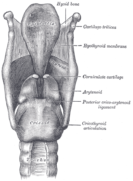
The Larynx, Ligaments of the larynx; Posterior view, Cartilago triticea, Hyothyroid membrane, Corniculate cartilage, Arytenoid, Posterior cricoarytenoid ligament, Cricothyroid articulation
Henry Vandyke Carter, Public Domain, via Wikimedia Commons
References
Constable JD, Bathala S, Ahmed JJ, McGlashan JA. Non-recurrent laryngeal nerve with a coexisting contralateral nerve demonstrating extralaryngeal branching. BMJ case reports. 2017 Mar 17:2017():. doi: 10.1136/bcr-2016-218280. Epub 2017 Mar 17 [PubMed PMID: 28314807]
Level 3 (low-level) evidenceYamamoto T, Flenner M, Schindler E. Complications associated with nasotracheal intubation and proposal of simple countermeasure. Anaesthesiology intensive therapy. 2019:51(1):72-73. doi: 10.5603/AIT.a2019.0002. Epub 2019 Feb 6 [PubMed PMID: 30723887]
Crilly SM, McElroy E, Ryan J, O'Donohue M, Lawler LP. "Mixed" trauma to the carotid artery in a mixed martial arts injury - A case report and review of the literature. Journal of radiology case reports. 2018 May:12(5):1-11. doi: 10.3941/jrcr.v12i5.3234. Epub 2018 May 31 [PubMed PMID: 30651908]
Level 3 (low-level) evidenceSethi RKV, Khatib D, Kligerman M, Kozin ED, Gray ST, Naunheim MR. Laryngeal fracture presentation and management in United States emergency rooms. The Laryngoscope. 2019 Oct:129(10):2341-2346. doi: 10.1002/lary.27790. Epub 2019 Jan 8 [PubMed PMID: 30623434]
Sperandio FA, Imamura R, Tsuji DH, Sennes LU. Surgical approach to the thyroarytenoid branch of the inferior laryngeal nerve through the thyroid cartilage. Acta cirurgica brasileira. 2016 Jul:31(7):442-7. doi: 10.1590/S0102-865020160070000003. Epub [PubMed PMID: 27487278]
Jaworek AJ, Earasi K, Lyons KM, Daggumati S, Hu A, Sataloff RT. Acute infectious laryngitis: A case series. Ear, nose, & throat journal. 2018 Sep:97(9):306-313 [PubMed PMID: 30273430]
Level 2 (mid-level) evidenceWalton C, Conway E, Blackshaw H, Carding P. Unilateral Vocal Fold Paralysis: A Systematic Review of Speech-Language Pathology Management. Journal of voice : official journal of the Voice Foundation. 2017 Jul:31(4):509.e7-509.e22. doi: 10.1016/j.jvoice.2016.11.002. Epub 2016 Dec 19 [PubMed PMID: 28007326]
Level 1 (high-level) evidenceGreco A, Rizzo MI, De Virgilio A, Gallo A, Fusconi M, Pagliuca G, Martellucci S, Turchetta R, De Vincentiis M. Cancer stem cells in laryngeal cancer: what we know. European archives of oto-rhino-laryngology : official journal of the European Federation of Oto-Rhino-Laryngological Societies (EUFOS) : affiliated with the German Society for Oto-Rhino-Laryngology - Head and Neck Surgery. 2016 Nov:273(11):3487-3495 [PubMed PMID: 26585332]