 Tools
Tools
 Kienbock Disease
Introduction
Kienböck disease refers to avascular necrosis of the lunate carpal bone, also known as lunatomalacia. Austrian radiologist Robert Kienböck first described the disorder in 1910.[1]
Relevant anatomy: Anatomically, the proximal carpal row of the midcarpal joint is largely responsible for wrist motion, whereas the distal carpal row is relatively fixed. The lunate is the central bone of the proximal row and articulates with the scaphoid, capitate, triquetrum, and occasionally with the hamate. Proximally, the lunate forms part of the radiocarpal joint and articulates with the ulna through the triangular fibrocartilage complex. Approximately 10% of the axial load across the radius, ulna, and carpus is transmitted through the triangular fibrocartilage complex to the ulna, whereas 35% is transmitted through the radiolunate articulation.[2][3]
Etiology
Register For Free And Read The Full Article
 Search engine and full access to all medical articles
- 10 free questions in your specialty
- Free CME/CE Activities
- Free daily question in your email
- Save favorite articles to your dashboard
- Emails offering discounts
Learn more about a Subscription to StatPearls Point-of-Care
Etiology
No consensus exists regarding the primary cause of Kienböck disease. The disorder is multifactorial and is associated with the following variables:
Negative ulnar variance: Negative ulnar variance, also known as ulna minus, refers to an ulna that is disproportionately shorter than the radius. A shortened ulna increases mechanical stress and repetitive microtrauma to the lunate from the relatively longer radius. Results from some studies showed that up to 78% of patients with Kienböck disease have this finding.[4]
Vascular supply to the lunate: The lunate receives blood from a variable number of dorsal and volar penetrating arteries that arise from the dorsal and palmar radiocarpal and intercarpal arches. Intraosseous collateral vessels are sparse. A lower number of penetrating arteries, particularly volar branches from the palmar radiocarpal arch, is associated with a greater likelihood of developing Kienböck disease.[5][6]
Lunate morphology: The risk of Kienböck disease increases as lunate size decreases, causing the bone to bear a greater axial load. The lunate may have a square or rectangular shape, classified as type 2 or type 3, or a more triangular shape without a medial articular facet, classified as type 1. The type 1 lunate has a weaker trabecular pattern and is associated with disease development and progression.[7]
Radial inclination angle: The radial inclination angle is formed by a horizontal line and a line extending from the ulnar edge of the radial articular surface to the tip of the radial styloid. The risk of Kienböck disease increases as the radial inclination angle decreases.[8]
Epidemiology
Kienböck disease is the second most common type of avascular necrosis affecting the carpal bones, after avascular necrosis of the scaphoid. The disorder most commonly affects men aged 20 to 40 years.[5]
History and Physical
Patients usually present with unilateral dorsal wrist pain, restricted wrist motion, weakness, or a combination of these findings. Wrist extension and axial loading typically worsen the pain. Symptoms range from mild to debilitating. Bilateral involvement is rare, and a history of trauma is often absent.[9] Physical examination commonly reveals wrist swelling, tenderness over the lunate, synovitis, and reduced grip strength.[9]
Evaluation
Kienböck disease is diagnosed through clinical assessment and imaging. Radiography, CT, and MRI are highly specific; however, MRI is the most sensitive modality and can detect radiographically occult disease.
Magnetic resonance imaging: A diffuse decrease in lunate bone marrow signal intensity on T1-weighted images is a hallmark of the disease. Signal changes on T2-weighted images or short tau inversion recovery images, which suppress fat signal, vary with disease progression and the extent of osteonecrosis. MRI also evaluates the integrity of the articular cartilage.[6][9]
Radiography: Radiographic findings may be normal early in the disease. When present, findings depend on the morphologic stage and may include diffuse lunate sclerosis, cystic changes, articular surface collapse, carpal collapse, and secondary midcarpal or radiocarpal arthrosis. Coronal fractures may occur in lunates with type 1 morphology.[6][9]
Computed tomography: CT is useful for surgical planning and is more sensitive than radiography for detecting subtle subchondral fractures, coronal lunate fractures, fragmentation, carpal instability, and the degree of trabecular disruption. Patients are frequently restaged after CT imaging.[10]
Nuclear scintigraphy: Findings are nonspecific. Nuclear scintigraphy was previously used as an adjunct for diagnosing early-stage disease but has largely been replaced by MRI.[6]
Treatment / Management
The goals of treating Kienböck disease are to relieve pain, preserve wrist motion, and maintain grip strength.[11] Treatment depends on the disease stage and contributing factors. Stage I disease is treated with splinting or cast immobilization. Stage II disease may also be treated with immobilization when necrosis is incomplete. Stage II disease with complete necrosis and stages III and IV generally require joint-leveling surgical procedures, sometimes combined with vascularized bone grafting or transfer of branches from adjacent arteries. Later-stage disease with lunate collapse and secondary degenerative wrist arthrosis may require proximal row carpectomy or intercarpal arthrodesis. Radial shortening osteotomy is the most common procedure used to unload the lunate in patients with coexisting negative ulnar variance. Treatment may improve symptoms and function without altering imaging findings in advanced disease.[6][9][11](B2)
Differential Diagnosis
Ulnar impaction syndrome: Ulnar impaction syndrome is more common in patients with positive ulnar variance and results from repetitive microtrauma to the lunate from a relatively long ulna. Similar to Kienböck disease, ulnar impaction syndrome causes decreased signal intensity on T1-weighted images and increased signal intensity on T2-weighted images when hyperemia is present or decreased T2-weighted signal intensity after progression to lunate sclerosis. However, signal abnormalities in Kienböck disease are more diffuse or more pronounced along the radial aspect of the lunate. Additionally, ulnar impaction syndrome affects the ulnar head and triquetrum, which remain intact in patients with Kienböck disease.[6][12]
Lunate intraosseous ganglion: Intraosseous ganglia are true cysts that demonstrate low signal intensity on T1-weighted images and high signal intensity on T2-weighted images. Sharp, smooth margins on MRI and radiography help distinguish these lesions from Kienböck disease.[6]
Bone contusion: A bone contusion may be difficult to distinguish from early-stage Kienböck disease on MRI. A history of recent trauma and associated wrist or hand injuries may help establish the diagnosis.[6]
Arthritis: Bone marrow signal changes in inflammatory or degenerative arthritides may mimic those seen in Kienböck disease. Distinguishing features include demographics, clinical presentation, and absence of ulna minus in arthritis.[6]
Osteoid osteoma: Osteoid osteoma of the carpal bones is rare, with only a few reported cases in the literature. The clinical presentation and identification of a lucent nidus surrounded by a sclerotic rim on CT help distinguish osteoid osteoma from Kienböck disease.[13]
Enostosis or bone island: An enostosis demonstrates low signal intensity on all imaging sequences while preserving normal bone morphology. The sclerotic area is typically stellate and interdigitates with the normal trabeculae.[6]
Staging
Staging of Kienböck disease is essential for treatment planning and includes morphologic and functional classification systems.
Morphologic Staging
Morphologic staging follows the Lichtman Classification, which is based on radiographic and MRI findings and has low interobserver variability.[6][9][14]
Stage 1: Normal radiograph, lunate signal intensity changes on MRI.
Stage 2: Radiography shows lunate sclerosis with or without fracture lines. The lunate maintains its normal shape.
Stage 3: The lunate articular surface has collapsed.
- 3A: Carpal alignment and height are preserved.
- 3B: Scaphoid flexion and loss of carpal height are present.
- 3C: A coronal lunate fracture is present.
Stage 4: Stage 3B findings are accompanied by radiocarpal or midcarpal arthrosis.
Functional Staging
Functional staging uses contrast-enhanced MRI to assess osseous perfusion and the extent of necrosis. This system helps guide revascularization procedures for morphologic stages 2 and 3A. Precontrast and postcontrast images are acquired using a T2-weighted fat-suppressed sequence.[6][15]
Stage 1: Intense homogeneous contrast enhancement reflects bone marrow ischemia and edema and a viable lunate (corresponds to morphologic stage 1).
Stage 2: Patchy, inhomogeneous enhancement reflects partial necrosis. The remaining viable portion is usually distal.
Stage 3: Absent contrast enhancement reflects complete necrosis.
Prognosis
Kienböck disease is invariably progressive, with joint destruction typically occurring within 3 to 5 years after onset.[6]
Prognosis depends on the following factors:
Functional staging: A greater proportion of viable bone is associated with a better prognosis.[6]
Negative ulnar variance: Greater negative variance is associated with more severe disease and a higher likelihood of progression.[16]
Age at diagnosis: Patients diagnosed at an older age are more likely to have advanced-stage disease and subsequent progression.[16]
Notably, symptom severity does not always correlate with the morphologic stage.
Complications
Kienböck disease can lead to scapholunate dissociation, secondary radiocarpal and midcarpal degenerative arthrosis, and triquetral malalignment.[5]
Enhancing Healthcare Team Outcomes
Kienböck disease is the second most common type of avascular necrosis affecting the carpal bones, after avascular necrosis of the scaphoid. The disorder most commonly affects men aged 20 to 40 years.[5] Kienböck disease is invariably progressive, with joint destruction typically occurring within 3 to 5 years after onset.[6] Prognosis depends on the following factors:
Functional staging: A greater proportion of viable bone is associated with a better prognosis.[6]
Negative ulnar variance: Greater negative variance is associated with more severe disease and a higher likelihood of progression.[16]
Age at diagnosis: Patients diagnosed at an older age are more likely to have advanced-stage disease and subsequent progression.[16]
Notably, symptom severity does not always correlate with morphologic stage.
Media
(Click Image to Enlarge)
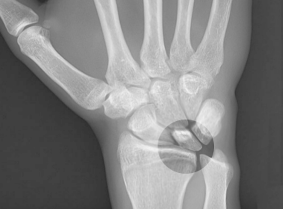
Kienbock Disease.
Contributed by S Bhimji, MD
References
Wagner JP, Chung KC. A historical report on Robert Kienböck (1871-1953) and Kienböck's Disease. The Journal of hand surgery. 2005 Nov:30(6):1117-21 [PubMed PMID: 16344165]
Viegas SF, Patterson RM, Todd PD, McCarty P. Load mechanics of the midcarpal joint. The Journal of hand surgery. 1993 Jan:18(1):14-8 [PubMed PMID: 8423300]
Schuind F, Cooney WP, Linscheid RL, An KN, Chao EY. Force and pressure transmission through the normal wrist. A theoretical two-dimensional study in the posteroanterior plane. Journal of biomechanics. 1995 May:28(5):587-601 [PubMed PMID: 7775494]
van Leeuwen WF, Oflazoglu K, Menendez ME, Ring D. Negative Ulnar Variance and Kienböck Disease. The Journal of hand surgery. 2016 Feb:41(2):214-8. doi: 10.1016/j.jhsa.2015.10.014. Epub 2015 Dec 11 [PubMed PMID: 26686062]
Murphey MD, Foreman KL, Klassen-Fischer MK, Fox MG, Chung EM, Kransdorf MJ. From the radiologic pathology archives imaging of osteonecrosis: radiologic-pathologic correlation. Radiographics : a review publication of the Radiological Society of North America, Inc. 2014 Jul-Aug:34(4):1003-28. doi: 10.1148/rg.344140019. Epub [PubMed PMID: 25019438]
Arnaiz J, Piedra T, Cerezal L, Ward J, Thompson A, Vidal JA, Canga A. Imaging of Kienböck disease. AJR. American journal of roentgenology. 2014 Jul:203(1):131-9. doi: 10.2214/AJR.13.11606. Epub [PubMed PMID: 24951206]
Rhee PC, Jones DB, Moran SL, Shin AY. The effect of lunate morphology in Kienböck disease. The Journal of hand surgery. 2015 Apr:40(4):738-44. doi: 10.1016/j.jhsa.2014.12.024. Epub 2015 Feb 18 [PubMed PMID: 25701489]
Level 2 (mid-level) evidenceThienpont E, Mulier T, Rega F, De Smet L. Radiographic analysis of anatomical risk factors for Kienböck's disease. Acta orthopaedica Belgica. 2004 Oct:70(5):406-9 [PubMed PMID: 15587027]
Level 2 (mid-level) evidenceLutsky K, Beredjiklian PK. Kienböck disease. The Journal of hand surgery. 2012 Sep:37(9):1942-52. doi: 10.1016/j.jhsa.2012.06.029. Epub [PubMed PMID: 22916868]
Quenzer DE, Linscheid RL, Vidal MA, Dobyns JH, Beckenbaugh RD, Cooney WP. Trispiral tomographic staging of Kienböck's disease. The Journal of hand surgery. 1997 May:22(3):396-403 [PubMed PMID: 9195446]
Level 2 (mid-level) evidenceIwasaki N, Minami A, Oizumi N, Suenaga N, Kato H, Minami M. Radial osteotomy for late-stage Kienböck's disease. Wedge osteotomy versus radial shortening. The Journal of bone and joint surgery. British volume. 2002 Jul:84(5):673-7 [PubMed PMID: 12188482]
Level 2 (mid-level) evidenceCerezal L, del Piñal F, Abascal F, García-Valtuille R, Pereda T, Canga A. Imaging findings in ulnar-sided wrist impaction syndromes. Radiographics : a review publication of the Radiological Society of North America, Inc. 2002 Jan-Feb:22(1):105-21 [PubMed PMID: 11796902]
Güner MD, Kamburoğlu HO, Bektaş U, Ay Ş. Osteoid osteoma of the lunatum mimicking Kienböck's disease. Case reports in plastic surgery & hand surgery. 2015:2(1):19-21. doi: 10.3109/23320885.2014.993647. Epub 2015 Jan 6 [PubMed PMID: 27252961]
Level 3 (low-level) evidenceGoldfarb CA, Hsu J, Gelberman RH, Boyer MI. The Lichtman classification for Kienböck's disease: an assessment of reliability. The Journal of hand surgery. 2003 Jan:28(1):74-80 [PubMed PMID: 12563641]
Schmitt R, Heinze A, Fellner F, Obletter N, Strühn R, Bautz W. Imaging and staging of avascular osteonecroses at the wrist and hand. European journal of radiology. 1997 Sep:25(2):92-103 [PubMed PMID: 9283837]
Level 3 (low-level) evidencevan Leeuwen WF, Tarabochia MA, Schuurman AH, Chen N, Ring D. Risk Factors of Lunate Collapse in Kienböck Disease. The Journal of hand surgery. 2017 Nov:42(11):883-888.e1. doi: 10.1016/j.jhsa.2017.06.107. Epub 2017 Sep 6 [PubMed PMID: 28888572]