 Tools
Tools
 Anatomy, Retroperitoneum: Nerve Supply to the Kidneys
Introduction
The kidneys are bilateral retroperitoneal organs located just below the diaphragm, exhibiting the highest degree of innervation after the adrenal glands.[1][2][3] The superior margin of the left kidney is usually near the T12 vertebra at approximately the level of the 11th rib, extending to L3, whereas the right kidney lies slightly lower, between the 12th rib and L3. The lower position of the right kidney reflects its placement beneath the liver.
Renal innervation comprises both afferent and efferent fibers forming the renal plexus (see Image. Renal and Adrenal Nerve Pathways).[4] Afferent fibers mediate nociception, enabling detection of renal pain and other sensory signals. Efferent fibers are predominantly sympathetic and play a central role in regulating renal blood flow, renin release, and systemic blood pressure.[5][6][7][8][9][10]
The renal plexus receives input from multiple contributing plexuses that accompany the renal artery and vein before entering the hilum of the kidney.[11] The clinical relevance of renal nervous system anatomy in blood pressure regulation has increased substantially with the development and U.S. Food and Drug Administration approval of catheter-based renal denervation devices for treating patients with drug-resistant hypertension.[12][13][14]
The critical role of renal innervation in regulating blood pressure and kidney function underscores its significance in managing cardiovascular and renal disorders. Advances in catheter-based renal arterial denervation therapies have highlighted the functional impact of the sympathetic perivascular nerve supply of the kidneys in patients with drug-resistant hypertension.[15][16] A comprehensive understanding of renal nerve anatomy and physiology enables clinicians to guide procedural strategy, optimize patient selection, and anticipate therapeutic outcomes (see Image. Kidney Anatomy).
Structure and Function
Register For Free And Read The Full Article
 Search engine and full access to all medical articles
- 10 free questions in your specialty
- Free CME/CE Activities
- Free daily question in your email
- Save favorite articles to your dashboard
- Emails offering discounts
Learn more about a Subscription to StatPearls Point-of-Care
Structure and Function
The kidney receives innervation from the renal plexus, a mixed neural network comprising sympathetic, visceral afferent, and parasympathetic fibers, with parasympathetic fibers absent from the renal parenchyma. Parasympathetic fibers have been identified in the renal pelvis and the arterial supply of the kidney, but evidence of parasympathetic innervation within the renal parenchyma remains lacking.[17][18][19] The renal plexus receives input from the celiac plexus, aortorenal ganglia, thoracic splanchnic nerves (T10-T12), lumbar splanchnic nerves (L1-L2), the intermesenteric plexus, and indirectly from the posterior vagal trunk via the celiac plexus.[20]
Renal afferent fibers originate predominantly from the renal pelvis, where their density is highest, and also from the renal cortex. These fibers project to central nervous system structures, including the subfornical organ, hypothalamus, and brainstem.[21] Pain arising from the kidney occurs in response to ischemia, infection, obstruction, or parenchymal injury. The sensation typically follows a dermatomal pattern along the anterior abdominal wall and flanks and is transmitted via visceral afferent fibers.[22][23][24] Afferent innervation is required for nociceptive signaling.
Efferent innervation of the kidney is primarily sympathetic and receives input from each contributing plexus. Sympathetic preganglionic fibers synapse in prevertebral and paravertebral ganglia before postganglionic axons join the renal plexus (see Image. Sympathetic Chain and Trunk).[25][26] The sympathetic supply is predominantly adrenergic and arises from the spinal cord (T8-L1), the paravertebral sympathetic chain, the least and lesser splanchnic nerves, and the upper lumbar splanchnic nerves.[27]
Renal sympathetic activity constitutes 1 of 3 physiological mechanisms involved in the regulation of blood pressure.[28] The other mechanisms are carotid baroreceptors, which monitor vascular hydrostatic pressure, and the juxtaglomerular apparatus, which regulates renal blood flow and glomerular filtration rate in response to changes in sodium concentration in the filtrate.[29][30]
Sympathetic stimulation of the kidney causes constriction of afferent arterioles and activates the juxtaglomerular apparatus, increasing plasma renin activity, triggering the renin-angiotensin-aldosterone system, and enhancing renal sodium reabsorption. Collectively, these effects contribute to the maintenance of systemic blood pressure.[31][32][33][34][35][36][37][38] Denervation of renal sympathetic nerves has been shown to reduce mean systolic blood pressure in most patients with drug-resistant hypertension and is recommended in guidelines for selected patients unresponsive to standard therapy.[39][40] For further details, refer to the StatPearls reference “Renal Denervation Therapy for Drug-Resistant Hypertension.”[41]
Embryology
Afferent renal innervation appears early in fetal development, likely preceding the formation of efferent fibers.[42] Evidence indicates that renal nerves play a critical role in proper embryological kidney development.[43][44] The kidney lacks innervation from the renal plexus during the initial embryonic period. Renal innervation begins on embryonic day 13.5, with nerves detectable around 9 weeks of gestation as branches of the immature autonomic abdominal plexus.[45][46] This development coincides with the formation of the renal veins, approximately 2 weeks after differentiation of the renal arteries.[47] Afferent input is required for normal kidney morphogenesis, regulating renal hemodynamics during early fetal life, modulating renin secretion during pregnancy, and supporting fluid and electrolyte homeostasis in late gestation.[48]
Blood Supply and Lymphatics
The kidney receives its primary arterial blood supply from the renal artery, a direct branch of the abdominal aorta.[49] Renal vascular anatomy may include multiple main arteries and accessory vessels.[50] All renal arteries function as end arteries, as the kidney lacks collateral arterial circulation.[51][52][53] Awareness of accessory renal vessels is important during aortic aneurysm repair, nephrectomy, partial nephrectomy, endovascular interventions, and renal denervation therapy.[54][55]
Accessory renal arteries occur in approximately 20% to 30% of the population, representing a significant consideration for patients undergoing kidney surgery.[56][57][58][59][60][61][62][63][64] Renal lymphatic drainage parallels the major renal vessels, originating in the cortex and ultimately draining into the paraaortic lymph nodes.[65]
Nerves
Sympathetic renal innervation arises from neurons in the intermediolateral cell columns of spinal cord segments T8 to L1, which give rise to preganglionic fibers forming the lesser and least splanchnic nerves.[66][67][68][69] These fibers synapse with postganglionic neurons in the celiac and aorticorenal ganglia, establishing the renal nerve plexus within the adventitia of the main and accessory renal arteries (see Image. Kidney Nerve Supply Diagram).[70][71][72]
Efferent sympathetic fibers extend throughout the renal cortex and outer medulla, encompassing the interlobar, arcuate, and interlobular arteries, as well as glomerular arterioles.[73] Sympathetic nerve density is highest in afferent glomerular arterioles, followed by efferent arterioles, thick ascending limbs, and distal convoluted tubules.[74][75][76] The inner renal medulla contains few sympathetic fibers.[77] Sympathetic renal nerves innervate the renal vasculature, juxtaglomerular apparatus, and renal tubules, increasing blood pressure by elevating norepinephrine levels, stimulating renin release, enhancing tubular sodium and water reabsorption, and inducing renal vasoconstriction.[78][79][80][81][82][83][84][85][86][87]
Activation of sympathetic fibers to the kidneys primarily reduces cortical blood flow, while intense stimulation also decreases medullary perfusion.[88] These nerves contribute to renovascular hypertension caused by renal artery stenosis or kidney irradiation.[89][90] Recent evidence indicates that sympathetic stimulation promotes renal fibrosis via activation of M2 macrophages, whereas sympathetic denervation reduces acute tubular necrosis, macrophage accumulation, and tubulointerstitial fibrosis induced by ureteral obstruction and ischemia/reperfusion.[91]
Sensory fibers originating from the dorsal root ganglia travel alongside sympathetic renal nerves.[92][93] These fibers transmit signals from mechanoreceptors and chemoreceptors in the renal pelvis to the spinal cord and brain, modulating the renorenal reflex and related functions.[94] Sensory fibers are densest in the renal pelvis. These nerves are present in the cortex and proximal ureter and absent from the renal medulla.[95][96][97][98][99] The fibers course adjacent to the renal artery to the dorsal root ganglia of T12 to L3, then ascend the spinal cord to the brainstem and hypothalamus, contributing to the regulation of sympathetic nerve activity and systemic blood pressure.[100]
Parasympathetic renal innervation originates from the dorsal motor nucleus of the vagus nerve in the medulla oblongata, the most inferior portion of the brainstem.[101][102] Preganglionic fibers travel via the vagus nerve (cranial nerve X) and form renal branches that join the plexus surrounding the renal arteries, providing parasympathetic input and promoting vasodilation upon stimulation.[103] Intrarenal parasympathetic fibers are sparse, although vagal afferent activation enhances renal dopamine release.[104]
Visceral afferent fibers transmit renal pain signals to spinal ganglia and cord segments T11 to L2. Pain may result from obstructive uropathy, including ureteral calculi, distension of the renal pelvis, infection, inflammation, or ischemia, corresponding to the affected dermatome. Nonobstructing and uninfected stones generally do not elicit flank or renal pain.[105][106][107][108]
Muscles
The left and right kidneys are positioned on the corresponding psoas major and quadratus lumborum muscles.[109] This anatomical relationship guides the course of sympathetic and afferent nerve fibers traveling to the renal plexus, thereby influencing renal innervation pathways and the patterns of referred pain.
Physiologic Variants
The renal plexus comprises fibers originating from the aorticorenal ganglia, lumbar splanchnic nerves, and the least splanchnic nerve, with additional contributions from the celiac, aortic, and intermesenteric plexuses.[110] These fibers join the renal arteries, including extrarenal branches, segmental vessels, and accessory arteries, at variable intervals and accompany the renal vessels into the parenchyma, providing both sympathetic efferent and afferent signaling.[111][112][113][114][115][116][117] Fibers associated with the celiac ganglion exhibit considerable variation in diameter, size, and number of ganglia.[118] Lumbar splanchnic nerves typically arise from spinal roots T12 to L1, although origins may extend to T11 or skip 1 or more spinal segments.[119]
Surgical Considerations
Renal transplantation disrupts the nerve fibers of the renal plexus, abolishing renal sympathetic activity. In the absence of sympathetic input, β1-adrenergic stimulation of juxtaglomerular cells is lost, thereby attenuating one mechanism of renin-angiotensin-aldosterone system activation. The transplanted kidney retains the ability to regulate systemic blood pressure via intrinsic autoregulatory mechanisms, including the myogenic response and macula densa-mediated sodium feedback.[120]
Clinical Significance
Renal sympathetic nerve activity is increased in patients with hypertension.[121] This elevation promotes sodium and water reabsorption, reduces renal blood flow, and stimulates renin release. Chronic sympathetic overactivity can contribute to impaired kidney function and cardiac hypertrophy.
Renal denervation therapy, achieved via ablation of the sympathetic nerve supply to the kidneys, is emerging as a viable option for selected patients with drug-resistant hypertension.[122][123][124][125][126] Unlike historical sympathectomy procedures, these approaches provide a safe, permanent, minimally invasive intervention without the adverse effect of postural hypotension. Renal denervation using endovascular ultrasound or radiofrequency devices has demonstrated efficacy in multiple sham-controlled, randomized clinical trials.[127][128][129][130][131][132][133][134][135] Such procedures are likely to become increasingly utilized in the future.
Media
(Click Image to Enlarge)
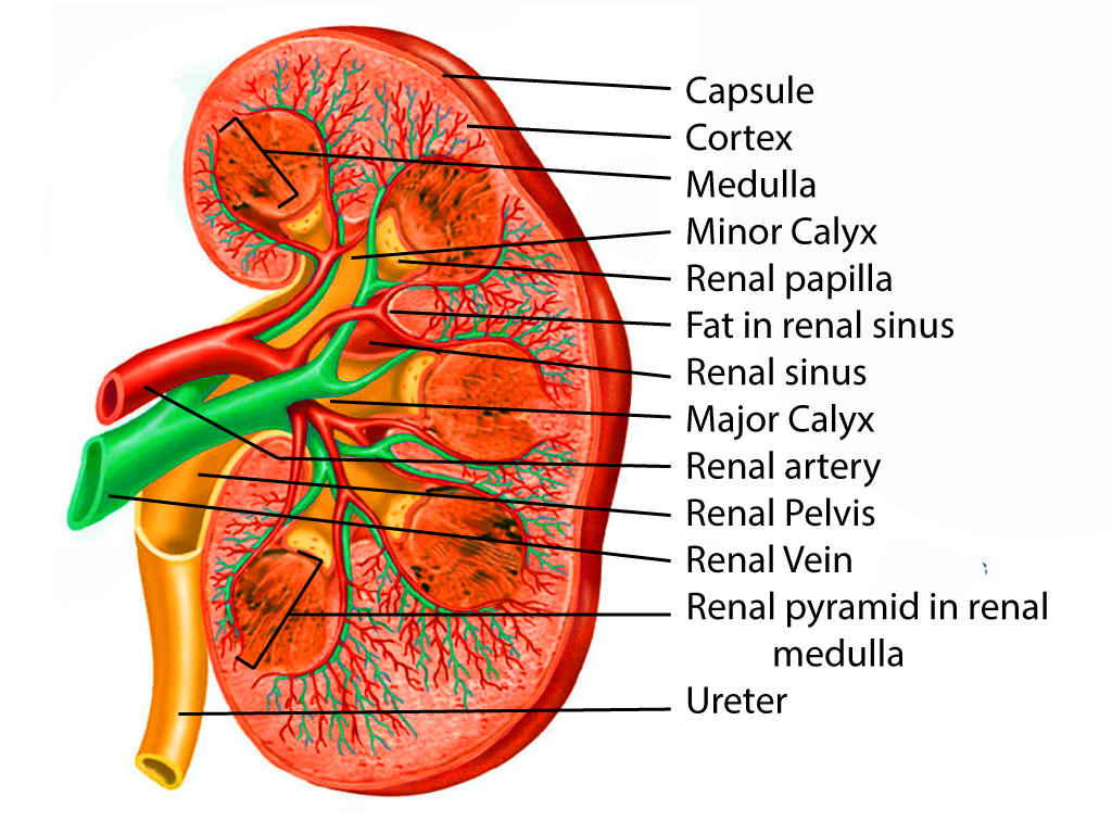
Kidney Anatomy. This illustration shows the internal structure of the kidney, highlighting key features such as the capsule, cortex, medulla, and renal pyramids. Other important labeled components include the minor and major calyces, the renal papilla, artery, vein, and pelvis, fat in the sinus, and the ureter. A clear view of the blood vessels and urine-collecting structures within the kidney is provided.
Contributed by S Dulebohn, MD
(Click Image to Enlarge)
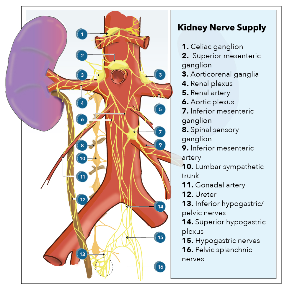
Kidney Innervation. The image illustrates the nerve supply to the kidney, showing connections from the celiac ganglion, superior mesenteric ganglion, and aorticorenal ganglia. Renal plexus fibers accompany the renal artery, passing through the aortic plexus and linking with both the inferior mesenteric ganglion and artery. Spinal sensory ganglia and the lumbar sympathetic trunk are positioned near the gonadal artery and ureter. Inferior and superior hypogastric plexuses converge with hypogastric and pelvic splanchnic nerves, which innervate regions near the pelvis. All major labeled anatomical structures are identified and interconnected by nerve pathways in this schematic.
Illustrated by K Humphreys
(Click Image to Enlarge)
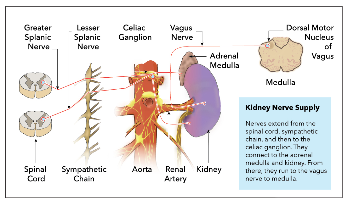
Renal and Adrenal Nerve Pathways. The diagram shows greater and lesser splanchnic nerves arising from the spinal cord and traveling through the sympathetic chain. These nerves reach the celiac ganglion, sending fibers to the aorta, renal artery, kidney, and adrenal medulla. The vagus nerve connects from the dorsal motor nucleus of the vagus in the medulla to both the kidney and adrenal gland, illustrating sympathetic and parasympathetic connections. Each labeled structure is represented by brief, direct neural pathways in the illustration.
Contributed by K Humphreys
(Click Image to Enlarge)
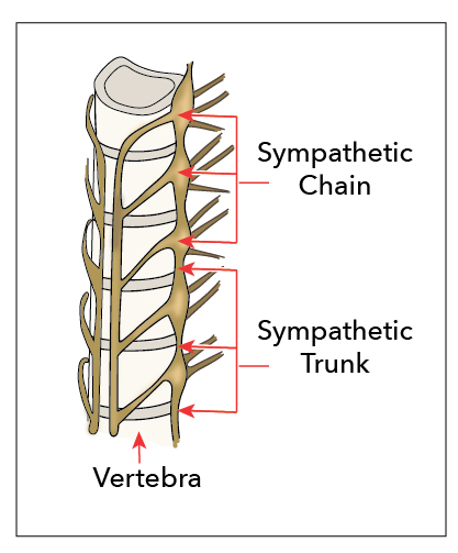
Sympathetic Chain and Trunk. This image depicts the anatomical relationship between the sympathetic chain and sympathetic trunk as they run alongside the vertebral column. The sympathetic chain consists of interconnected ganglia forming a vertical structure, whereas the sympathetic trunk refers to the paired nerve cords that extend along the vertebral bodies. The vertebra is shown for anatomical orientation to highlight the proximity of these sympathetic nervous system components to the spinal column.
Contributed by K Humphreys
References
Lambert G, Samra NS. Anatomy, Abdomen and Pelvis, Retroperitoneum. StatPearls. 2026 Jan:(): [PubMed PMID: 31751047]
Garza FA, Leslie SW. Anatomy, Abdomen and Pelvis: Kidneys. StatPearls. 2026 Jan:(): [PubMed PMID: 29494007]
Nakamura Y, Inoue T. Neuroimmune Communication in the Kidney. JMA journal. 2020 Jul 15:3(3):164-174. doi: 10.31662/jmaj.2020-0024. Epub 2020 Jul 8 [PubMed PMID: 33150250]
DiBona GF, Kopp UC. Neural control of renal function. Physiological reviews. 1997 Jan:77(1):75-197 [PubMed PMID: 9016301]
Level 3 (low-level) evidenceSata Y, Head GA, Denton K, May CN, Schlaich MP. Role of the Sympathetic Nervous System and Its Modulation in Renal Hypertension. Frontiers in medicine. 2018:5():82. doi: 10.3389/fmed.2018.00082. Epub 2018 Mar 29 [PubMed PMID: 29651418]
Díaz-Morales N, Baranda-Alonso EM, Martínez-Salgado C, López-Hernández FJ. Renal sympathetic activity: A key modulator of pressure natriuresis in hypertension. Biochemical pharmacology. 2023 Feb:208():115386. doi: 10.1016/j.bcp.2022.115386. Epub 2022 Dec 17 [PubMed PMID: 36535529]
Kim GH. Primary Role of the Kidney in Pathogenesis of Hypertension. Life (Basel, Switzerland). 2024 Jan 14:14(1):. doi: 10.3390/life14010119. Epub 2024 Jan 14 [PubMed PMID: 38255734]
Osborn JW, Tyshynsky R, Vulchanova L. Function of Renal Nerves in Kidney Physiology and Pathophysiology. Annual review of physiology. 2021 Feb 10:83():429-450. doi: 10.1146/annurev-physiol-031620-091656. Epub [PubMed PMID: 33566672]
Grisk O, Rettig R. Interactions between the sympathetic nervous system and the kidneys in arterial hypertension. Cardiovascular research. 2004 Feb 1:61(2):238-46 [PubMed PMID: 14736540]
Grisk O. Sympatho-renal interactions in the determination of arterial pressure: role in hypertension. Experimental physiology. 2005 Mar:90(2):183-7 [PubMed PMID: 15604108]
Becker BK, Zhang D, Soliman R, Pollock DM. Autonomic nerves and circadian control of renal function. Autonomic neuroscience : basic & clinical. 2019 Mar:217():58-65. doi: 10.1016/j.autneu.2019.01.003. Epub 2019 Jan 10 [PubMed PMID: 30704976]
Iyer A, Roh S, Muhammad H, Hammad A, Kumar V, Patel D, Sayanlar J. Renal denervation for resistant hypertension: A review of current literature. Cardiovascular revascularization medicine : including molecular interventions. 2025 Aug:77():122-127. doi: 10.1016/j.carrev.2025.05.014. Epub 2025 May 13 [PubMed PMID: 40450401]
Sharp ASP, Fisher NDL. Renal Denervation to Treat Hypertension: A Clinical State-of-the-Art Review. American journal of hypertension. 2025 Jul 23:():. pii: hpaf134. doi: 10.1093/ajh/hpaf134. Epub 2025 Jul 23 [PubMed PMID: 40702973]
Zhang J, Belford PM, Stouffer GA. Renal Denervation After USA FDA Approval: An Update from an Interventional Cardiologist's Perspective. Journal of clinical medicine. 2025 May 19:14(10):. doi: 10.3390/jcm14103554. Epub 2025 May 19 [PubMed PMID: 40429549]
Level 3 (low-level) evidenceSingh SP, Varghese KJ, Qureshi FM, Anderson MC, Foxworth J, Knuepfer MM. Catheter-based renal sympathetic nerve denervation on hypertension management outcomes. World journal of radiology. 2022 Jul 28:14(7):238-248. doi: 10.4329/wjr.v14.i7.238. Epub [PubMed PMID: 36160631]
Azeez GA, Thirunagari M, Fatima N, Anand A, Palvia AR, Kaur A, Nassar ST. The Efficacy of Renal Denervation in Treating Resistant Hypertension: A Systematic Review. Cureus. 2024 Aug:16(8):e67007. doi: 10.7759/cureus.67007. Epub 2024 Aug 16 [PubMed PMID: 39286705]
Level 1 (high-level) evidencevan Amsterdam WA, Blankestijn PJ, Goldschmeding R, Bleys RL. The morphological substrate for Renal Denervation: Nerve distribution patterns and parasympathetic nerves. A post-mortem histological study. Annals of anatomy = Anatomischer Anzeiger : official organ of the Anatomische Gesellschaft. 2016 Mar:204():71-9. doi: 10.1016/j.aanat.2015.11.004. Epub 2015 Nov 23 [PubMed PMID: 26617159]
Maeda S, Kuwahara-Otani S, Tanaka K, Hayakawa T, Seki M. Origin of efferent fibers of the renal plexus in the rat autonomic nervous system. The Journal of veterinary medical science. 2014 May:76(5):763-5 [PubMed PMID: 24430660]
Level 3 (low-level) evidenceCheng X, Zhang Y, Chen R, Qian S, Lv H, Liu X, Zeng S. Anatomical Evidence for Parasympathetic Innervation of the Renal Vasculature and Pelvis. Journal of the American Society of Nephrology : JASN. 2022 Dec:33(12):2194-2210. doi: 10.1681/ASN.2021111518. Epub 2022 Oct 17 [PubMed PMID: 36253054]
Level 2 (mid-level) evidenceCandal R, Reddy V, Samra NS. Anatomy, Abdomen and Pelvis: Celiac Ganglia. StatPearls. 2025 Jan:(): [PubMed PMID: 30844156]
Ciriello J. Afferent renal inputs onto subfornical organ neurons responsive to angiotensin II. The American journal of physiology. 1997 May:272(5 Pt 2):R1684-9 [PubMed PMID: 9176365]
Level 3 (low-level) evidencePatti L, Leslie SW. Acute Renal Colic. StatPearls. 2026 Jan:(): [PubMed PMID: 28613743]
Sikandar S, Dickenson AH. Visceral pain: the ins and outs, the ups and downs. Current opinion in supportive and palliative care. 2012 Mar:6(1):17-26. doi: 10.1097/SPC.0b013e32834f6ec9. Epub [PubMed PMID: 22246042]
Level 3 (low-level) evidenceJin Q, Chang Y, Lu C, Chen L, Wang Y. Referred pain: characteristics, possible mechanisms, and clinical management. Frontiers in neurology. 2023:14():1104817. doi: 10.3389/fneur.2023.1104817. Epub 2023 Jun 28 [PubMed PMID: 37448749]
Torres H, Huesing C, Burk DH, Molinas AJR, Neuhuber WL, Berthoud HR, Münzberg H, Derbenev AV, Zsombok A. Sympathetic innervation of the mouse kidney and liver arising from prevertebral ganglia. American journal of physiology. Regulatory, integrative and comparative physiology. 2021 Sep 1:321(3):R328-R337. doi: 10.1152/ajpregu.00079.2021. Epub 2021 Jul 7 [PubMed PMID: 34231420]
Level 2 (mid-level) evidenceGauthier MM, Hayoz S, Banek CT. Neuroimmune interplay in kidney health and disease: Role of renal nerves. Autonomic neuroscience : basic & clinical. 2023 Dec:250():103133. doi: 10.1016/j.autneu.2023.103133. Epub 2023 Nov 30 [PubMed PMID: 38061177]
Sharabi AF, Carey FJ. Anatomy, Abdomen and Pelvis, Splanchnic Nerves. StatPearls. 2025 Jan:(): [PubMed PMID: 32809339]
Burke M, Pabbidi MR, Farley J, Roman RJ. Molecular mechanisms of renal blood flow autoregulation. Current vascular pharmacology. 2014:12(6):845-58 [PubMed PMID: 24066938]
Level 3 (low-level) evidenceArmstrong M, Moore RA. Physiology, Baroreceptors. StatPearls. 2025 Jan:(): [PubMed PMID: 30844199]
Dalal R, Bruss ZS, Sehdev JS. Physiology, Renal Blood Flow and Filtration. StatPearls. 2026 Jan:(): [PubMed PMID: 29489242]
Kaur J, Rout P. Physiology, Renin Angiotensin System. StatPearls. 2026 Jan:(): [PubMed PMID: 29261862]
Keeton TK, Campbell WB. The pharmacologic alteration of renin release. Pharmacological reviews. 1980 Jun:32(2):81-227 [PubMed PMID: 7012850]
Trerattanavong K, Chen J. Biochemistry, Renin. StatPearls. 2025 Jan:(): [PubMed PMID: 32310516]
Broeker KAE, Schrankl J, Fuchs MAA, Kurtz A. Flexible and multifaceted: the plasticity of renin-expressing cells. Pflugers Archiv : European journal of physiology. 2022 Aug:474(8):799-812. doi: 10.1007/s00424-022-02694-8. Epub 2022 May 5 [PubMed PMID: 35511367]
Briggs JP, Schnermann JB. Whys and wherefores of juxtaglomerular apparatus function. Kidney international. 1996 Jun:49(6):1724-6 [PubMed PMID: 8743485]
Higashihara E, Harada T, Fukuhara H. Juxtaglomerular apparatus-mediated homeostatic mechanisms: therapeutic implication for chronic kidney disease. Expert opinion on pharmacotherapy. 2024 May:25(7):819-832. doi: 10.1080/14656566.2024.2357188. Epub 2024 May 22 [PubMed PMID: 38773961]
Level 3 (low-level) evidenceAtlas SA. The renin-angiotensin aldosterone system: pathophysiological role and pharmacologic inhibition. Journal of managed care pharmacy : JMCP. 2007 Oct:13(8 Suppl B):9-20. doi: 10.18553/jmcp.2007.13.s8-b.9. Epub [PubMed PMID: 17970613]
Level 3 (low-level) evidenceCorbin JM, McNulty MJ, Macharoen K, McDonald KA, Nandi S. Technoeconomic analysis of semicontinuous bioreactor production of biopharmaceuticals in transgenic rice cell suspension cultures. Biotechnology and bioengineering. 2020 Oct:117(10):3053-3065. doi: 10.1002/bit.27475. Epub 2020 Jul 14 [PubMed PMID: 32592492]
Cluett JL, Blazek O, Brown AL, East C, Ferdinand KC, Fisher NDL, Ford CD, Griffin KA, Mena-Hurtado CI, Sarathy H, Vongpatanasin W, Townsend RR, American Heart Association Council on Hypertension; Council on Cardiovascular and Stroke Nursing; Council on the Kidney in Cardiovascular Disease; and Council on Peripheral Vascular Disease. Renal Denervation for the Treatment of Hypertension: A Scientific Statement From the American Heart Association. Hypertension (Dallas, Tex. : 1979). 2024 Oct:81(10):e135-e148. doi: 10.1161/HYP.0000000000000240. Epub 2024 Aug 5 [PubMed PMID: 39101202]
Fisher NDL, Kirtane AJ. Renal denervation for hypertension. Nature reviews. Cardiology. 2025 Sep:22(9):664-674. doi: 10.1038/s41569-024-01104-z. Epub 2025 Jan 2 [PubMed PMID: 39743561]
Rosendorff C, Lackland DT, Allison M, Aronow WS, Black HR, Blumenthal RS, Cannon CP, de Lemos JA, Elliott WJ, Findeiss L, Gersh BJ, Gore JM, Levy D, Long JB, O'Connor CM, O'Gara PT, Ogedegbe G, Oparil S, White WB, American Heart Association, American College of Cardiology, and American Society of Hypertension. Treatment of hypertension in patients with coronary artery disease: a scientific statement from the American Heart Association, American College of Cardiology, and American Society of Hypertension. Circulation. 2015 May 12:131(19):e435-70. doi: 10.1161/CIR.0000000000000207. Epub 2015 Mar 31 [PubMed PMID: 25829340]
Robillard JE, Guillery EN, Segar JL, Merrill DC, Jose PA. Influence of renal nerves on renal function during development. Pediatric nephrology (Berlin, Germany). 1993 Oct:7(5):667-71 [PubMed PMID: 8251342]
Rehman S, Ahmed D. Embryology, Kidney, Bladder, and Ureter. StatPearls. 2026 Jan:(): [PubMed PMID: 31613527]
Donovan MF, Cascella M. Embryology, Weeks 6-8. StatPearls. 2026 Jan:(): [PubMed PMID: 33085328]
N'Guetta PY, McLarnon SR, Tassou A, Geron M, Shirvan S, Hill RZ, Scherrer G, O'Brien LL. Comprehensive mapping of sensory and sympathetic innervation of the developing kidney. Cell reports. 2024 Oct 22:43(10):114860. doi: 10.1016/j.celrep.2024.114860. Epub 2024 Oct 15 [PubMed PMID: 39412983]
Tarnick J, Elhendawi M, Holland I, Chang Z, Davies JA. Innervation of the developing kidney in vivo and in vitro. Biology open. 2023 Aug 15:12(8):. doi: 10.1242/bio.060001. Epub 2023 Jul 31 [PubMed PMID: 37439314]
Mompeó B, Maranillo E, García-Touchard A, Sanudo JR. The morphogenesis of the renal plexus: Renal artery and sympathetic fibers. Clinical anatomy (New York, N.Y.). 2019 Mar:32(2):272-276. doi: 10.1002/ca.23297. Epub 2018 Oct 31 [PubMed PMID: 30300460]
Page WV, Perlman S, Smith FG, Segar JL, Robillard JE. Renal nerves modulate kidney renin gene expression during the transition from fetal to newborn life. The American journal of physiology. 1992 Mar:262(3 Pt 2):R459-63 [PubMed PMID: 1373041]
Leslie SW, Sajjad H. Anatomy, Abdomen and Pelvis, Renal Artery. StatPearls. 2025 Jan:(): [PubMed PMID: 29083626]
Pradhay G, Gopidas GS, Karumathil Pullara S, Mathew G, Mathew AJ, Sukumaran TT, Pavikuttan N, Sudhakaran R Sr. Prevalence and Relevance of Multiple Renal Arteries: A Radioanatomical Perspective. Cureus. 2021 Oct:13(10):e18957. doi: 10.7759/cureus.18957. Epub 2021 Oct 21 [PubMed PMID: 34815903]
Level 3 (low-level) evidencePAUL RE Jr, ETTINGER A, FAINSINGER MH, CALLOW AD, KAHN PC, INKER LH. ANGIOGRAPHIC VISUALIZATION OF RENAL COLLATERAL CIRCULATION AS A MEANS OF DETECTING AND DELINEATING RENAL ISCHEMIA. Radiology. 1965 Jun:84():1013-21 [PubMed PMID: 14296456]
Rani N, Singh S, Dhar P, Kumar R. Surgical importance of arterial segments of human kidneys: an angiography and corrosion cast study. Journal of clinical and diagnostic research : JCDR. 2014 Mar:8(3):1-3. doi: 10.7860/JCDR/2014/7396.4086. Epub 2014 Mar 15 [PubMed PMID: 24783063]
Aquino RV, Rhee RY, Muluk SC, Tzeng EY, Carrol NM, Makaroun MS. Exclusion of accessory renal arteries during endovascular repair of abdominal aortic aneurysms. Journal of vascular surgery. 2001 Nov:34(5):878-84 [PubMed PMID: 11700490]
VonAchen P, Hamann J, Houghland T, Lesser JR, Wang Y, Caye D, Rosenthal K, Garberich RF, Daniels M, Schwartz RS. Accessory renal arteries: Prevalence in resistant hypertension and an important role in nonresponse to radiofrequency renal denervation. Cardiovascular revascularization medicine : including molecular interventions. 2016 Oct-Nov:17(7):470-473. doi: 10.1016/j.carrev.2016.07.009. Epub 2016 Jul 21 [PubMed PMID: 27493150]
Kasprzycki K, Petkow-Dimitrow P, Krawczyk-Ożóg A, Bartuś S, Rajtar-Salwa R. Anatomic Variations of Renal Arteries as an Important Factor in the Effectiveness of Renal Denervation in Resistant Hypertension. Journal of cardiovascular development and disease. 2023 Aug 29:10(9):. doi: 10.3390/jcdd10090371. Epub 2023 Aug 29 [PubMed PMID: 37754800]
Bhatt DL, Kandzari DE, O'Neill WW, D'Agostino R, Flack JM, Katzen BT, Leon MB, Liu M, Mauri L, Negoita M, Cohen SA, Oparil S, Rocha-Singh K, Townsend RR, Bakris GL, SYMPLICITY HTN-3 Investigators. A controlled trial of renal denervation for resistant hypertension. The New England journal of medicine. 2014 Apr 10:370(15):1393-401. doi: 10.1056/NEJMoa1402670. Epub 2014 Mar 29 [PubMed PMID: 24678939]
Rimoldi SF, Scheidegger N, Scherrer U, Farese S, Rexhaj E, Moschovitis A, Windecker S, Meier B, Allemann Y. Anatomical eligibility of the renal vasculature for catheter-based renal denervation in hypertensive patients. JACC. Cardiovascular interventions. 2014 Feb:7(2):187-192. doi: 10.1016/j.jcin.2013.10.013. Epub 2014 Jan 15 [PubMed PMID: 24440022]
Okada T, Pellerin O, Savard S, Curis E, Monge M, Frank M, Bobrie G, Yamaguchi M, Sugimoto K, Plouin PF, Azizi M, Sapoval M. Eligibility for renal denervation: anatomical classification and results in essential resistant hypertension. Cardiovascular and interventional radiology. 2015 Feb:38(1):79-87. doi: 10.1007/s00270-014-0865-6. Epub 2014 Mar 6 [PubMed PMID: 24595661]
Sato Y, Kawakami R, Jinnouchi H, Sakamoto A, Cornelissen A, Mori M, Kawai K, Guo L, Coleman L, Nash S, Claude L, Barman NC, Romero M, Kolodgie FD, Virmani R, Finn AV. Comprehensive Assessment of Human Accessory Renal Artery Periarterial Renal Sympathetic Nerve Distribution. JACC. Cardiovascular interventions. 2021 Feb 8:14(3):304-315. doi: 10.1016/j.jcin.2020.09.043. Epub 2020 Nov 25 [PubMed PMID: 33541541]
Gulas E, Wysiadecki G, Szymański J, Majos A, Stefańczyk L, Topol M, Polguj M. Morphological and clinical aspects of the occurrence of accessory (multiple) renal arteries. Archives of medical science : AMS. 2018 Mar:14(2):442-453. doi: 10.5114/aoms.2015.55203. Epub 2016 Mar 17 [PubMed PMID: 29593819]
Jamkar AA, Khan B, Joshi DS. Anatomical study of renal and accessory renal arteries. Saudi journal of kidney diseases and transplantation : an official publication of the Saudi Center for Organ Transplantation, Saudi Arabia. 2017 Mar-Apr:28(2):292-297. doi: 10.4103/1319-2442.202760. Epub [PubMed PMID: 28352010]
Glodny B, Cromme S, Wörtler K, Winde G. A possible explanation for the frequent concomitance of arterial hypertension and multiple renal arteries. Medical hypotheses. 2001 Feb:56(2):129-33 [PubMed PMID: 11425275]
Wu F, Yuan X, Sun K, Zhang Y, Zhu L, Bai C, Cheng Y, Lu Y, Jiang Y, Song W. Effect of Accessory Renal Arteries on Essential Hypertension and Related Mechanisms. Journal of the American Heart Association. 2024 Feb 20:13(4):e030427. doi: 10.1161/JAHA.123.030427. Epub 2024 Feb 13 [PubMed PMID: 38348775]
Gulas E, Wysiadecki G, Cecot T, Majos A, Stefańczyk L, Topol M, Polguj M. Accessory (multiple) renal arteries - Differences in frequency according to population, visualizing techniques and stage of morphological development. Vascular. 2016 Oct:24(5):531-7. doi: 10.1177/1708538116631223. Epub 2016 Mar 4 [PubMed PMID: 26945775]
Donnan MD, Kenig-Kozlovsky Y, Quaggin SE. The lymphatics in kidney health and disease. Nature reviews. Nephrology. 2021 Oct:17(10):655-675. doi: 10.1038/s41581-021-00438-y. Epub 2021 Jun 22 [PubMed PMID: 34158633]
LeBouef T, Yaker Z, Whited L. Physiology, Autonomic Nervous System. StatPearls. 2025 Jan:(): [PubMed PMID: 30860751]
Scott-Solomon E, Boehm E, Kuruvilla R. The sympathetic nervous system in development and disease. Nature reviews. Neuroscience. 2021 Nov:22(11):685-702. doi: 10.1038/s41583-021-00523-y. Epub 2021 Oct 1 [PubMed PMID: 34599308]
Glebova NO, Ginty DD. Growth and survival signals controlling sympathetic nervous system development. Annual review of neuroscience. 2005:28():191-222 [PubMed PMID: 16022594]
Huang J, Chowhdury SI, Weiss ML. Distribution of sympathetic preganglionic neurons innervating the kidney in the rat: PRV transneuronal tracing and serial reconstruction. Autonomic neuroscience : basic & clinical. 2002 Jan 10:95(1-2):57-70 [PubMed PMID: 11871786]
Tanaka S, Okusa MD. Crosstalk between the nervous system and the kidney. Kidney international. 2020 Mar:97(3):466-476. doi: 10.1016/j.kint.2019.10.032. Epub 2019 Nov 22 [PubMed PMID: 32001065]
Augustyniak RA, Picken MM, Leonard D, Zhou XJ, Zhang W, Victor RG. Sympathetic nerves and the progression of chronic kidney disease during 5/6 nephrectomy: studies in sympathectomized rats. Clinical and experimental pharmacology & physiology. 2010 Jan:37(1):12-8. doi: 10.1111/j.1440-1681.2009.05253.x. Epub 2009 Jun 29 [PubMed PMID: 19566818]
Mompeo B, Maranillo E, Garcia-Touchard A, Larkin T, Sanudo J. The gross anatomy of the renal sympathetic nerves revisited. Clinical anatomy (New York, N.Y.). 2016 Jul:29(5):660-4. doi: 10.1002/ca.22720. Epub 2016 Apr 19 [PubMed PMID: 27090982]
DiBona GF. Nervous kidney. Interaction between renal sympathetic nerves and the renin-angiotensin system in the control of renal function. Hypertension (Dallas, Tex. : 1979). 2000 Dec:36(6):1083-8 [PubMed PMID: 11116129]
Barajas L, Liu L, Powers K. Anatomy of the renal innervation: intrarenal aspects and ganglia of origin. Canadian journal of physiology and pharmacology. 1992 May:70(5):735-49 [PubMed PMID: 1423018]
Ciriello J, de Oliveira CV. Renal afferents and hypertension. Current hypertension reports. 2002 Apr:4(2):136-42 [PubMed PMID: 11884269]
Barajas L, Wang P. Localization of tritiated norepinephrine in the renal arteriolar nerves. The Anatomical record. 1979 Nov:195(3):525-34 [PubMed PMID: 507406]
Cowley AW Jr, Roman RJ, Mattson DL, Franchini KG, O'Connor PM, Makino A, Taylor NE, Evans LC, Mori T, Dickhout JG, Jin C, Miyata N, Nakanishi K, Szentiványi M Jr, Park F, Skelton MM, Kurth T, Shimada S. Renal Medulla in Hypertension. Hypertension (Dallas, Tex. : 1979). 2024 Dec:81(12):2383-2394. doi: 10.1161/HYPERTENSIONAHA.124.21711. Epub 2024 Sep 30 [PubMed PMID: 39344517]
Bello-Reuss E, Colindres RE, Pastoriza-Muñoz E, Mueller RA, Gottschalk CW. Effects of acute unilateral renal denervation in the rat. The Journal of clinical investigation. 1975 Jul:56(1):208-17 [PubMed PMID: 1141432]
DiBona GF, Sawin LL. Renal nerves in renal adaptation to dietary sodium restriction. The American journal of physiology. 1983 Sep:245(3):F322-8 [PubMed PMID: 6614170]
Bradley T, Hjemdahl P. Further studies on renal nerve stimulation induced release of noradrenaline and dopamine from the canine kidney in situ. Acta physiologica Scandinavica. 1984 Nov:122(3):369-79 [PubMed PMID: 6516886]
Johansson M, Friberg P. Role of the sympathetic nervous system in human renovascular hypertension. Current hypertension reports. 2000 Jun:2(3):319-26 [PubMed PMID: 10981166]
Kopp U, Bradley T, Hjemdahl P. Renal venous outflow and urinary excretion of norepinephrine, epinephrine, and dopamine during graded renal nerve stimulation. The American journal of physiology. 1983 Jan:244(1):E52-60 [PubMed PMID: 6849382]
Morgunov N, Baines AD. Renal nerves and catecholamine excretion. The American journal of physiology. 1981 Jan:240(1):F75-81 [PubMed PMID: 7457605]
Barajas L. Anatomy of the juxtaglomerular apparatus. The American journal of physiology. 1979 Nov:237(5):F333-43 [PubMed PMID: 386808]
Level 3 (low-level) evidenceBarajas L, Müller J. The innervation of the juxtaglomerular apparatus and surrounding tubules: a quantitative analysis by serial section electron microscopy. Journal of ultrastructure research. 1973 Apr:43(1):107-32 [PubMed PMID: 4349957]
Johns EJ, Kopp UC, DiBona GF. Neural control of renal function. Comprehensive Physiology. 2011 Apr:1(2):731-67. doi: 10.1002/cphy.c100043. Epub [PubMed PMID: 23737201]
Level 3 (low-level) evidenceDiBona GF. Neural regulation of renal tubular sodium reabsorption and renin secretion. Federation proceedings. 1985 Oct:44(13):2816-22 [PubMed PMID: 2995141]
Kharazmi F, Hosseini-Dastgerdi H, Pourshanazari AA, Nematbakhsh M. The denervation or activation of renal sympathetic nerve and renal blood flow. Journal of research in medical sciences : the official journal of Isfahan University of Medical Sciences. 2023:28():76. doi: 10.4103/jrms.jrms_216_23. Epub 2023 Oct 26 [PubMed PMID: 38152073]
Johansson M, Elam M, Rundqvist B, Eisenhofer G, Herlitz H, Lambert G, Friberg P. Increased sympathetic nerve activity in renovascular hypertension. Circulation. 1999 May 18:99(19):2537-42 [PubMed PMID: 10330385]
Cai X, Shen Y, Yang Y, Wang W, Qian L, Cai J, Chi R, Yu S, Li K, Wei Z, Chen T, Fei Y, Han Y, Chen X, Liu M, Liu Y, Wang D, Jiang M, Li YG. Renal Denervation by Noninvasive Stereotactic Radiotherapy Induces Persistent Reduction of Sympathetic Activity in a Hypertensive Swine Model. Journal of the American Heart Association. 2021 Aug 17:10(16):e020068. doi: 10.1161/JAHA.120.020068. Epub 2021 Aug 7 [PubMed PMID: 34369204]
Level 2 (mid-level) evidenceLiu L, Deng Y, Li Q, Cai Y, Zhang C, Zhang T, Xu G, Han M. Sympathetic nerve promotes renal fibrosis by activating M2 macrophages through β2-AR-Gsa. Clinical immunology (Orlando, Fla.). 2025 Jan:270():110397. doi: 10.1016/j.clim.2024.110397. Epub 2024 Nov 22 [PubMed PMID: 39580043]
N'Guetta PY, McLarnon SR, Tassou A, Geron M, Shirvan S, Hill RZ, Scherrer G, O'Brien LL. Comprehensive mapping of sensory and sympathetic innervation of the developing kidney. bioRxiv : the preprint server for biology. 2024 Mar 7:():. pii: 2023.11.15.567276. doi: 10.1101/2023.11.15.567276. Epub 2024 Mar 7 [PubMed PMID: 38496522]
Briasoulis A, Bakris GL. A clinician's perspective of the role of renal sympathetic nerves in hypertension. Frontiers in physiology. 2015:6():75. doi: 10.3389/fphys.2015.00075. Epub 2015 Mar 25 [PubMed PMID: 25859218]
Level 3 (low-level) evidenceMarfurt CF, Echtenkamp SF. Sensory innervation of the rat kidney and ureter as revealed by the anterograde transport of wheat germ agglutinin-horseradish peroxidase (WGA-HRP) from dorsal root ganglia. The Journal of comparative neurology. 1991 Sep 15:311(3):389-404 [PubMed PMID: 1720146]
Level 2 (mid-level) evidenceKopp UC, Role of renal sensory nerves in physiological and pathophysiological conditions. American journal of physiology. Regulatory, integrative and comparative physiology. 2015 Jan 15; [PubMed PMID: 25411364]
Level 3 (low-level) evidenceConverse RL Jr, Jacobsen TN, Toto RD, Jost CM, Cosentino F, Fouad-Tarazi F, Victor RG. Sympathetic overactivity in patients with chronic renal failure. The New England journal of medicine. 1992 Dec 31:327(27):1912-8 [PubMed PMID: 1454086]
Recordati G, Moss NG, Genovesi S, Rogenes P. Renal chemoreceptors. Journal of the autonomic nervous system. 1981 Apr:3(2-4):237-51 [PubMed PMID: 7276433]
Iheanacho F, Vellipuram AR. Physiology, Mechanoreceptors. StatPearls. 2025 Jan:(): [PubMed PMID: 31082112]
Frame AA, Carmichael CY, Wainford RD. Renal Afferents. Current hypertension reports. 2016 Sep:18(9):69. doi: 10.1007/s11906-016-0676-z. Epub [PubMed PMID: 27595156]
Zheng H, Patel KP. Integration of renal sensory afferents at the level of the paraventricular nucleus dictating sympathetic outflow. Autonomic neuroscience : basic & clinical. 2017 May:204():57-64. doi: 10.1016/j.autneu.2016.08.008. Epub 2016 Aug 6 [PubMed PMID: 27527558]
Baker E, Lui F. Neuroanatomy, Vagal Nerve Nuclei. StatPearls. 2023 Jan:(): [PubMed PMID: 31424793]
Iordanova R, Reddivari AKR. Neuroanatomy, Medulla Oblongata. StatPearls. 2025 Jan:(): [PubMed PMID: 31869070]
Kenny BJ, Bordoni B. Neuroanatomy, Cranial Nerve 10 (Vagus Nerve). StatPearls. 2025 Jan:(): [PubMed PMID: 30725856]
Morgunov N, Baines AD. Vagal afferent activity and renal nerve release of dopamine. Canadian journal of physiology and pharmacology. 1985 Jun:63(6):636-41 [PubMed PMID: 4042002]
Glazer K, Brea IJ, Leslie SW, Vaitla P. Ureterolithiasis. StatPearls. 2025 Jan:(): [PubMed PMID: 32809509]
Rakowska M, Królikowska K, Jobs K, Placzyńska M, Kalicki B. Pathophysiology and symptoms of renal colic in children - a case report. Developmental period medicine. 2018:22(3):265-269 [PubMed PMID: 30281523]
Level 3 (low-level) evidenceLim RT, Moll NR, Bryan AC, Pais VM Jr. Pain Management Strategies for Patients with Kidney Stones. The Urologic clinics of North America. 2025 Aug:52(3):429-439. doi: 10.1016/j.ucl.2025.04.008. Epub 2025 May 15 [PubMed PMID: 40610088]
Szymanski J, Chlosta M, Dudek P, Rajwa P, Krajewski W, Bryniarski P, Krzanowska K, Chlosta P, Przydacz M. Prevalence, correlates, and treatment behaviors for urolithiasis and renal colic-like pain symptoms at the population level in Poland. Scientific reports. 2025 Mar 28:15(1):10827. doi: 10.1038/s41598-025-95504-x. Epub 2025 Mar 28 [PubMed PMID: 40155525]
Siccardi MA, Tariq MA, Valle C. Anatomy, Bony Pelvis and Lower Limb: Psoas Major. StatPearls. 2025 Jan:(): [PubMed PMID: 30571039]
Kirkpatrick JJ, Foutz S, Leslie SW. Anatomy, Abdomen and Pelvis: Kidney Nerves. StatPearls. 2025 Jan:(): [PubMed PMID: 29083631]
Weber MA, Osborn JW. Improved Understanding of Renal Nerve Anatomy: An Opportunity to Enhance Denervation Treatment of Hypertension. JACC. Cardiovascular interventions. 2021 Feb 8:14(3):316-318. doi: 10.1016/j.jcin.2020.11.003. Epub [PubMed PMID: 33541542]
Level 3 (low-level) evidenceTellez A, Rousselle S, Palmieri T, Rate WR 4th, Wicks J, Degrange A, Hyon CM, Gongora CA, Hart R, Grundy W, Kaluza GL, Granada JF. Renal artery nerve distribution and density in the porcine model: biologic implications for the development of radiofrequency ablation therapies. Translational research : the journal of laboratory and clinical medicine. 2013 Dec:162(6):381-9. doi: 10.1016/j.trsl.2013.07.002. Epub 2013 Jul 30 [PubMed PMID: 23911638]
Tzafriri AR, Keating JH, Markham PM, Spognardi AM, Stanley JR, Wong G, Zani BG, Highsmith D, O'Fallon P, Fuimaono K, Mahfoud F, Edelman ER. Arterial microanatomy determines the success of energy-based renal denervation in controlling hypertension. Science translational medicine. 2015 Apr 29:7(285):285ra65. doi: 10.1126/scitranslmed.aaa3236. Epub [PubMed PMID: 25925684]
Imnadze G, Balzer S, Meyer B, Neumann J, Krech RH, Thale J, Franz N, Warnecke H, Awad K, Hayek SS, Devireddy C. Anatomic Patterns of Renal Arterial Sympathetic Innervation: New Aspects for Renal Denervation. Journal of interventional cardiology. 2016 Dec:29(6):594-600. doi: 10.1111/joic.12343. Epub 2016 Sep 29 [PubMed PMID: 27687514]
Sakakura K, Ladich E, Cheng Q, Otsuka F, Yahagi K, Fowler DR, Kolodgie FD, Virmani R, Joner M. Anatomic assessment of sympathetic peri-arterial renal nerves in man. Journal of the American College of Cardiology. 2014 Aug 19:64(7):635-43. doi: 10.1016/j.jacc.2014.03.059. Epub [PubMed PMID: 25125292]
Struthoff H, Lauder L, Hohl M, Hermens A, Tzafriri AR, Edelman ER, Kunz M, Böhm M, Tschernig T, Mahfoud F. Histological examination of renal nerve distribution, density, and function in humans. EuroIntervention : journal of EuroPCR in collaboration with the Working Group on Interventional Cardiology of the European Society of Cardiology. 2023 Sep 18:19(7):612-620. doi: 10.4244/EIJ-D-23-00264. Epub [PubMed PMID: 37501502]
Roy AK, Fabre A, Cunningham M, Buckley U, Crotty T, Keane D. Post mortem study of the depth and circumferential location of sympathetic nerves in human renal arteries--implications for renal denervation catheter design. Catheterization and cardiovascular interventions : official journal of the Society for Cardiac Angiography & Interventions. 2015 Aug:86(2):E32-7. doi: 10.1002/ccd.26035. Epub 2015 May 27 [PubMed PMID: 26012883]
Loukas M, Klaassen Z, Merbs W, Tubbs RS, Gielecki J, Zurada A. A review of the thoracic splanchnic nerves and celiac ganglia. Clinical anatomy (New York, N.Y.). 2010 Jul:23(5):512-22. doi: 10.1002/ca.20964. Epub [PubMed PMID: 20235178]
McCausland C, Carey FJ, Sajjad H. Anatomy, Back, Splanchnic Nerve. StatPearls. 2025 Jan:(): [PubMed PMID: 31751046]
Carlström M, Wilcox CS, Arendshorst WJ. Renal autoregulation in health and disease. Physiological reviews. 2015 Apr:95(2):405-511. doi: 10.1152/physrev.00042.2012. Epub [PubMed PMID: 25834230]
Santos M, Carvalho H. Renal sympathetic denervation in resistant hypertension. World journal of cardiology. 2013 Apr 26:5(4):94-101. doi: 10.4330/wjc.v5.i4.94. Epub [PubMed PMID: 23675555]
Li J, Chiang J, Bockus L, Yang E. Renal Denervation for Hypertension: Current Evidence and Clinical Perspectives. Current atherosclerosis reports. 2025 Aug 12:27(1):79. doi: 10.1007/s11883-025-01326-7. Epub 2025 Aug 12 [PubMed PMID: 40794217]
Level 3 (low-level) evidenceGonçalves OR, Kelly FA, Maia JG, de Oliveira Macena Lôbo A, Tsuchiya Sano VK, Cavalcanti Souza ME, de Moraes FCA, Farid N, Bispo da Silva Júnior A, da Silva AA. Assessing the efficacy of renal denervation in patients with resistant arterial hypertension : Systematic review and meta-analysis. Herz. 2025 Feb:50(1):34-41. doi: 10.1007/s00059-024-05268-9. Epub 2024 Sep 10 [PubMed PMID: 39254858]
Level 1 (high-level) evidenceParati G, Pengo MF, Avolio A, Azizi M, Bothe TL, Burnier M, Cappuccio FP, Sierra A, Fava C, Gironacci MM, Hoshide S, Kario K, Kollias A, Lombardi C, Maiolino G, Maule S, Narkiewicz K, Ohkubo T, Palatini P, Pepin JL, Sarafidis P, Schutte AE, Silvani A, Stergiou G, Verdecchia P, Mancia G, Bilo G, ESH Working Group on Blood Pressure Monitoring and Cardiovascular Variability. Nocturnal blood pressure: pathophysiology, measurement and clinical implications. Position paper of the European Society of Hypertension. Journal of hypertension. 2025 Aug 1:43(8):1296-1318. doi: 10.1097/HJH.0000000000004053. Epub 2025 Jun 12 [PubMed PMID: 40509714]
Wang BX. Diagnosis and Management of Hypertensive Heart Disease: Incorporating 2023 European Society of Hypertension and 2024 European Society of Cardiology Guideline Updates. Journal of cardiovascular development and disease. 2025 Jan 26:12(2):. doi: 10.3390/jcdd12020046. Epub 2025 Jan 26 [PubMed PMID: 39997480]
Barbato E, Azizi M, Schmieder RE, Lauder L, Böhm M, Brouwers S, Bruno RM, Dudek D, Kahan T, Kandzari DE, Lüscher TF, Parati G, Pathak A, Ribichini FL, Schlaich MP, Sharp ASP, Sudano I, Volpe M, Tsioufis C, Wijns W, Mahfoud F. Renal denervation in the management of hypertension in adults. A clinical consensus statement of the ESC Council on Hypertension and the European Association of Percutaneous Cardiovascular Interventions (EAPCI). EuroIntervention : journal of EuroPCR in collaboration with the Working Group on Interventional Cardiology of the European Society of Cardiology. 2023 Mar 20:18(15):1227-1243. doi: 10.4244/EIJ-D-22-00723. Epub [PubMed PMID: 36789560]
Level 3 (low-level) evidenceMufarrih SH, Qureshi NQ, Khan MS, Kazimuddin M, Secemsky E, Bloch MJ, Giri J, Cohen D, Swaminathan RV, Feldman DN, Alaswad K, Kirtane A, Kandzari D, Aronow HD. Randomized Trials of Renal Denervation for Uncontrolled Hypertension: An Updated Meta-Analysis. Journal of the American Heart Association. 2024 Aug 20:13(16):e034910. doi: 10.1161/JAHA.124.034910. Epub 2024 Aug 14 [PubMed PMID: 39140334]
Level 1 (high-level) evidenceAzizi M, Schmieder RE, Mahfoud F, Weber MA, Daemen J, Davies J, Basile J, Kirtane AJ, Wang Y, Lobo MD, Saxena M, Feyz L, Rader F, Lurz P, Sayer J, Sapoval M, Levy T, Sanghvi K, Abraham J, Sharp ASP, Fisher NDL, Bloch MJ, Reeve-Stoffer H, Coleman L, Mullin C, Mauri L, RADIANCE-HTN Investigators. Endovascular ultrasound renal denervation to treat hypertension (RADIANCE-HTN SOLO): a multicentre, international, single-blind, randomised, sham-controlled trial. Lancet (London, England). 2018 Jun 9:391(10137):2335-2345. doi: 10.1016/S0140-6736(18)31082-1. Epub 2018 May 23 [PubMed PMID: 29803590]
Level 1 (high-level) evidenceOgoyama Y, Abe M, Okamura K, Tada K, Katsurada K, Shibata S, Kai H, Rakugi H, Node K, Yokoi H, Kario K, Arima H. Effects of renal denervation on blood pressure in patients with hypertension: a latest systematic review and meta-analysis of randomized sham-controlled trials. Hypertension research : official journal of the Japanese Society of Hypertension. 2024 Oct:47(10):2745-2759. doi: 10.1038/s41440-024-01739-y. Epub 2024 Jun 3 [PubMed PMID: 38831091]
Level 1 (high-level) evidenceBöhm M, Kario K, Kandzari DE, Mahfoud F, Weber MA, Schmieder RE, Tsioufis K, Pocock S, Konstantinidis D, Choi JW, East C, Lee DP, Ma A, Ewen S, Cohen DL, Wilensky R, Devireddy CM, Lea J, Schmid A, Weil J, Agdirlioglu T, Reedus D, Jefferson BK, Reyes D, D'Souza R, Sharp ASP, Sharif F, Fahy M, DeBruin V, Cohen SA, Brar S, Townsend RR, SPYRAL HTN-OFF MED Pivotal Investigators. Efficacy of catheter-based renal denervation in the absence of antihypertensive medications (SPYRAL HTN-OFF MED Pivotal): a multicentre, randomised, sham-controlled trial. Lancet (London, England). 2020 May 2:395(10234):1444-1451. doi: 10.1016/S0140-6736(20)30554-7. Epub 2020 Mar 29 [PubMed PMID: 32234534]
Level 1 (high-level) evidenceMahfoud F, Kandzari DE, Kario K, Townsend RR, Weber MA, Schmieder RE, Tsioufis K, Pocock S, Dimitriadis K, Choi JW, East C, D'Souza R, Sharp ASP, Ewen S, Walton A, Hopper I, Brar S, McKenna P, Fahy M, Böhm M. Long-term efficacy and safety of renal denervation in the presence of antihypertensive drugs (SPYRAL HTN-ON MED): a randomised, sham-controlled trial. Lancet (London, England). 2022 Apr 9:399(10333):1401-1410. doi: 10.1016/S0140-6736(22)00455-X. Epub 2022 Apr 4 [PubMed PMID: 35390320]
Level 1 (high-level) evidenceAzizi M, Saxena M, Wang Y, Jenkins JS, Devireddy C, Rader F, Fisher NDL, Schmieder RE, Mahfoud F, Lindsey J, Sanghvi K, Todoran TM, Pacella J, Flack J, Daemen J, Sharp ASP, Lurz P, Bloch MJ, Weber MA, Lobo MD, Basile J, Claude L, Reeve-Stoffer H, McClure CK, Kirtane AJ, RADIANCE II Investigators and Collaborators. Endovascular Ultrasound Renal Denervation to Treat Hypertension: The RADIANCE II Randomized Clinical Trial. JAMA. 2023 Feb 28:329(8):651-661. doi: 10.1001/jama.2023.0713. Epub [PubMed PMID: 36853250]
Level 1 (high-level) evidenceAzizi M, Mahfoud F, Weber MA, Sharp ASP, Schmieder RE, Lurz P, Lobo MD, Fisher NDL, Daemen J, Bloch MJ, Basile J, Sanghvi K, Saxena M, Gosse P, Jenkins JS, Levy T, Persu A, Kably B, Claude L, Reeve-Stoffer H, McClure C, Kirtane AJ, RADIANCE-HTN Investigators. Effects of Renal Denervation vs Sham in Resistant Hypertension After Medication Escalation: Prespecified Analysis at 6 Months of the RADIANCE-HTN TRIO Randomized Clinical Trial. JAMA cardiology. 2022 Dec 1:7(12):1244-1252. doi: 10.1001/jamacardio.2022.3904. Epub [PubMed PMID: 36350593]
Level 1 (high-level) evidenceAzizi M, Sanghvi K, Saxena M, Gosse P, Reilly JP, Levy T, Rump LC, Persu A, Basile J, Bloch MJ, Daemen J, Lobo MD, Mahfoud F, Schmieder RE, Sharp ASP, Weber MA, Sapoval M, Fong P, Pathak A, Lantelme P, Hsi D, Bangalore S, Witkowski A, Weil J, Kably B, Barman NC, Reeve-Stoffer H, Coleman L, McClure CK, Kirtane AJ, RADIANCE-HTN investigators. Ultrasound renal denervation for hypertension resistant to a triple medication pill (RADIANCE-HTN TRIO): a randomised, multicentre, single-blind, sham-controlled trial. Lancet (London, England). 2021 Jun 26:397(10293):2476-2486. doi: 10.1016/S0140-6736(21)00788-1. Epub 2021 May 16 [PubMed PMID: 34010611]
Level 1 (high-level) evidenceJiang X, Mahfoud F, Li W, Dong H, Yu J, Yu S, Chen X, Wang P, Li Z, Lauder L, Wang Z, Ji Z, Dong Y, Han B, Zhu Z, Chen Y, Xu J, Zhao X, Fan W, Xie W, Hubbard B, Hu X, Kario K, Gao R. Efficacy and Safety of Catheter-Based Radiofrequency Renal Denervation in Chinese Patients With Uncontrolled Hypertension: The Randomized, Sham-Controlled, Multi-Center Iberis-HTN Trial. Circulation. 2024 Nov 12:150(20):1588-1598. doi: 10.1161/CIRCULATIONAHA.124.069215. Epub 2024 Sep 4 [PubMed PMID: 39229700]
Level 1 (high-level) evidence