 Tools
Tools
 Kaposi Varicelliform Eruption
Introduction
Kaposi varicelliform eruption (KVE), also known as eczema herpeticum, is a disseminated skin infection usually caused by herpes simplex virus that typically leads to localized vesicular eruptions in patients with an underlying cutaneous disease. Although rare, it is a potentially life-threatening disorder.[1][2][3] Most cases occur in patients with atopic dermatitis.[4][5] However, Kaposi varicelliform eruption has been associated with other skin conditions such as pemphigus foliaceus, ichthyosis vulgaris, bullous pemphigoid, Darier disease,[6] Grover disease, Hailey-Hailey disease, dyskeratosis follicularis, mycosis fungoides, Sézary syndrome, psoriasis, pityriasis rubra pilaris, rosacea, seborrheic dermatitis, allergic and irritant contact dermatitis, second-degree burns, and skin grafts.[7] Clinical manifestations of Kaposi varicelliform eruption include widespread clusters of umbilicated vesicles and pustules that evolve into crusted skin erosions (see Image. Kaposi Varicelliform Eruption).
- The most frequently affected sites are the trunk, neck, and head.
- Diagnosis is made primarily on clinical findings.
- In inconclusive cases, the Tzanck smear, viral cultures, skin biopsy, or detection of viral DNA by polymerase chain reaction may be useful.
- Antiviral therapy may be effective, but it should be initiated promptly after diagnosis to reduce morbidity and mortality.
Etiology
Register For Free And Read The Full Article
 Search engine and full access to all medical articles
- 10 free questions in your specialty
- Free CME/CE Activities
- Free daily question in your email
- Save favorite articles to your dashboard
- Emails offering discounts
Learn more about a Subscription to StatPearls Point-of-Care
Etiology
Kaposi varicelliform eruption is primarily caused by herpes simplex virus type 1; however, other viral agents, such as herpes simplex virus type 2, coxsackie virus A16, vaccinia virus, varicella zoster virus, and variola virus (smallpox), have also been implicated in its pathogenesis.
Epidemiology
Kaposi varicelliform eruption is a rare condition first described in 1887 by Moritz Kaposi. This condition occurs more frequently in children due to its relationship with atopic dermatitis; however, adult cases are also reported. The incidence is not precisely known due to its rarity and the absence of a large case series.[8][9] The disease equally affects men and women and does not appear to have a specific ethnic predominance.
Pathophysiology
Kaposi varicelliform eruption was initially assumed to be secondary to a fungal infection. However, the finding of cytoplasmic inclusion bodies on histological examination suggests a viral origin. The mechanisms underlying the pathogenesis of viral reactivation in Kaposi varicelliform eruption remain incompletely understood. A defective skin barrier in conjunction with an immune deficiency contributes to the development of the disease. Both cell-mediated and humoral immunity dysfunction are implicated. In addition, cytokines released by type 2 helper T cells in atopic dermatitis also contribute.[10] Results from recent studies indicate a genetic contribution to the pathogenesis. Reports describe KVE occurring in sun-damaged or barrier-damaged skin, including peeling or injured areas.[11][12]
Histopathology
A skin biopsy is not required to confirm a diagnosis; however, if performed, histological findings may include intraepidermal blisters, acantholysis, multinucleated giant cells with intranuclear inclusions, and ballooning degeneration of keratinocytes.
History and Physical
Patients with Kaposi varicelliform eruption present with a sudden skin eruption of painful clusters of umbilicated vesicles and pustules. These vesiculopustules often evolve into crusted, hemorrhagic, punched-out skin erosions that may enlarge and coalesce into extensive denuded areas, susceptible to bacterial infection. The distribution of affected skin reflects skin barrier impairment since the eruption typically begins in areas of underlying dermatosis. This initial topographic distribution can delay diagnosis because the eruption is often mistaken for another condition. Kaposi varicelliform eruption may be associated with systemic symptoms, such as malaise, fever, and lymphadenopathy. In addition, the disease can be complicated by multiorgan involvement, mainly of the central nervous system, liver, lungs, gastrointestinal tract, and adrenal glands.
Evaluation
The diagnosis of Kaposi varicelliform eruption is primarily based on clinical examination, although several laboratory tests can be helpful.[13][14] A Tzanck smear, performed by scraping the floor of an opened vesicle and staining with Wright-Giemsa stain, may demonstrate multinucleated giant cells. The test is inexpensive, easily applicable, and rapid to perform. However, this method has low sensitivity and does not differentiate between herpes simplex virus types 1 and 2, or varicella-zoster virus.
Viral culture and direct fluorescent antibody staining on Tzanck smear are the most reliable techniques for herpes simplex virus detection. A skin biopsy or polymerase chain reaction may be performed in case of atypical, equivocal, or chronic lesions. Histological examination may confirm the diagnosis in uncertain cases, whereas the polymerase chain reaction detects viral DNA.
Treatment / Management
Treatment of Kaposi varicelliform eruption must be immediate since it is a potentially life-threatening disease. Antiviral therapy reduces morbidity and prevents complications. Nucleoside analogs are the antiviral agents most commonly used because they inhibit viral DNA replication.[15][16][17] Acyclovir is the most widely studied and prescribed drug for Kaposi varicelliform eruption. High-dose intravenous acyclovir is often necessary for disease control. The severity of the disease determines whether oral or intravenous treatment is required. The following dosing regimen is preferred:(B3)
- Oral acyclovir: 200 mg 5 times daily or 400 mg 3 times daily for mild to moderate disease
- Pediatric dosing: 40–80 mg/kg/day divided into 3–4 doses (maximum 1 g/day)
- Intravenous acyclovir: 10–15 mg/kg every 8 hours for severe or systemic disease
Most patients experience resolution of the skin lesions within several days. Prophylactic treatment with systemic antibiotics is recommended to prevent secondary bacterial infection.
Differential Diagnosis
Kaposi varicelliform eruption may resemble several other skin conditions, including:
- Varicella (chickenpox)
- Impetigo
- Allergic contact dermatitis
Prognosis
Kaposi varicelliform eruption is a serious condition that could be fatal. The disease may occur as a primary or a recurrent infection. The primary form primarily affects children, is usually disseminated, and is associated with systemic symptoms and life-threatening complications such as bacterial sepsis, viremia, and multiorgan involvement. The recurrent form occurs in adults and is usually a milder, more localized form that generally presents without viremia. Septicemia from secondary bacterial infection of cutaneous lesions also increases morbidity and mortality.
The most common species isolated from patients with Kaposi varicelliform eruption are Staphylococcus aureus, group A β-hemolytic streptococcus, Peptostreptococcus, and Pseudomonas aeruginosa. Ocular disease can occur if herpes simplex virus–associated Kaposi varicelliform eruption affects the face. Ocular manifestations include uveitis, conjunctivitis, keratitis, and blepharitis. Herpetic keratitis is the most serious ophthalmological sequela, which may lead to vision loss from corneal scarring.
Complications
The principal cutaneous complications of Kaposi varicelliform eruption are secondary bacterial infections, most commonly due to Staphylococcus aureus or Streptococcus pyogenes. Such superinfections may evolve into cellulitis, abscess formation, or systemic bacteremia, increasing morbidity and prolonging the disease course. Extensive erosions and chronic ulceration often delay reepithelialization and may give rise to postinflammatory dyspigmentation and permanent scarring, particularly in cases of widespread involvement. Ocular complications represent an additional and clinically significant concern; periocular extension can precipitate herpetic keratoconjunctivitis, which, in the absence of urgent intervention, may culminate in irreversible visual impairment.
Systemic dissemination of herpes simplex virus in the context of KVE, although uncommon, is a life-threatening complication. Hematogenous spread may produce herpetic meningoencephalitis, fulminant hepatitis, or multiorgan failure, with mortality rates of up to 10% in untreated individuals. Patients at most significant risk include infants, immunocompromised hosts, and those with profound cutaneous barrier dysfunction, most notably in association with atopic dermatitis, pemphigus, or other blistering disorders. Importantly, even with appropriate systemic antiviral therapy, recurrent episodes may occur, especially among patients with uncontrolled inflammatory dermatoses or persistent immunosuppression.
Deterrence and Patient Education
Effective prevention of Kaposi varicelliform eruption requires optimizing management of underlying dermatoses and educating patients and caregivers. Individuals with chronic conditions such as atopic dermatitis, Darier disease, pemphigus, or erythroderma should be counseled about their elevated risk for KVE, especially during outbreaks of herpes simplex virus. Epidemiologic data indicate that approximately 1.4% to 3% of patients with atopic dermatitis may develop eczema herpeticum (KVE form), particularly in pediatric populations. Registry data from Germany show that 21.8% of patients with moderate to severe atopic dermatitis experienced at least 1 KVE episode, and more than half (54.9%) had multiple episodes.
Patient education should emphasize early recognition of prodromal signs, such as the rapid onset of monomorphic vesiculopustules, fever, or malaise, and the critical need for immediate medical evaluation. Barrier-protective measures, such as avoiding contact with active herpes simplex lesions, rigorous hygiene, and judicious use of immunosuppressants, are key deterrents to the spread of herpes simplex virus. Clinicians should personalize instruction for high-risk groups, including patients with immunosuppression and children living in the same household, and encourage prompt systemic antiviral treatment when symptoms arise. Evidence-based counseling enhances adherence, reduces morbidity, and may prevent life-threatening systemic complications.
Pearls and Other Issues
Kaposi varicelliform eruption should be accurately diagnosed because delayed recognition can lead to adverse outcomes. Although there is no consensus on the optimal therapeutic approach, early initiation of antiviral therapy in combination with systemic antibiotics is crucial.
Enhancing Healthcare Team Outcomes
A patient with Kaposi varicelliform eruption may present to any interprofessional team member and should be referred immediately to a dermatologist. If ocular involvement is present, an ophthalmology consult is necessary. Treatment of Kaposi varicelliform eruption must be immediate because it is a potentially life-threatening disease. Antiviral therapy is effective in reducing morbidity and preventing complications. Acyclovir is the most widely studied and prescribed agent for Kaposi varicelliform eruption.
High-dose intravenous acyclovir is often necessary for disease control, and a pharmacist should be involved to verify dosing and perform medication reconciliation. Most patients achieve resolution of the skin lesions over several days. Prophylactic systemic antibiotics are recommended to prevent secondary bacterial infection. Clinicians need to be aware of the signs of adverse drug reactions and closely monitor treatment response. These patients need close monitoring until the lesions have resolved, and management by an interprofessional team is the optimal approach.[18]
Media
(Click Image to Enlarge)
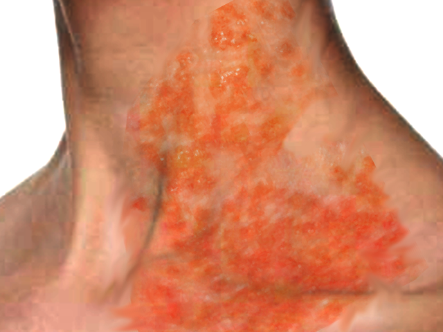
Kaposi Varicelliform Eruption in a Patient With Atopic Dermatitis. Kaposi varicelliform eruption, also known as eczema herpeticum, is a disseminated viral skin infection that occurs in patients with an underlying cutaneous disease. This condition is most commonly caused by the herpes simplex virus and presents with localized vesicular eruptions. Although uncommon, the condition can be potentially life-threatening if not treated promptly.
Contributed by O Chaigasame, MD
References
Skrek SV, Timoshchuk EA, Sidikov AA, Zaslavsky DV, Megna M. Kaposi varicelliform eruption induced by methotrexate in an adult atopic dermatitis patient. Dermatologic therapy. 2019 Mar:32(2):e12826. doi: 10.1111/dth.12826. Epub 2019 Feb 7 [PubMed PMID: 30659717]
Miller DM, Trowbridge RM, Desai A, Drews RE. Kaposi's varicelliform eruption in a patient with metastatic melanoma and primary cutaneous anaplastic large cell lymphoma treated with talimogene laherparepvec and nivolumab. Journal for immunotherapy of cancer. 2018 Nov 19:6(1):122. doi: 10.1186/s40425-018-0437-4. Epub 2018 Nov 19 [PubMed PMID: 30454071]
Gottesman SP, Rosen JR, Geller JD, Freeman BB. Atypical Varicella-Zoster Kaposi Varicelliform Eruption in Sézary Syndrome. The American Journal of dermatopathology. 2018 Dec:40(12):920-923. doi: 10.1097/DAD.0000000000001264. Epub [PubMed PMID: 30211729]
Dazé R, Canales J, Ross R. Kaposi varicelliform eruption in a patient with atopic dermatitis. Journal of osteopathic medicine. 2021 Jul 15:121(10):825-826. doi: 10.1515/jom-2021-0170. Epub 2021 Jul 15 [PubMed PMID: 34261200]
Martín-Galache M, Escalona-Gil AM, Posado-Domínguez L, Jiménez-Domínguez A, Arévalo-Cenzual A, López-Ávila FJ, Consuegra-Llapur E. Kaposi's Varicelliform Eruption: A Potentially Life-Threatening Complication of Atopic Dermatitis. European journal of case reports in internal medicine. 2024:11(5):004392. doi: 10.12890/2024_004392. Epub 2024 Mar 28 [PubMed PMID: 38715890]
Siddiqui S, Sanchez S, Cavazos A, Abdelnabi M. Kaposi varicelliform eruption due to disseminated herpes simplex in a patient with known Darier disease. The American journal of the medical sciences. 2023 Sep:366(3):e35-e36. doi: 10.1016/j.amjms.2023.02.001. Epub 2023 Feb 10 [PubMed PMID: 36773694]
Azmi M, Nasim A, Dodani S, Laiq SM, Mehdi SH, Mubarak M. Kaposi Varicelliform Eruption Associated With Chickenpox in a Liver Transplant Recipient. Experimental and clinical transplantation : official journal of the Middle East Society for Organ Transplantation. 2020 Apr:18(2):252-254. doi: 10.6002/ect.2017.0282. Epub 2018 Jun 28 [PubMed PMID: 29957162]
Gruhl RR, Wu A, Niermann M, Olson A. Not all that vesicles is herpes. Diagnosis (Berlin, Germany). 2017 Nov 27:4(4):261-264. doi: 10.1515/dx-2017-0033. Epub [PubMed PMID: 29536938]
Okamoto M, Takahagi S, Tanaka A, Ogawa A, Nobuki H, Hide M. A case of Kaposi varicelliform eruption progressing to herpes simplex virus hepatitis in an immunocompetent patient. Clinical and experimental dermatology. 2018 Jul:43(5):636-638. doi: 10.1111/ced.13405. Epub 2018 Feb 15 [PubMed PMID: 29446473]
Level 3 (low-level) evidenceSato E, Hiromatsu K, Murata K, Imafuku S. Loss of ATP2A2 Allows Herpes Simplex Virus 1 Infection of a Human Epidermis Model by Disrupting Innate Immunity and Barrier Function. The Journal of investigative dermatology. 2018 Dec:138(12):2540-2549. doi: 10.1016/j.jid.2018.05.019. Epub 2018 Jun 2 [PubMed PMID: 29870688]
Rogel-Vence M, González-Ruiz L, Franco-Muñoz M, Garrido-Martín JA. Kaposi Varicelliform Eruption on Sunburned Skin. Actas dermo-sifiliograficas. 2021 Jan:112(1):71. doi: 10.1016/j.ad.2018.12.013. Epub 2020 Sep 25 [PubMed PMID: 32986970]
Adams JL, Schnaubelt ER, Hewlett AL, Georgesen CJ, Lauer SR, Wysong A, Lawler JV. Kaposi Varicelliform Eruption of Mpox in a Peeling Sunburn. Cutis. 2023 Mar:111(3):E7-E8. doi: 10.12788/cutis.0733. Epub [PubMed PMID: 37224489]
Sun D, Ong PY. Infectious Complications in Atopic Dermatitis. Immunology and allergy clinics of North America. 2017 Feb:37(1):75-93. doi: 10.1016/j.iac.2016.08.015. Epub [PubMed PMID: 27886912]
Lehman JS, el-Azhary RA. Kaposi varicelliform eruption in patients with autoimmune bullous dermatoses. International journal of dermatology. 2016 Mar:55(3):e136-40. doi: 10.1111/ijd.13091. Epub 2015 Oct 24 [PubMed PMID: 26500144]
Lyons JJ, Milner JD, Stone KD. Atopic dermatitis in children: clinical features, pathophysiology, and treatment. Immunology and allergy clinics of North America. 2015 Feb:35(1):161-83. doi: 10.1016/j.iac.2014.09.008. Epub 2014 Nov 21 [PubMed PMID: 25459583]
Wollenberg A, Seba A, Antal AS. Immunological and molecular targets of atopic dermatitis treatment. The British journal of dermatology. 2014 Jul:170 Suppl 1():7-11. doi: 10.1111/bjd.12975. Epub 2014 May 9 [PubMed PMID: 24720588]
Reed JL, Scott DE, Bray M. Eczema vaccinatum. Clinical infectious diseases : an official publication of the Infectious Diseases Society of America. 2012 Mar:54(6):832-40. doi: 10.1093/cid/cir952. Epub 2012 Jan 30 [PubMed PMID: 22291103]
Level 3 (low-level) evidenceSohail M, Khan FA, Shami HB, Bashir MM. Management of eczema herpeticum in a Burn Unit. JPMA. The Journal of the Pakistan Medical Association. 2016 Nov:66(11):1357-1361 [PubMed PMID: 27812048]