 Tools
Tools
 Anatomy, Abdomen and Pelvis: Ileocolic Artery
Introduction
The ileocolic artery, the most inferior branch of the superior mesenteric artery (SMA), is a midgut-derived vessel of mesodermal origin supplying the distal ileum, cecum, appendix, and proximal ascending colon (see Image. Angiogram of the Superior Mesenteric Artery). The distribution of this vessel parallels the myenteric plexus spanning these bowel segments. Lymphatic drainage mirrors arterial territories. Common variants include differing origins of the right colic (RCA), middle colic (MCA), and ileocolic arteries, including shared trunks or separate origins. In many individuals, the artery courses anterior to the superior mesenteric vein (SMV).
Major surgical and clinical significance is attributed to the ileocolic artery and its branches for several reasons. Ligation of this blood vessel or its branches is required during appendectomy or Meckel diverticulectomy to achieve hemostasis. Ischemia with possible necrosis of the visceral organs supplied by the ileocolic artery may occur in the setting of intussusception. Ischemia within the ileocolic distribution may also occur secondary to SMA thromboembolism. Careful attention is required during colon cancer resection at sites of vascular anastomoses, including those involving the RCA and the ileocolic artery, to ensure adequate hemostasis. Lymphatic drainage parallels arterial supply, and comprehension of anatomic variation remains essential for successful oncologic surgery. Understanding of gastrointestinal vascular anatomy is essential for the diagnosis and management of gastrointestinal bleeding (see Image. Anatomy of the Ileocolic Artery).
Structure and Function
Register For Free And Read The Full Article
 Search engine and full access to all medical articles
- 10 free questions in your specialty
- Free CME/CE Activities
- Free daily question in your email
- Save favorite articles to your dashboard
- Emails offering discounts
Learn more about a Subscription to StatPearls Point-of-Care
Structure and Function
The major branches of the SMA include the RCA, MCA, and the ileocolic and inferior pancreaticoduodenal arteries. Additional jejunal and ileal branches arise from the left aspect of the SMA.
The MCA is the 1st colic branch of the SMA and supplies the proximal 2/3 of the transverse colon. The RCA is the 2nd colic branch and supplies the right colon, predominantly the ascending colon.
The ileocolic artery is the most inferior branch of the SMA, often described as the terminal branch. After arising from the SMA, the ileocolic artery courses inferiorly in a retroperitoneal plane toward the right iliac fossa. Division into superior and inferior branches occurs in this region.
The superior branch of the ileocolic artery anastomoses with the RCA and supplies the terminal ileum, cecum, and appendix. The inferior branch gives rise to the anterior and posterior cecal arteries, the ileal branch of the ileocolic artery, and the appendicular artery. The anterior and posterior cecal arteries course anterior and posterior to the terminal ileum, respectively.
The ileal branch of the ileocolic artery forms an anastomosis with the last intestinal (ileal) artery. The ileocolic artery also anastomoses superiorly with the RCA. The appendicular artery courses within the mesoappendix and is located posterior to the terminal ileum.
Normal anatomic variations of the right colonic vasculature and SMA branches are well described, with the most common configuration consisting of separate origins of the RCA, MCA, and ileocolic artery, as previously described. Additional frequent variants include a common origin of the MCA and RCA with a separate origin of the ileocolic artery, as well as a common origin of the RCA and ileocolic artery with a separate origin of the MCA.
Arteries of the midgut and hindgut form an anastomotic arcade known as the marginal artery of Drummond. This blood vessel consists of an anastomosis between the terminal branches of the SMA and the inferior mesenteric artery (IMA), coursing along the inner colonic margin within the mesentery. Small colonic branches arise from this arcade and are termed "vasa recta."[1][2]][3][4][5][6]
The ileocolic artery and its branches supply the distal ileum, ileocecal valve, cecum, vermiform appendix, and proximal ascending colon. The ileocolic artery courses anterior to the right gonadal vessels and ureter. The anterior cecal artery lies anterior to the terminal ileum and cecum, while the posterior cecal artery is situated posterior to these structures. The appendicular artery traverses posterior to the terminal ileum. An anastomosis occurs between the ileocolic artery and the RCA. Terminal branches of the SMA and IMA form an anastomosis via the marginal artery of Drummond.
Embryology
The ileocolic artery is of midgut origin, arising as the most inferior branch of the SMA and supplying midgut derivatives. Consistent with general vascular development, the ileocolic artery is derived from mesoderm.[7]
Blood Supply and Lymphatics
The blood supply to the ileocolic artery is derived from the SMA, the 2nd major branch of the abdominal aorta, located just distal to the celiac trunk. The celiac trunk supplies the stomach, proximal small bowel, liver, spleen, and pancreas. The IMA arises from the abdominal aorta and supplies the hindgut. Lymphatic drainage associated with the ileocolic artery involves lymph nodes within the mesentery of the mesoileum, mesocecum, mesoappendix, and mesocolon.[8]
Nerves
The ileocolic artery supplies the portion of the myenteric plexus corresponding to its visceral distribution. The myenteric plexus extends along the territory supplied by the ileocolic artery from the distal ileum to the proximal ascending colon.[9]
Muscles
Muscles supplied by the ileocolic artery are located within the bowel walls. Adequate perfusion of the muscular layer, composed of inner circular and outer longitudinal layers, is required for peristalsis. In addition to the bowel wall musculature, the ileocolic artery supplies the intrinsic sphincter muscle of the ileocecal valve. The proximal portion of the taeniae coli also receives vascular supply, extending from their convergence at the vermiform appendix to the proximal ascending colon at the level of anastomosis with the RCA.[10]
Physiologic Variants
The ileocolic artery is present in more than 95% of individuals. In approximately 50% of patients, this blood vessel courses anterior and superior to the SMV. Common variations of the right colonic vasculature include a common origin of the RCA and MCA with a separate origin of the ileocolic artery, as well as a common origin of the RCA and ileocolic artery with a distinct origin of the MCA. An additional variation includes the absence of the RCA.[11][12]
Surgical Considerations
The appendicular artery courses within the mesoappendix, lying close to its free margin. The blood vessel terminates in multiple small branches supplying the appendix. Ligation of the inferior branch of the ileocolic artery at the base of the appendix is required during an appendectomy. Postoperative bleeding may result from inadequate vessel ligation. Complete hemostasis should be achieved, and mesoappendiceal integrity must be confirmed before closure. By the same principle, ileocolic arterial branches should be ligated during Meckel diverticulectomy to ensure hemostasis.[13]
Clinical Significance
Ileocecal intussusception may result in the occlusion of the ileocolic artery as the terminal ileum telescopes into the cecum. Progressive invagination of a bowel segment into another may compress the vascular supply, sufficient to occlude the ileocolic artery and produce ischemia within its distribution. The extent of ischemic involvement depends on the level of vascular compression and may include proximal or distal segments or individual arterial branches. Ischemia may involve a limited section of bowel or the full vascular territory of the ileocolic artery. Prolonged or recurrent intussusception may progress to necrosis and perforation, resulting in an acute abdomen and surgical emergency.
Intussusception occurs most frequently in young children, typically aged between 2 and 3 years. Clinical presentation may include intermittent abdominal pain, vomiting, a right upper quadrant mass, and bloody stools. A lead point is rarely found in young children, although commonly identified in older children and adults. These lead points include benign and malignant neoplasms. Management in most pediatric cases may include water-soluble contrast or air enema reduction. Surgical backup is required due to the risk of perforation. Recurrence rates as high as 30% have been reported.[14][15][16][17][18]
Ischemia may occur following SMA thromboembolism. Occlusion of the SMA can impair blood flow throughout its entire vascular territory. Smaller thromboemboli may lodge within distal SMA branches, including the inferior terminal branch and the ileocolic artery. Similar to intussusception, the extent of ischemia or necrosis depends on the level of vascular occlusion.[19][20]
A thorough understanding of right colonic vascular anatomy and its anatomic variations is essential for surgical intervention. Clinical contexts include appendicitis, Meckel diverticulitis, inflammatory bowel disease, colon cancer, and symptomatic benign neoplasms of the bowel or mesentery. Accurate anatomic knowledge supports effective hemostasis and optimal healing of bowel anastomoses. Anastomotic sites within the colon carry an increased risk of ischemia following tumor or bowel resection. Adequate vascular supply is particularly critical in patients who are of advanced age or have diabetes to support normal anastomotic healing.
Traumatic injury may rarely result in an isolated rupture of the ileocolic artery. This condition may occur in the setting of blunt abdominal trauma.[21]
Other Issues
Colic vessels include the ileocolic artery, and postcancer resection status may serve as a marker of the extent of resection. Arterial stumps identified on routine portal venous computed tomography have demonstrated potential as in vivo markers of surgical quality and the extent of mesenteric resection.[22]
Media
(Click Image to Enlarge)
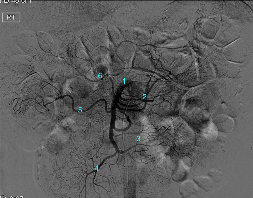
Angiogram of the Superior Mesenteric Artery. This conventional angiogram highlights the vascular anatomy of the midgut, originating from the proximal superior mesenteric artery (1). Key branches include the jejunal (2) and ileal (3) arteries, alongside the ileocolic (4), right colic (5), and middle colic (6) vessels.
Contributed by DW Byerly, MD, PhD
(Click Image to Enlarge)
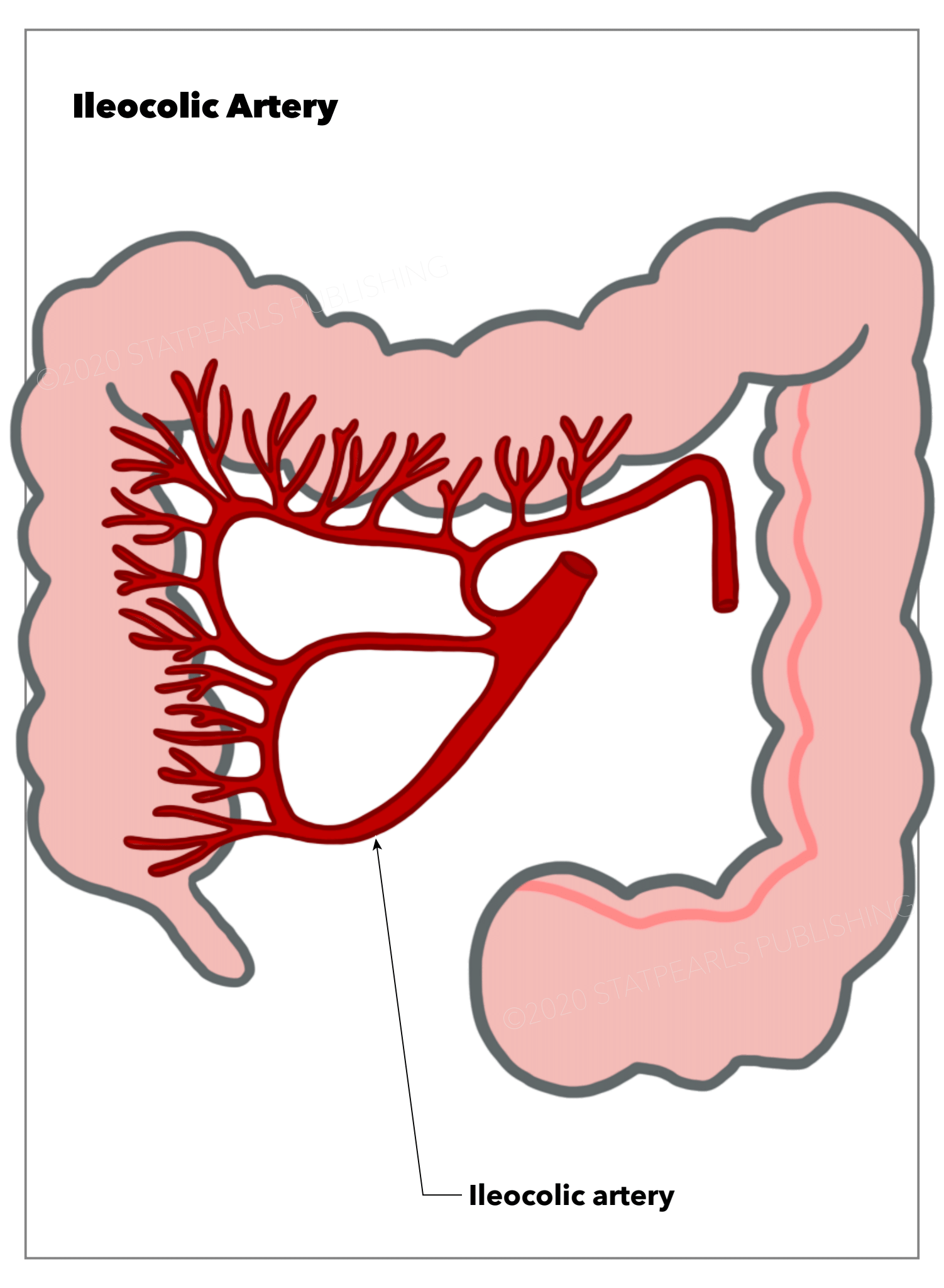
Anatomy of the Ileocolic Artery. This illustration demonstrates the path and distribution of the ileocolic artery as it supplies the terminal ileum, cecum, and ascending colon. The diagram specifically highlights how this vessel branches from the superior mesenteric artery to nourish the transition between the small and large intestines.
Illustration by E Gregory
References
Charnsangavej C, DuBrow RA, Varma DG, Herron DH, Robinson TJ, Whitley NO. CT of the mesocolon. Part 1. Anatomic considerations. Radiographics : a review publication of the Radiological Society of North America, Inc. 1993 Sep:13(5):1035-45 [PubMed PMID: 8210588]
Kuzu MA, İsmail E, Çelik S, Şahin MF, Güner MA, Hohenberger W, Açar Hİ. Variations in the Vascular Anatomy of the Right Colon and Implications for Right-Sided Colon Surgery. Diseases of the colon and rectum. 2017 Mar:60(3):290-298. doi: 10.1097/DCR.0000000000000777. Epub [PubMed PMID: 28177991]
Mike M, Kano N. Reappraisal of the vascular anatomy of the colon and consequences for the definition of surgical resection. Digestive surgery. 2013:30(4-6):383-92. doi: 10.1159/000343156. Epub 2013 Oct 10 [PubMed PMID: 24135859]
Du K, Ren J, Zheng G, Li S, Chen L, Hou W, Duan W, Huang D, Zhang H, Feng F, Zheng J. Variation of the ileocolic artery and superior mesenteric artery in a patient with right-sided colon cancer with Lynch syndrome: a case report. Annals of translational medicine. 2022 Sep:10(17):939. doi: 10.21037/atm-22-3012. Epub [PubMed PMID: 36172100]
Level 3 (low-level) evidenceBalcerzak A, Tubbs RS, Waśniewska-Włodarczyk A, Rapacka E, Olewnik Ł. Classification of the superior mesenteric artery. Clinical anatomy (New York, N.Y.). 2022 May:35(4):501-511. doi: 10.1002/ca.23841. Epub 2022 Mar 7 [PubMed PMID: 35088464]
Nigah S, Patra A, Chumber S. Analysis of the Variations in the Colic Branching Pattern of the Superior Mesenteric Artery: A Cadaveric Study With Proposal to Modify Its Current Anatomical Classification. Cureus. 2022 May:14(5):e25025. doi: 10.7759/cureus.25025. Epub 2022 May 15 [PubMed PMID: 35719766]
Bhatia A, Shatanof RA, Bordoni B. Embryology, Gastrointestinal. StatPearls. 2026 Jan:(): [PubMed PMID: 30725857]
Sun HL, Wang W, Yao L, Chen SX, Ren A, Hu YY, Xu YY. [Preoperative evaluation of mesenteric vascular anatomy using 256 multi-slice computed tomography before laparoscopic surgery]. Zhonghua wei chang wai ke za zhi = Chinese journal of gastrointestinal surgery. 2011 Nov:14(11):855-8 [PubMed PMID: 22116719]
Timmermans JP, Hens J, Adriaensen D. Outer submucous plexus: an intrinsic nerve network involved in both secretory and motility processes in the intestine of large mammals and humans. The Anatomical record. 2001 Jan 1:262(1):71-8 [PubMed PMID: 11146430]
Level 3 (low-level) evidencePollard MF, Thompson-Fawcett MW, Stringer MD. The human ileocaecal junction: anatomical evidence of a sphincter. Surgical and radiologic anatomy : SRA. 2012 Jan:34(1):21-9. doi: 10.1007/s00276-011-0865-z. Epub 2011 Aug 24 [PubMed PMID: 21863224]
Haywood M, Molyneux C, Mahadevan V, Srinivasaiah N. Right colic artery anatomy: a systematic review of cadaveric studies. Techniques in coloproctology. 2017 Dec:21(12):937-943. doi: 10.1007/s10151-017-1717-6. Epub 2017 Dec 2 [PubMed PMID: 29196959]
Level 1 (high-level) evidenceGamo E, Jiménez C, Pallares E, Simón C, Valderrama F, Sañudo JR, Arrazola J. The superior mesenteric artery and the variations of the colic patterns. A new anatomical and radiological classification of the colic arteries. Surgical and radiologic anatomy : SRA. 2016 Jul:38(5):519-27. doi: 10.1007/s00276-015-1608-3. Epub 2016 Jan 4 [PubMed PMID: 26728989]
Pan K. [Measures to anatomic variations of the colonic vessels in laparoscopic operations]. Zhonghua wei chang wai ke za zhi = Chinese journal of gastrointestinal surgery. 2013 Oct:16(10):944-6 [PubMed PMID: 24158864]
Liu X, Yu HK, Gu LX, Chen JK, Wen ZB. Atropine Premedication Facilitates Ultrasound-Guided Reduction by Saline Enema in Children With Intussusception. Frontiers in pharmacology. 2019:10():43. doi: 10.3389/fphar.2019.00043. Epub 2019 Jan 31 [PubMed PMID: 30766486]
Gollub MJ. Colonic intussusception: clinical and radiographic features. AJR. American journal of roentgenology. 2011 May:196(5):W580-5. doi: 10.2214/AJR.10.5112. Epub [PubMed PMID: 21512048]
Level 2 (mid-level) evidenceApplegate KE. Clinically suspected intussusception in children: evidence-based review and self-assessment module. AJR. American journal of roentgenology. 2005 Sep:185(3 Suppl):S175-83 [PubMed PMID: 16120899]
Level 3 (low-level) evidenceKlein EJ, Kapoor D, Shugerman RP. The diagnosis of intussusception. Clinical pediatrics. 2004 May:43(4):343-7 [PubMed PMID: 15118777]
Level 2 (mid-level) evidenceSoto Castro AE, Escoto Alemán OJ, Soto Castro TR, Valadez Almaraz KC, Delgado Gaheta D, Muñoz Ornelas D, Andrade Lara JA, May Rodriguez J. Laparoscopic Right Hemicolectomy With D2 Lymphadenectomy for Adult Ileocecal Intussusception Secondary to Cecal Adenocarcinoma: A Case Report. Cureus. 2025 Nov:17(11):e97702. doi: 10.7759/cureus.97702. Epub 2025 Nov 24 [PubMed PMID: 41306816]
Level 3 (low-level) evidenceKamiya C, Deguchi J, Kitaoka T, Suzuki J, Abe K, Sato O. Obstruction of the superior mesenteric artery due to emboli from the thoracic aorta in a patient with thromboangiitis obliterans. Annals of vascular diseases. 2014:7(3):320-4. doi: 10.3400/avd.cr.14-00035. Epub 2014 Jul 30 [PubMed PMID: 25298837]
Level 3 (low-level) evidenceSakai A, Hara H, Nakamura M. Successful endovascular treatment for simultaneous multiple thromboemboli following myocardial infarction. The Journal of invasive cardiology. 2013 Jun:25(6):E133-5 [PubMed PMID: 23735368]
Level 3 (low-level) evidenceBastuev NV, Bekenov BM, Ivankov VT. [Isolated rupture of the ileocolic artery in blunt trauma to the abdomen]. Sudebno-meditsinskaia ekspertiza. 1976 Oct-Dec:19(4):47 [PubMed PMID: 1014059]
Level 3 (low-level) evidenceKaye TL, West NP, Jayne DG, Tolan DJ. CT assessment of right colonic arterial anatomy pre and post cancer resection - a potential marker for quality and extent of surgery? Acta radiologica (Stockholm, Sweden : 1987). 2016 Apr:57(4):394-400. doi: 10.1177/0284185115583033. Epub 2015 May 4 [PubMed PMID: 25940063]
Level 2 (mid-level) evidence