 Tools
Tools
 Horner Syndrome
Introduction
Horner syndrome is a rare condition classically presenting with partial ptosis (drooping of the upper eyelid), miosis (pupillary constriction), and facial anhidrosis (loss of sweating) due to disruption of the sympathetic nerve supply. The disorder is usually acquired following injury to the sympathetic pathway, although rare congenital forms occur. Therefore, treatment focuses on identifying and appropriately treating the underlying cause. The syndrome has several names, including Bernard-Horner syndrome in French-speaking countries, Horner syndrome in English-speaking countries, oculosympathetic palsy, and von Passow syndrome, which refers to Horner syndrome associated with iris heterochromia.
The syndrome was first described by François Pourfour du Petit in 1727 after an animal experiment involving resection of the intercostal nerves produced changes in the ipsilateral eye and face.[1] French physiologist Claude Bernard provided a more detailed description in 1852, followed by several clinicians who offered different interpretations. Swiss ophthalmologist Johann Friedrich Horner formally described the condition in 1869, and the syndrome was subsequently named after him.[2][3][4][5]
Anatomy
Understanding the sympathetic innervation of the eye is essential for recognizing the clinical features of Horner syndrome. The nerve supply consists of a 3-neuron pathway extending from the posterolateral hypothalamus to the long ciliary nerves, which innervate the iris dilator and superior tarsal muscle, also known as the Müller muscle.[6] The first-order neurons originate in the hypothalamus and descend uncrossed through the midbrain and pons, terminating in the intermediolateral cell columns at the C8 through T2 levels of the spinal cord, known as the ciliospinal center of Budge. Second-order preganglionic neurons exit the spinal cord at the T1 level and enter the cervical sympathetic chain, where they ascend to synapse in the superior cervical ganglion at the C3 through C4 levels.[7] Third-order postganglionic fibers divide into sudomotor and vasomotor branches that follow the external carotid artery and innervate the sweat glands and blood vessels of the face. The remaining fibers ascend within the carotid plexus along the internal carotid artery and enter the cavernous sinus, where they join the abducens nerve (cranial nerve VI). After leaving the cavernous sinus, the fibers enter the orbit through the superior orbital fissure and travel with the ophthalmic division of the trigeminal nerve (cranial nerve V) before continuing as the long ciliary nerves.
Etiology
Register For Free And Read The Full Article
 Search engine and full access to all medical articles
- 10 free questions in your specialty
- Free CME/CE Activities
- Free daily question in your email
- Save favorite articles to your dashboard
- Emails offering discounts
Learn more about a Subscription to StatPearls Point-of-Care
Etiology
Horner syndrome is primarily an acquired condition secondary to systemic or local disorders or iatrogenic causes, although rare congenital and hereditary forms occur.[8][9] Sympathetic fibers follow an extensive course and can be interrupted during their extracranial, intracranial, or intraorbital traversal.[10][11][12] Disruption may occur centrally between the hypothalamus and the point at which the fibers exit the spinal cord at C8 through T2 or peripherally within the cervical sympathetic chain, superior cervical ganglion, or carotid artery pathway.[10] Preganglionic Horner syndrome can be an ominous sign because of its association with pulmonary malignant neoplasms. Overall, the causes of Horner syndrome can be categorized according to the anatomical location of the disruption. First-order neurons are mostly affected by intracranial conditions and include the following:
- Cerebrovascular accident
- Multiple sclerosis
- Arnold-Chiari malformation
- Encephalitis
- Meningitis
- Lateral medullary syndrome
- Syringomyelia
- Intracranial tumors (pituitary or basal skull)
- Spinal trauma above the T2 through T3 levels
- Spinal cord tumors [13][14][15]
Second-order neurons traverse the thoracic region and are affected by the following:
- Malignant neoplasms involving the lung apex, including Pancoast tumors
- Cervical rib causing tractional injury
- Subclavian artery lesions, including aneurysms
- Mediastinal lymphadenopathy
- Trauma to the brachial plexus [11]
- Neuroblastoma of the paravertebral sympathetic chain [15]
- A dental abscess involving the mandibular region
- Iatrogenic injury associated with thyroidectomy, radical neck dissection, tonsillectomy, coronary artery bypass grafting, or carotid angiography [12][13][12][16][17]
Third-order neurons are in close proximity to the internal carotid artery and cavernous sinus and are affected by the following:
- Carotid-cavernous fistula
- Internal carotid artery dissection or an aneurysm [18]
- Cluster headaches or migraine
- Raeder paratrigeminal syndrome, characterized by unilateral facial pain, headache, and Horner syndrome
- Herpes zoster infection
- Temporal arteritis
Epidemiology
Horner syndrome is uncommon, with an estimated frequency of 1 in 6000 individuals. The condition may occur at any age and in any racial or ethnic group.[15][19]
Pathophysiology
Horner syndrome results from disruption of the sympathetic pathway. Clinical manifestations depend on the location of the lesion, whereas severity depends on the degree of denervation.[9] The superior tarsal muscle helps elevate the upper eyelid and receives sympathetic innervation. Denervation of this muscle causes ptosis, which is milder than the ptosis associated with oculomotor nerve palsy because the oculomotor nerve innervates the levator palpebrae superioris. The superior tarsal muscle maintains elevation of the upper eyelid after the levator palpebrae superioris raises it, accounting for the partial ptosis seen in Horner syndrome. The lower eyelid may be slightly elevated because of denervation of the lower eyelid muscle, which is analogous to the superior tarsal muscle.[20]
The sympathetic nervous system mediates pupillary dilation. Disruption of this pathway leaves parasympathetic innervation unopposed, resulting in pupillary constriction. Pupillary responses to light and accommodation remain normal because these systems do not depend on sympathetic innervation.[21] Ipsilateral anhidrosis, another classic manifestation, depends on the level of sympathetic interruption. First-order neuron lesions may cause anhidrosis involving the ipsilateral side of the body because these fibers arise centrally.[22] Second-order neuron lesions typically affect the ipsilateral face. Third-order postganglionic neuron lesions produce limited facial anhidrosis, usually confined to the area adjacent to the ipsilateral brow, because the vasomotor and sudomotor fibers have already branched from the pathway. Iris heterochromia, characterized by reduced pigmentation of the iris on the affected side, may occur in children younger than 2 years with congenital Horner syndrome.[9]
History and Physical
Accurate localization of the lesion in Horner syndrome is crucial for guiding subsequent evaluation and treatment. Therefore, a detailed history and physical examination are essential. Clinical assessment should address the following features:
- Balance impairment, hearing loss, sensory changes, or swallowing difficulties may indicate a central lesion involving first-order neurons.
- A history of trauma or a surgical procedure involving the head, face, neck, shoulder, or back may indicate second-order neuron involvement.[17]
- A detailed medical and medication history should assess for exposure to miotic or mydriatic agents.
- Headache, diplopia, facial numbness, or facial pain may indicate third-order neuron involvement.
- The presence and distribution of anhidrosis may assist with lesion localization.
- The characteristics and duration of any headache should be documented.
- Longstanding, stable symptoms may suggest a more benign cause, whereas recently progressive symptoms accompanied by weight loss, hemoptysis, low-grade fever, or lymphadenopathy raise concern for a serious underlying disorder.[23]
- Facial or orbital pain accompanied by miosis and ptosis may indicate Raeder paratrigeminal syndrome.[24]
- A history of skin lesions or previously diagnosed herpes zoster infection should be obtained.
- The severity and location of pain should be documented.
A detailed ocular examination is warranted and should include the following assessments:
- Pupillary reactivity to light and accommodation
- Measurement of pupillary diameter in dim and bright light
- Examination of the upper eyelid for ptosis and fatigability
- Evaluation of extraocular movements
- Assessment of visual acuity, color vision, and visual fields
- Slit-lamp examination for structural details
- Evaluation of the presence of nystagmus
A detailed ocular examination may reveal a round, constricted pupil. The affected pupil demonstrates dilation lag, and anisocoria is typically more pronounced in darkness than in light. The ciliospinal reflex may also be absent. Additional findings may include partial ptosis, iris heterochromia, apparent enophthalmos, contralateral eyelid retraction, conjunctival injection, and unchanged or transiently reduced intraocular pressure.[17][25][17] A comprehensive systemic examination should also focus on the neurologic, pulmonary, and cardiovascular systems while accounting for the relevant differential diagnoses.
Evaluation
Laboratory Testing
- The initial workup should include a complete blood count, erythrocyte sedimentation rate, and serum chemistry panel.
- Urine or blood cultures may be ordered if an infectious agent is suspected.
- Testing for suspected neurosyphilis, HIV, thyroid function, vitamin B12, and folate levels may be ordered if indicated following a detailed history and examination.
- Urine testing for elevated metabolic catecholamine metabolites is important in the pediatric population with suspected neuroblastoma.
- Purified protein derivative is warranted in suspected tuberculosis.
Imaging
- Chest radiography followed by CT of the chest should be performed when a pulmonary malignant neoplasm is suspected.
- Head CT and MRI are recommended when stroke is suspected.
- MRI is warranted and preferred over ultrasonography when carotid artery dissection is a possibility (particularly in painful Horner syndrome).[26]
Pharmacological Testing
Pharmacologic testing is the most useful modality for confirming Horner syndrome and assisting with lesion localization.[27] Pharmacologic testing for Horner syndrome includes the following:
- Topical cocaine test: Cocaine acts as an indirect sympathomimetic by inhibiting norepinephrine reuptake from the synaptic cleft. Cocaine solution at a concentration of 2% to 10% is instilled into both eyes. Pupillary responses are assessed after at least 30 min. Denervation causes the affected pupil to dilate poorly compared with the unaffected pupil. Anisocoria of 0.8 mm or more is considered diagnostic. However, the test does not localize the lesion. Additional disadvantages include the relatively high cost of cocaine, prolonged testing time, potentially ambiguous results, and detectable urinary cocaine metabolites.[28]
- Topical hydroxyamphetamine test: This test is particularly helpful for localizing the lesion. Hydroxyamphetamine stimulates the release of stored norepinephrine from postganglionic nerve terminals into the synapse. The test differentiates postganglionic third-order neuron lesions from first- or second-order neuron lesions. Two drops of a 1% hydroxyamphetamine solution are instilled into each eye. With a third-order neuron lesion, the affected pupil dilates less than the unaffected pupil. In patients with intact postganglionic fibers and first- or second-order neuron lesions, the affected pupil dilates equally or more than the unaffected pupil. Disadvantages include the inability to perform the test on the same day as the cocaine test and the possibility of false-negative results.[28]
- Topical apraclonidine test: This test is considered the preferred pharmacologic test because of its sensitivity and practicality. Apraclonidine acts as a weak α1-adrenergic agonist and strong α2-adrenergic agonist and is categorized as an ocular hypotensive agent. Upregulation of α1-adrenergic receptors in Horner syndrome produces denervation supersensitivity and an exaggerated response of the iris dilator muscle to apraclonidine.[29] A 0.5% to 1% solution is instilled into both eyes. The affected eye develops mydriasis, whereas the unaffected eye shows minimal dilation or constriction. Apraclonidine therefore reverses the anisocoria because its α2-adrenergic activity predominates in the unaffected eye, whereas α1-adrenergic receptor supersensitivity produces dilation in the affected eye. Disadvantages include false-negative results in acute cases, systemic adverse effects in children, inability to localize the lesion, and the relatively long half-life of the drug.
Other proposed pharmacologic agents have limited clinical relevance:
- Pholedrine: Pholedrine, an N-methyl derivative of hydroxyamphetamine, is approximately half as potent as hydroxyamphetamine and has produced comparable results. However, limited availability restricts its use.
- Epinephrine: Epinephrine is a direct-acting sympathomimetic with poor corneal penetration and variable sensitivity.
- Phenylephrine: Phenylephrine is a selective α1-adrenergic agonist with poor corneal penetration and variable results depending on the degree of denervation.[30]
Treatment / Management
Treatment depends on identifying and treating the underlying cause. Timely diagnosis is critical and should be followed by referral to the appropriate specialist. Healthcare professionals should incorporate comprehensive ocular examinations into clinical practice when Horner syndrome is suspected. The indication for a surgical procedure and the appropriate approach largely depend on the underlying disorder. Surgical treatment options include the following:
- Neurosurgical evaluation and treatment for Horner syndrome associated with an aneurysm
- Vascular surgical evaluation and treatment for carotid artery dissection or aneurysm
Differential Diagnosis
Pain accompanying Horner syndrome may indicate a serious underlying cause and warrants thorough evaluation. Systemic symptoms such as weight loss and progressive fatigue may suggest an underlying malignant neoplasm. Furthermore, Horner syndrome can be an early manifestation of neuroblastoma in children. Carotid artery dissection may present with unilateral headache and facial or neck pain. Suspected carotid artery dissection requires urgent diagnostic evaluation and treatment. Raeder paratrigeminal syndrome may also present with headache but is accompanied by trigeminal nerve (cranial nerve V) impairment. Anisocoria or ptosis may result from numerous disorders, including Holmes-Adie syndrome, neurosyphilis with an Argyll Robertson pupil, oculomotor nerve palsy, and optic neuritis.
Prognosis
The long-term prognosis of idiopathic Horner syndrome appears to be benign. Symptoms generally do not worsen, and approximately half of affected patients no longer experience anisocoria. Almost one-third of patients with ptosis report spontaneous improvement or complete resolution after a mean of 7.9 years following diagnosis. In unilateral cases, the oculosympathetic defect does not progress to bilateral involvement. New medical disorders may develop 3 to 20 years after the diagnosis of Horner syndrome; however, these conditions are generally unrelated to the syndrome.[31]
Complications
Horner syndrome is almost always secondary to an underlying cause, such as a tumor, trauma, or infection, and is only rarely idiopathic. The complications associated with Horner syndrome are primarily related to the underlying cause. For instance, Horner syndrome resulting from multiple sclerosis may be associated with muscle stiffness or spasms, impaired bladder, bowel, or sexual function, and seizures. Similarly, ocular disorders associated with Horner syndrome may cause temporary or permanent visual impairment. Severe visual involvement may result in partial or complete vision loss.
Consultations
Appropriate treatment of Horner syndrome and its underlying cause may require consultation with the following specialists:
- Pulmonology
- Neurology
- Neurosurgery
- Interventional radiology
- Oncology
- Neuro-ophthalmology
Deterrence and Patient Education
Patients should be educated about the possible manifestations of Horner syndrome. Parents of infants born with congenital Horner syndrome should be informed about treatment options and appropriate follow-up care. Patients with Horner syndrome should understand that the syndrome does not damage the eye or cause vision loss directly but may indicate injury to structures along the sympathetic nerve pathway. Clinicians must evaluate the extent and cause of this injury because the underlying disorder may be serious. In addition, patients may report a gritty sensation and have an increased risk of ocular infection. Artificial tears and ointment are often sufficient; however, a surgical procedure may be needed in severe or complicated cases.
Enhancing Healthcare Team Outcomes
Horner syndrome has numerous potential causes and is best treated by an interprofessional team. The key is to determine the location of the sympathetic pathway lesion. A detailed systemic examination should focus on the neurologic, pulmonary, and cardiovascular systems while considering the relevant differential diagnoses. Clinical outcomes depend on the underlying cause.
Media
(Click Image to Enlarge)
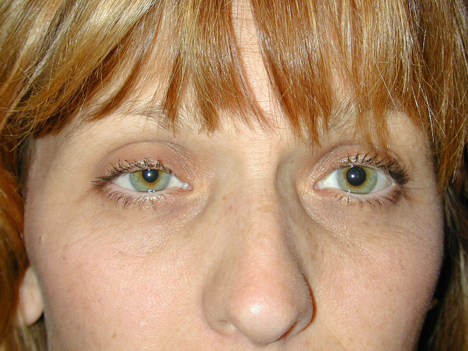
Right sided ptosis and miosis i.e. horners syndrome Contributed by Geoffrey Kwitko M.D.
References
van der Wiel HL. Johann Friedrich Horner (1831-1886). Journal of neurology. 2002 May:249(5):636-7 [PubMed PMID: 12021960]
Thompson HS. Johann Friedrich Horner (1831-1886). American journal of ophthalmology. 1986 Dec 15:102(6):792-5 [PubMed PMID: 3538884]
. Johann Friedrich Horner, (1831-1886) "a form of ptosis". JAMA. 1969 Jun 9:208(10):1899-900 [PubMed PMID: 4890333]
Mitchell SW, Morehouse GR, Keen WW. Gunshot wounds and other injuries of nerves. 1864. Clinical orthopaedics and related research. 2007 May:458():35-9 [PubMed PMID: 17473596]
DURHAM DG. Congenital hereditary Horner's syndrome. A.M.A. archives of ophthalmology. 1958 Nov:60(5):939-40 [PubMed PMID: 13582337]
Amonoo-Kuofi HS. Horner's syndrome revisited: with an update of the central pathway. Clinical anatomy (New York, N.Y.). 1999:12(5):345-61 [PubMed PMID: 10462732]
Lee JH, Lee HK, Lee DH, Choi CG, Kim SJ, Suh DC. Neuroimaging strategies for three types of Horner syndrome with emphasis on anatomic location. AJR. American journal of roentgenology. 2007 Jan:188(1):W74-81 [PubMed PMID: 17179330]
Biousse V, Touboul PJ, D'Anglejan-Chatillon J, Lévy C, Schaison M, Bousser MG. Ophthalmologic manifestations of internal carotid artery dissection. American journal of ophthalmology. 1998 Oct:126(4):565-77 [PubMed PMID: 9780102]
Level 3 (low-level) evidenceReede DL, Garcon E, Smoker WR, Kardon R. Horner's syndrome: clinical and radiographic evaluation. Neuroimaging clinics of North America. 2008 May:18(2):369-85, xi. doi: 10.1016/j.nic.2007.11.003. Epub [PubMed PMID: 18466837]
Lyrer PA, Brandt T, Metso TM, Metso AJ, Kloss M, Debette S, Leys D, Caso V, Pezzini A, Bonati LH, Thijs V, Bersano A, Touzé E, Gensicke H, Martin JJ, Lichy C, Tatlisumak T, Engelter ST, Grond-Ginsbach C, Cervical Artery Dissection and Ischemic Stroke Patients (CADISP) Study Group. Clinical import of Horner syndrome in internal carotid and vertebral artery dissection. Neurology. 2014 May 6:82(18):1653-9. doi: 10.1212/WNL.0000000000000381. Epub 2014 Apr 11 [PubMed PMID: 24727317]
Level 2 (mid-level) evidenceJadon A. Horner's syndrome and weakness of upper limb after epidural anaesthesia for caesarean section. Indian journal of anaesthesia. 2014 Jul:58(4):464-6. doi: 10.4103/0019-5049.139012. Epub [PubMed PMID: 25197119]
Ying X, Dandan G, Bin C. Postoperative Horner's syndrome after video-assisted thyroidectomy: a report of two cases. World journal of surgical oncology. 2013 Dec 30:11():315. doi: 10.1186/1477-7819-11-315. Epub 2013 Dec 30 [PubMed PMID: 24378178]
Level 3 (low-level) evidenceFoss-Skiftesvik J, Hougaard MG, Larsen VA, Hansen K. Clinical Reasoning: Partial Horner syndrome and upper right limb symptoms following chiropractic manipulation. Neurology. 2015 May 26:84(21):e175-80. doi: 10.1212/WNL.0000000000001616. Epub [PubMed PMID: 26009566]
Sandoval MA, Cabungcal AC. Horner syndrome after radical neck surgery for anaplastic thyroid carcinoma. BMJ case reports. 2015 Apr 9:2015():. doi: 10.1136/bcr-2015-209324. Epub 2015 Apr 9 [PubMed PMID: 25858945]
Level 3 (low-level) evidenceSmith SJ, Diehl N, Leavitt JA, Mohney BG. Incidence of pediatric Horner syndrome and the risk of neuroblastoma: a population-based study. Archives of ophthalmology (Chicago, Ill. : 1960). 2010 Mar:128(3):324-9. doi: 10.1001/archophthalmol.2010.6. Epub [PubMed PMID: 20212203]
Level 2 (mid-level) evidenceKucur C, Ozbay I, Oghan F, Yildirim N, Zeybek Sivas Z, Canbaz Kabay S. A Rare Complication of Radiofrequency Tonsil Ablation: Horner Syndrome. Case reports in otolaryngology. 2015:2015():570520. doi: 10.1155/2015/570520. Epub 2015 May 7 [PubMed PMID: 26064747]
Level 3 (low-level) evidenceAllen AY, Meyer DR. Neck procedures resulting in Horner syndrome. Ophthalmic plastic and reconstructive surgery. 2009 Jan-Feb:25(1):16-8. doi: 10.1097/IOP.0b013e318191febf. Epub [PubMed PMID: 19273916]
Level 3 (low-level) evidencePirouzian A, Holz HA, Ip KC, Sudesh R. Acquired infantile Horner syndrome and spontaneous internal carotid artery dissection: a case report and review of literature. Journal of AAPOS : the official publication of the American Association for Pediatric Ophthalmology and Strabismus. 2010 Apr:14(2):172-4. doi: 10.1016/j.jaapos.2009.12.169. Epub [PubMed PMID: 20451860]
Level 3 (low-level) evidenceGuglielmi V, Visser J, Arnold M, Sarikaya H, van den Berg R, Nederkoorn PJ, Leys D, Calvet D, Kloss M, Pezzini A, Tatlisumak T, Schilling S, Debette S, Coutinho JM. Triple and quadruple cervical artery dissections: a systematic review of individual patient data. Journal of neurology. 2019 Jun:266(6):1383-1388. doi: 10.1007/s00415-019-09269-1. Epub 2019 Mar 23 [PubMed PMID: 30904955]
Level 1 (high-level) evidenceKong YX, Wright G, Pesudovs K, O'Day J, Wainer Z, Weisinger HS. Horner syndrome. Clinical & experimental optometry. 2007 Sep:90(5):336-44 [PubMed PMID: 17697179]
Level 3 (low-level) evidenceWalker L, French S. Horner's Syndrome: A Case Report and Review of the Pathophysiology and Clinical Features. The West Indian medical journal. 2014 Jun:63(3):278-80. doi: 10.7727/wimj.2014.004. Epub 2014 Jul 17 [PubMed PMID: 25314289]
Level 3 (low-level) evidenceOfri A, Malka V, Lodh S. Horner's syndrome in traumatic first rib fracture without carotid injury; review of anatomy and pathophysiology. Trauma case reports. 2017 Apr:8():1-4. doi: 10.1016/j.tcr.2017.01.007. Epub 2017 Jan 7 [PubMed PMID: 29644305]
Level 3 (low-level) evidenceMorrison DA, Bibby K, Woodruff G. The "harlequin" sign and congenital Horner's syndrome. Journal of neurology, neurosurgery, and psychiatry. 1997 Jun:62(6):626-8 [PubMed PMID: 9219751]
Level 3 (low-level) evidenceCho TT, Da Costa JL. Raeder's paratrigeminal syndrome. Singapore medical journal. 1968 Jun:9(2):92-7 [PubMed PMID: 4300458]
González-Aguado R, Morales-Angulo C, Obeso-Agüera S, Longarela-Herrero Y, García-Zornoza R, Acle Cervera L. Horner's syndrome after neck surgery. Acta otorrinolaringologica espanola. 2012 Jul-Aug:63(4):299-302 [PubMed PMID: 22502736]
Level 2 (mid-level) evidenceAlmog Y, Gepstein R, Kesler A. Diagnostic value of imaging in horner syndrome in adults. Journal of neuro-ophthalmology : the official journal of the North American Neuro-Ophthalmology Society. 2010 Mar:30(1):7-11. doi: 10.1097/WNO.0b013e3181ce1a12. Epub [PubMed PMID: 20182199]
Level 2 (mid-level) evidenceSmit DP. Pharmacologic testing in Horner's syndrome - a new paradigm. South African medical journal = Suid-Afrikaanse tydskrif vir geneeskunde. 2010 Nov 9:100(11):738-40 [PubMed PMID: 21081027]
Mughal M, Longmuir R. Current pharmacologic testing for Horner syndrome. Current neurology and neuroscience reports. 2009 Sep:9(5):384-9 [PubMed PMID: 19664368]
Bremner F. Apraclonidine Is Better Than Cocaine for Detection of Horner Syndrome. Frontiers in neurology. 2019:10():55. doi: 10.3389/fneur.2019.00055. Epub 2019 Jan 31 [PubMed PMID: 30804875]
Koc F, Kavuncu S, Kansu T, Acaroglu G, Firat E. The sensitivity and specificity of 0.5% apraclonidine in the diagnosis of oculosympathetic paresis. The British journal of ophthalmology. 2005 Nov:89(11):1442-4 [PubMed PMID: 16234449]
Bellégo C, Borruat FX, Kawasaki A. Long-term prognosis of patients with idiopathic Horner syndrome. Journal of neurology. 2022 May:269(5):2781-2783. doi: 10.1007/s00415-021-10916-9. Epub 2021 Nov 28 [PubMed PMID: 34839389]