 Tools
Tools
 Histoplasmosis
Introduction
Histoplasma capsulatum is a dimorphic fungus found worldwide, primarily in soil enriched with bird or bat droppings within humid, river-valley environments. Although widely endemic throughout Africa and parts of Asia, most infections occur in the Americas. In the United States, endemic regions include the central and eastern areas, particularly within the Ohio, Mississippi, and Missouri river valleys.[1]
As a soil-borne fungus, H capsulatum releases conidia into the air when contaminated soil becomes disturbed, allowing inhalation and subsequent infection. Many exposures result in asymptomatic infection; however, H capsulatum can cause acute or chronic pulmonary disease as well as infections involving other organ systems. Disseminated disease occurs primarily among immunocompromised individuals and can involve widespread systemic manifestations.[2]
Etiology
Register For Free And Read The Full Article
 Search engine and full access to all medical articles
- 10 free questions in your specialty
- Free CME/CE Activities
- Free daily question in your email
- Save favorite articles to your dashboard
- Emails offering discounts
Learn more about a Subscription to StatPearls Point-of-Care
Etiology
In 1905, an American pathologist, Samuel Darling, observed encapsulated organisms on autopsy from a deceased individual from Martinique who was working in the Panama Canal Zone. He mistakenly believed the organism was a parasite and named it H capsulatum, as the pathogen appeared to possess a protective capsule. In 1912, Brazilian tropical disease specialist Henrique da Rocha-Lima re-examined Darling's samples and correctly concluded that the organism was a yeast-like fungus. H capsulatum was successfully isolated and cultured in 1934, revealing its dimorphic nature for the first time.[3]
H capsulatum exhibits thermal dimorphism. At 37 °C, H capsulatum exists as a yeast, but at ambient temperatures it exists as a mold.[4] H capsulatum prefers moist soil, particularly with decaying guano. Bats carry the fungus in their gastrointestinal tract, and birds carry H capsulatum on their feathers.[5] Birds are not affected by H capsulatum due to their high body temperatures (40 °C). Outbreaks have occurred in endemic regions with intense construction activity.[6][7][8][9] H capsulatum is a heterothallic fungus and requires 2 different, compatible mating types to undergo sexual reproduction. The mating types are bipolar and genetically designated as MAT1-1 and MAT1-2.[10]
Molecular techniques have identified at least 8 clades of H capsulatum, distributed across different parts of the world comprising 2 North American clades (North American Class 1 and North American Class 2), 2 South American clades (Latin American Group A, which includes Histoplasma suramericanum, and Latin American Group B), 1 Australian clade (Australian Clade), 1 Indonesian clade (Indonesian/Netherlands Clade), 1 African clade (African Clade which includes Histoplasma capsulatum var. duboisii), and 1 Eurasian clade (Eurasian Clade).[11]
Epidemiology
H capsulatum demonstrates a global distribution and has been identified on every continent, including Antarctica.[12] The organism remains widely endemic in Africa and parts of Asia, although the majority of infections occur in the Americas. Seroprevalence studies estimate that histoplasmosis prevalence exceeds 10% in the Americas, Southeast Asia, and Africa.[13] More than 20% of individuals in Central and South America have evidence of prior H capsulatum infection,[14] with exposure rates reaching up to 80% in certain regions of Brazil and Mexico.[14][15][14] In Africa and Asia, seroprevalence rates vary substantially among countries and occupational groups.[13][15] Worldwide, approximately 50 million individuals are estimated to harbor latent H capsulatum infection, with approximately 500,000 new infections occurring annually.[16]
Estimating the true prevalence and incidence of H capsulatum infection in the United States remains challenging because histoplasmosis is reportable in only 14 states. Previous seroprevalence studies indicate that approximately 60% to 90% of individuals living in highly endemic regions of the United States, including the Ohio, Mississippi, and Missouri river valleys, experience exposure to H capsulatum during their lifetimes. The United States Centers for Disease Control and Prevention (CDC) estimates a national incidence of approximately 1 to 2 histoplasmosis cases per 100,000 people, with substantially higher rates occurring in endemic areas.
Sporadic histoplasmosis outbreaks continue to occur. Between 1938 and 2013, 105 outbreaks involving 2,850 individuals were reported across 26 United States states and Puerto Rico. Common exposure settings included chicken coops, buildings, and other structures undergoing renovation or demolition. Exposure to birds, bats, or their droppings contributed to 77% of outbreaks, while workplace-related exposures accounted for 41%.[17] Specific occupations increase the risk of H capsulatum exposure, including bridge workers, construction and demolition workers, farmers, landscapers, tree removal workers, and microbiology laboratory personnel.[18]
Pathophysiology
H capsulatum conidia typically enter the human body through the lungs and reach the alveoli, where they transform from a mold form into pathogenic budding yeast cells. Alveolar macrophages phagocytose the yeast cells, but H capsulatum can survive inside the macrophage by blocking the acidification process. Infected macrophages disseminate over the ensuing weeks. Once the adaptive immune response is initiated, macrophages and dendritic cells present antigen to T cells, triggering a cell-mediated immune response that leads to granuloma formation, walling off the yeast in healthy individuals.[16]
Whether an individual develops symptoms is largely determined by the number of conidia inhaled and overall immune status. If a low level of conidia is inhaled, an otherwise healthy individual will likely remain asymptomatic. If a large number of conidia are inhaled or the individual is immunocompromised, symptomatic disease is more likely to develop.[2]
Histopathology
Histopathology is characterized by both caseating and non-caseating granulomatous inflammation. In severely immunocompromised individuals, granulomas may fail to form, and tissue will demonstrate diffuse infiltrates of macrophages packed with yeast. In tissue sections, H capsulatum appears as a small, ovoid, narrow-based budding yeast (Image. Tissue Specimen of H capsulatum), often inside macrophages or histiocytes.
History and Physical
Although less than 5% of individuals exposed to a low-level inoculum of H capsulatum will develop clinical symptoms, up to 75% of individuals exposed to a high-level inoculum will. The incubation period is typically 3 to 17 days. Furthermore, those exposed to virulent strains or who are immunocompromised are more likely to develop clinical symptoms or disseminated disease.[19] Most commonly, individuals who develop symptomatic disease will develop either acute or chronic pulmonary histoplasmosis. Dissemination can occur, particularly in immunocompromised individuals, and can affect the central nervous system (CNS). Pericarditis can occur in individuals with pulmonary disease, and immune-driven complications can occur, including erythema nodosum, arthritis, and fibrosing mediastinitis.[2]
Acute Pulmonary Histoplasmosis
The major determinant of clinical symptoms is the inoculum size. The most common symptoms are fever, chills, dry cough, headache, chest pain, fatigue, and myalgia.[19] The chest pain is typically nonpleuritic and anterior, believed to be due to enlargement of mediastinal or hilar lymph nodes. Most of the symptoms resolve within 10 days. Rheumatologic manifestations can develop in about 5 percent of individuals, and erythema nodosum and erythema multiforme, which are more common in women, can also occur.[2]
On physical exam, fever and rales on pulmonary auscultation may be noted. Pulmonary imaging with chest x-ray or computed tomography (CT) scan of the chest typically reveals enlarged hilar or mediastinal lymph nodes as well as patchy or nodular pulmonary infiltrates.
Chronic Pulmonary Histoplasmosis
Chronic pulmonary histoplasmosis most commonly occurs in individuals with underlying lung disease. Individuals typically present with a combination of productive cough, weight loss, weakness, fatigue, fever, chest pain, hemoptysis, and dyspnea. Unlike acute pulmonary histoplasmosis, individuals with chronic pulmonary histoplasmosis are more likely to have cavitary disease on chest x-ray or CT scan, occurring in around 30% of cases, most often in the upper lobes.[20]
Because chronic pulmonary histoplasmosis tends to occur in individuals with underlying lung disease (eg, chronic obstructive pulmonary disease), many of these individuals have a significant smoking history. Nodules are commonly seen on imaging and can mimic pulmonary malignancy. For individuals with pulmonary nodules that do not respond well to treatment or for individuals who develop new pulmonary nodules during treatment, consideration should be made to evaluate them for lung cancer. In addition, symptoms and lung imaging in chronic pulmonary aspergillosis can be similar to those of pulmonary tuberculosis and sarcoidosis. Therefore, individuals should be evaluated for sarcoidosis, histoplasmosis, and tuberculosis frequently, particularly in the appropriate clinical and epidemiologic setting.
Disseminated Histoplasmosis
From a pathogenesis perspective, all histoplasmosis infections can become disseminated because macrophages are constantly trafficking between the lungs and other organs, although dissemination typically occurs in immunocompromised individuals. Individuals with disseminated histoplasmosis typically present with fever, weight loss, malaise, and anorexia. On physical exam, they may have hepatosplenomegaly and lymphadenopathy. If pancytopenia is present, they may have pallor and petechiae.[2]
Chest imaging may be normal or show diffuse reticulonodular infiltrates, findings similar to sarcoidosis. Individuals with severe disease may present with hypotension, disseminated intravascular coagulation, renal failure, and acute respiratory distress. Infiltration of the adrenal glands by H capsulatum may result in Addison's disease, a manifestation unique to histoplasmosis. Individuals may present with fever, malaise, orthostatic hypotension, nausea, and vomiting. CT scan shows enlarged adrenal glands with central necrosis.[2] Disseminated histoplasmosis can involve every organ system, although symptoms related to the involved organ system are not always present.[2]
Evaluation
Pulmonary histoplasmosis should be suspected in all individuals with community-acquired pneumonia (CAP) who have symptoms that do not improve following empiric antibiotics for CAP and live in or have traveled to an area endemic for H capsulatum.[21] Testing can also be considered on initial presentation of CAP in individuals who have any of the following:
- An extensive exposure to bird or bat droppings (eg, caving, demolition, or remediation of buildings with bird droppings)
- A chest x-ray demonstrating new nodules or lymphadenopathy consistent with histoplasmosis
- An epidemiologic link to a histoplasmosis outbreak
Both Histoplasma antigen and antibody tests have extensive cross-reactivity with Blastomyces, as well as Coccidioides, Paracoccidioides, Talaromyces, Sporothrix, and Candida.[22][23]
Antigen Detection
In most cases, initial testing with a Histoplasma antigen detection enzyme immunoassay (EIA) from urine is the preferred initial test, as it provides rapid diagnosis, which is particularly important in individuals who are severely ill or have disseminated infection. Antigen testing can also be performed on blood and bronchoalveolar lavage fluid (BALF). Clinicians should consider obtaining a concurrent immunodiffusion or complement fixation antibody test to increase sensitivity. Immunodiffusion antibody positivity typically wanes within a few years following infection, so a positive Histoplasma EIA and immunodiffusion is more likely to indicate recent infection.[21] Antigen testing is highly sensitive and specific, with sensitivity ranging from 82% to 95% and specificity from 97% to 99%.[24]
Antigen testing has lower sensitivity in individuals with subacute pulmonary histoplasmosis than in those with disseminated disease, due to the lower fungal burden. The sensitivity of the Histoplasma urine antigen assay drops to around 30%.[12][25]
Histopathology
Histopathology typically reveals granulomas and can reveal H capsulatum with special staining. Yeast-phase H capsulatum are ovoid, measure 2 to 4 microns in diameter, and exhibit narrow-based budding. While histopathology allows for rapid diagnosis of histoplasmosis, its sensitivity is low, ranging from 9% to 50%, and it requires highly skilled laboratory personnel and invasive tissue sampling.[24]
Fungal Culture
Culture is most useful for individuals with subacute and chronic pulmonary histoplasmosis. The sensitivity ranges from 40% to 74% and increases when multiple specimens are submitted (eg, multiple BALF specimens for culture).[24] The sensitivity of respiratory cultures for acute pulmonary histoplasmosis is lower. One drawback of culture is the slow growth of H capsulatum, which may take as long as 5 or 6 weeks to result.
Serology Testing
Antibody testing includes complement fixation, immunodiffusion, and EIA. Background seropositivity is not a major limitation of serologic testing, as fewer than 1% of individuals living in endemic areas are seropositive by immunodiffusion and fewer than 5% are seropositive by complement fixation assays.[26] Overall, antibody tests have a sensitivity between 54% and 95% and a specificity between 58% and 100%.[24] Immunodiffusion assays are more specific than complement fixation assays. Complement fixation antibodies against H capsulatum typically appear within 3 to 6 weeks following infection, and can be detected as early as 2 weeks.[27] The sensitivity of immunodiffusion ranges from 70% to 100%, with a specificity of nearly 100%.[28]
Complement fixation antibody testing is less specific than immunodiffusion, which detects the presence of M and H precipitin bands. The antigen extract of the Histoplasma mycelial form is histoplasmin. The antibodies to histoplasmin are C, H, and M. The C antigen is a carbohydrate (galactomannan) that is largely responsible for the cross-reactions observed with other fungal species. An M band develops with acute infection, persists for months to years, and is also present in chronic forms. The H band appears after the M band and may disappear early. Thus, the H band is almost always present with the M band, and the presence of both bands suggests active histoplasmosis. By 6 weeks, between 75% and 95% of individuals have a positive complement fixation test. A single high titer of 1:32 or a 4-fold rise in titer is used to diagnose acute infection.
Antibodies may be falsely negative in severely immunocompromised individuals, as they are unable to mount an appropriate antibody response.
Polymerase Chain Reaction
Polymerase chain reaction (PCR) is becoming more widely used for the diagnosis of histoplasmosis, despite a lack of standardization among reference laboratories (a common drawback of PCR testing in general). PCR testing is highly sensitive and specific, with sensitivity ranging from 86% to 98% and specificity approaching 100%.[24] Results are typically available rapidly, which is another advantage of PCR testing.
Treatment / Management
The management of histoplasmosis depends on the presence and degree of clinical symptoms and underlying immune status.
Asymptomatic Pulmonary Nodules
Asymptomatic individuals with only calcific pulmonary nodules do not require treatment. In addition, most individuals with only a few noncalcified nodules do not require treatment. Those with highly immunocompromising conditions should be followed for clinical or radiological changes; if these occur, treatment should be considered. The treatment of pregnant women should be considered only after carefully weighing the potential benefits and harms of treatment, ideally between a maternal-fetal medicine specialist and an infectious diseases specialist. If treatment is necessary, liposomal amphotericin B is typically used, given the potential teratogenicity of azoles, particularly in the first trimester.[29](A1)
Acute Pulmonary Histoplasmosis
Most individuals with mild or moderate acute pulmonary histoplasmosis do not require treatment. Treatment should be considered in immunocompromised individuals and individuals whose symptoms have not improved after 4 weeks, with progression of pulmonary infiltrates, or with enlarging hilar or mediastinal adenopathy. In adults, itraconazole is preferred, with a loading dose of 200 mg orally 3 times daily for 3 days followed by 200 mg orally twice daily for 6 to 12 weeks. For children, the dose is 5 mg/kg (maximum dose 200mg/dose) orally 3 times daily for 3 days, followed by 5mg/kg orally twice daily (maximum dose 400mg/dose) for 6 to 12 weeks. Therapeutic drug monitoring (TDM) should be performed when possible for individuals receiving itraconazole.[29] If individuals cannot tolerate itraconazole, posaconazole is a good alternative. (A1)
In individuals with severe pulmonary histoplasmosis, eg, severe dyspnea, hypoxia, or acute respiratory distress syndrome (ARDS), initial treatment with amphotericin B is preferred, plus consideration of concurrent methylprednisolone, which has shown clinical benefit with severe disease. Liposomal amphotericin B is preferred and should be dosed at 3 mg/kg/day intravenously. If liposomal amphotericin B is unavailable, Amphotericin B deoxycholate should be used, dosed at 0.7 to 1 mg/kg/day intravenously.
For adults, amphotericin B is typically given for 1 to 2 weeks, followed by itraconazole 200 mg orally 3 times daily for 3 days, then 200 mg orally twice daily for a minimum of 12 weeks.[30] In children, after 1 to 2 weeks of amphotericin B, itraconazole 5 mg/kg (maximum dose 200 mg/dose) orally 3 times daily should be given for 3 days, followed by 5 mg/kg orally twice daily (maximum dose 400mg/dose) for at least 12 weeks. Treatment should continue until the individual's symptoms have resolved and radiographic evidence of the pneumonia has resolved. (A1)
Chronic Pulmonary Histoplasmosis
All individuals with chronic cavitary pulmonary histoplasmosis require treatment, as this condition tends to be progressive without treatment. Itraconazole is preferred, with a loading dose of 200 mg orally 3 times daily for 3 days, followed by 200 mg twice daily for at least 12 months for adults. Some experts prefer 18 to 24 months of treatment, given the risk for relapse. For children, the dose is 5 mg/kg (maximum dose 200 mg/dose) orally 3 times daily for 3 days, followed by 5mg/kg orally twice daily (maximum dose 400 mg/dose). The standard treatment duration in children is a minimum of 12 to 24 months. TDM should be obtained 2 weeks after itraconazole initiation.[30] Treatment should be continued until all radiologic findings have stabilized. Once treatment is stopped, up to 10 to 20 percent of individuals will relapse within a year, so a chest x-ray or CT scan should be obtained every 6 months for the first year after treatment discontinuation and then annually or with a recurrence of symptoms. (A1)
Disseminated Histoplasmosis
Moderately severe to severe disseminated histoplasmosis should be treated with liposomal amphotericin B 3 mg/kg intravenously daily for 1 to 2 weeks, followed by oral itraconazole 200 mg 3 times daily for 3 days, then 200 mg twice daily for at least 12 months. For mild to moderate disseminated histoplasmosis, itraconazole 200 mg orally 3 times daily for 3 days, followed by 200 mg orally twice daily for at least 12 months, is preferred. Lifelong suppressive therapy with itraconazole 200 mg orally daily may be required in immunosuppressed individuals if the immunosuppression cannot be reversed. TDM should be obtained at 2 weeks following initiation of treatment.
In children, amphotericin B deoxycholate is usually well-tolerated and should be initiated at 1.0 mg/kg/day and continued for 4 to 6 weeks or amphotericin B 1.0 mg/kg/day for 2 to 4 weeks, followed by itraconazole 5 mg/kg (maximum dose 200 mg/dose) orally 3 times daily for 3 days, followed by 5mg/kg orally twice daily (maximum dose 400 mg/dose) for a total treatment duration of 3 months.[30] For the treatment of other clinical syndromes caused by histoplasmosis that were not discussed here, please refer to the 2007 Infectious Diseases Society of America (IDSA) Clinical Practice Guidelines for the Management of Patients with Histoplasmosis.[30] (A1)
| Pause and Reflect |
A 58-year-old patient presents with 3 weeks of fever, fatigue, dry cough, and anterior chest discomfort. The patient recently completed empiric antibiotics for community-acquired pneumonia without improvement. Chest imaging reveals new hilar lymphadenopathy and patchy pulmonary infiltrates. The patient reports recent extensive exposure to bird droppings while renovating an old structure in an area endemic for Histoplasma capsulatum. The clinician is considering additional diagnostic evaluation and management strategies.
|
Differential Diagnosis
The differential diagnosis for histoplasmosis includes the following:
- Infectious
- Tuberculosis
- Atypical bacterial pneumonia (eg, Mycoplasma pneumoniae, Chlamydial pneumoniae, Legionella pneumoniae)
- Other dimorphic fungi (eg, Coccidioides and Blastomyces)
- Pneumocystis jiroveci pneumonia (PJP), particularly in immunocompromised individuals
- On histopathology or cytology, H capsulatum can resemble other bacteria and fungi (eg, Candida glabrata, Cryptococcus neoformans, Talaromyces marneffei, Pneumocystis jirovecii, Toxoplasma gondii, Leishmania donovani)
- Inflammatory and autoimmune diseases
- Sarcoidosis (given identical radiologic findings, including hilar and mediastinal lymphadenopathy)
- Malignancy
- Lung cancer (eg, small cell lung cancer and carcinoid tumors
- Lymphoma (eg, mediastinal lymphoma)
- Metastatic cancer (eg, particularly if adrenal involvement is present)
Pertinent Studies and Ongoing Trials
As of June 2026, the following clinical trials were ongoing:
- NCT07261150 is a multi-center, randomized, open-label study evaluating the safety and efficacy of a single high-dose (10 mg/kg) of intravenous liposomal amphotericin B compared to the standard-of-care daily dosing of liposomal amphotericin B (3mg/kg) as induction therapy for moderate to severe histoplasmosis. This study will also evaluate the safety and efficacy of oral posaconazole 300 mg orally 3 times daily for 2 days, followed by once daily dosing for consolidation therapy, compared to standard-of-care oral itraconazole 200 mg 3 times daily for 3 days, followed by twice daily dosing for moderate to severe histoplasmosis. Lastly, it will assess the safety and efficacy of 6 months of consolidation therapy compared to the standard-of-care 12 months in persons with human immunodeficiency virus (HIV) on appropriate antiretroviral therapy.[ClincalTrials.gov. Histoplasmosis Induction and Consolidation Therapy Factorial Randomized Clinical Trial (Histo-FACT). 2026]
- NCT05814432 is a multicenter, phase III, randomized, open-label study evaluating the safety and efficacy of a single high-dose (10mg/kg) of intravenous liposomal amphotericin B as induction for disseminated histoplasmosis in persons with acquired immunodeficiency syndrome (AIDS), compared to standard therapy of liposomal amphotericin B at 3 mg/kg for 2 weeks.[ClincalTrials.gov. Efficacy and Safety of High-dose Liposomal Amphotericin B for Disseminated Histoplasmosis in AIDS. 2026]
Prognosis
The prognosis for histoplasmosis is very favorable for immunocompetent individuals, with survival rates above 95%. For immunocompetent individuals, mortality rates are much higher. A recent systematic review found mortality rates between 21% and 53% for people living with HIV/AIDS. One study of immunosuppressed individuals, including those living with HIV, transplant recipients, and individuals on Tumor Necrosis Factor (TNF)-α inhibitors, found a mortality rate of 9%. Other studies found a mortality rate of 11% for solid organ transplant recipients, with the majority of deaths occurring within the first month after diagnosis. Histoplasmosis of the CNS was found to have a mortality rate between 20% and 44%.[31]
Complications
Lymph nodes and pulmonary granulomas calcify within several years following infection. They can rarely erode into the adjacent bronchi, causing chronic cough, wheezing, fevers, chills, and even hemoptysis. In addition, mediastinal lymph nodes can enlarge, compressing adjacent mediastinal structures, resulting in mediastinal lymphadenitis. Mediastinal granulomas can also develop, resulting in the compression of adjacent structures or even fistula formation. Rarely, histoplasmosis can cause extensive mediastinal fibrosis, leading to cough, dyspnea, hemoptysis, night sweats, and chest pain.
Around 6% of individuals with symptomatic pulmonary histoplasmosis will develop pericarditis.[32][33] In addition, around 5% of symptomatic individuals with pulmonary histoplasmosis will develop arthritis or arthralgias. This appears to be more common in women and results from a systemic inflammatory reaction rather than from the dissemination of H capsulatum.[32][34]
Deterrence and Patient Education
Certain occupations place individuals at greater risk of exposure to H capsulatum, including bridge workers, construction or demolition workers, farmers, landscapers, tree removal workers, and microbiology laboratory workers.[18] Per the CDC, these individuals should be counseled in key protective measures when at risk of inhaling aerosolized Histoplasma, including wearing a National Institute for Occupational Safety and Health (NIOSH)-approved respiratory (N95 or higher), dampening contaminated soil with water to suppress dust, and using personal protective equipment like disposable gowns, eye protection, and protective coveralls.
Enhancing Healthcare Team Outcomes
Histoplasma capsulatum infection represents a globally distributed endemic fungal disease acquired through inhalation of airborne conidia from contaminated soil, particularly environments containing bird or bat droppings. After reaching the lungs, the organism converts from a mold to a yeast form, survives within macrophages, and may disseminate through infected immune cells. Clinical manifestations range from asymptomatic infection to acute pulmonary disease, chronic cavitary lung infection, and disseminated histoplasmosis, with severe disease occurring primarily in immunocompromised individuals. Diagnosis requires integration of exposure history, clinical findings, imaging, and laboratory testing, including antigen detection, antibody testing, culture, histopathology, and molecular assays. Management depends on disease severity and host factors, with observation appropriate for selected asymptomatic patients and antifungal therapy, including itraconazole or amphotericin B, indicated for more severe or progressive disease.
Interprofessional collaboration improves outcomes by promoting early recognition, accurate diagnosis, appropriate treatment selection, and coordinated monitoring. Physicians, primary care clinicians, and advanced practitioners evaluate risk factors, interpret diagnostic findings, initiate guideline-based management, and coordinate referrals to infectious diseases specialists, pulmonologists, or other consultants when complications arise. Nurses support patient education, medication adherence, symptom monitoring, and recognition of clinical deterioration. Pharmacists optimize antifungal selection and dosing, assess drug interactions, support therapeutic drug monitoring, and reinforce adherence strategies. Laboratory professionals contribute accurate and timely diagnostic testing, while radiologists assist with interpretation of imaging findings that may overlap with malignancy or other inflammatory diseases. Effective communication among healthcare professionals and shared decision-making with patients help reduce diagnostic delays, prevent complications, and improve long-term outcomes through coordinated, systems-based care.
Media
(Click Image to Enlarge)
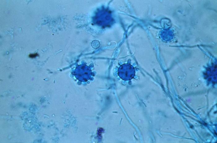
Histoplasma capsulatum Contributed by Dr. Libero Ajello, CDC (Public Domain
References
Gandhi P, Hebert B, Yun A, Bradley J, Moldoveanu B. Histoplasmosis around the world: A global perspective on the presentation, virulence factors, and treatment of histoplasmosis. The American journal of the medical sciences. 2024 Oct:368(4):287-299. doi: 10.1016/j.amjms.2024.06.011. Epub 2024 Jun 15 [PubMed PMID: 38885929]
Level 3 (low-level) evidenceKauffman CA. Histoplasmosis: a clinical and laboratory update. Clinical microbiology reviews. 2007 Jan:20(1):115-32 [PubMed PMID: 17223625]
BAUM GL, SCHWARZ J. The history of histoplasmosis, 1906 to 1956. The New England journal of medicine. 1957 Feb 7:256(6):253-8 [PubMed PMID: 13400245]
Beyhan S, Sil A. Sensing the heat and the host: Virulence determinants of Histoplasma capsulatum. Virulence. 2019 Dec:10(1):793-800. doi: 10.1080/21505594.2019.1663596. Epub [PubMed PMID: 31560240]
Souza DC, Rodrigues-Neto AA, Monnerat GMB, Sardou M, Hottz PL, Oliveira J, McBenedict B, Vilte RMCV, Fonseca SC, Martins EB. Acute pulmonary histoplasmosis: a case series from an outbreak in Southeastern Brazil. Revista do Instituto de Medicina Tropical de Sao Paulo. 2023:65():e45. doi: 10.1590/S1678-9946202365045. Epub 2023 Aug 18 [PubMed PMID: 37610941]
Level 2 (mid-level) evidenceBourque DL, Solomon DA, Sax PE. Health Considerations for HIV-Infected International Travelers. Current infectious disease reports. 2019 Apr 12:21(5):16. doi: 10.1007/s11908-019-0672-y. Epub 2019 Apr 12 [PubMed PMID: 30980287]
Miller R, Assi M, AST Infectious Diseases Community of Practice. Endemic fungal infections in solid organ transplant recipients-Guidelines from the American Society of Transplantation Infectious Diseases Community of Practice. Clinical transplantation. 2019 Sep:33(9):e13553. doi: 10.1111/ctr.13553. Epub 2019 Apr 23 [PubMed PMID: 30924967]
Rickerts V. [Climate change and systemic fungal infections]. Bundesgesundheitsblatt, Gesundheitsforschung, Gesundheitsschutz. 2019 May:62(5):646-651. doi: 10.1007/s00103-019-02931-z. Epub [PubMed PMID: 30923845]
Prakash K, Richman D. A case report of disseminated histoplasmosis and concurrent cryptococcal meningitis in a patient treated with ruxolitinib. BMC infectious diseases. 2019 Mar 27:19(1):287. doi: 10.1186/s12879-019-3922-6. Epub 2019 Mar 27 [PubMed PMID: 30917797]
Level 3 (low-level) evidenceMuniz MM, Sousa CN, Evangelista Oliveira MM, Pizzini CV, Almeida MA, Rodríguez-Arellanes G, Taylor ML, Zancopé-Oliveira RM. Sexual variability in Histoplasma capsulatum and its possible distribution: what is going on? Revista iberoamericana de micologia. 2014 Jan-Mar:31(1):7-10. doi: 10.1016/j.riam.2013.10.002. Epub 2013 Nov 18 [PubMed PMID: 24262630]
Teixeira Mde M, Patané JS, Taylor ML, Gómez BL, Theodoro RC, de Hoog S, Engelthaler DM, Zancopé-Oliveira RM, Felipe MS, Barker BM. Worldwide Phylogenetic Distributions and Population Dynamics of the Genus Histoplasma. PLoS neglected tropical diseases. 2016 Jun:10(6):e0004732. doi: 10.1371/journal.pntd.0004732. Epub 2016 Jun 1 [PubMed PMID: 27248851]
Eichenberger EM, Little JS, Baddley JW. Histoplasmosis. Infectious disease clinics of North America. 2025 Mar:39(1):145-161. doi: 10.1016/j.idc.2024.11.009. Epub 2024 Dec 18 [PubMed PMID: 39701897]
Brooks RA, Yan K, Head JR. Assessment of Prior Infection With Histoplasmosis Using Skin Test and Serological Surveys: A Global Systematic Review. Open forum infectious diseases. 2025 Aug:12(8):ofaf437. doi: 10.1093/ofid/ofaf437. Epub 2025 Jul 23 [PubMed PMID: 40765713]
Level 1 (high-level) evidenceGiusiano G, Gómez BL. Trends in the epidemiology of systemic endemic mycoses in Latin America. Medical mycology. 2025 Dec 1:63(12):. pii: myaf112. doi: 10.1093/mmy/myaf112. Epub [PubMed PMID: 41334817]
Ashraf N, Kubat RC, Poplin V, Adenis AA, Denning DW, Wright L, McCotter O, Schwartz IS, Jackson BR, Chiller T, Bahr NC. Re-drawing the Maps for Endemic Mycoses. Mycopathologia. 2020 Oct:185(5):843-865. doi: 10.1007/s11046-020-00431-2. Epub 2020 Feb 10 [PubMed PMID: 32040709]
Mittal J, Ponce MG, Gendlina I, Nosanchuk JD. Histoplasma Capsulatum: Mechanisms for Pathogenesis. Current topics in microbiology and immunology. 2019:422():157-191. doi: 10.1007/82_2018_114. Epub [PubMed PMID: 30043340]
Benedict K, Mody RK. Epidemiology of Histoplasmosis Outbreaks, United States, 1938-2013. Emerging infectious diseases. 2016 Mar:22(3):370-8. doi: 10.3201/eid2203.151117. Epub [PubMed PMID: 26890817]
de Perio MA, Benedict K, Williams SL, Niemeier-Walsh C, Green BJ, Coffey C, Di Giuseppe M, Toda M, Park JH, Bailey RL, Nett RJ. Occupational Histoplasmosis: Epidemiology and Prevention Measures. Journal of fungi (Basel, Switzerland). 2021 Jun 26:7(7):. doi: 10.3390/jof7070510. Epub 2021 Jun 26 [PubMed PMID: 34206791]
Barros N, Wheat JL, Hage C. Pulmonary Histoplasmosis: A Clinical Update. Journal of fungi (Basel, Switzerland). 2023 Feb 10:9(2):. doi: 10.3390/jof9020236. Epub 2023 Feb 10 [PubMed PMID: 36836350]
Baker J, Kosmidis C, Rozaliyani A, Wahyuningsih R, Denning DW. Chronic Pulmonary Histoplasmosis-A Scoping Literature Review. Open forum infectious diseases. 2020 May:7(5):ofaa119. doi: 10.1093/ofid/ofaa119. Epub 2020 Apr 6 [PubMed PMID: 32411810]
Level 2 (mid-level) evidenceSmith DJ, Free RJ, Thompson GR 3rd, Baddley JW, Pappas PG, Benedict K, Gold JAW, Endemic Mycoses Diagnostic Algorithm Subject Matter Expert Group, Tushla LA, Chiller T, Jackson BR, Toda M. Clinical Testing Guidance for Coccidioidomycosis, Histoplasmosis, and Blastomycosis in Patients With Community-Acquired Pneumonia for Primary and Urgent Care Providers. Clinical infectious diseases : an official publication of the Infectious Diseases Society of America. 2024 Jun 14:78(6):1559-1563. doi: 10.1093/cid/ciad619. Epub [PubMed PMID: 37802909]
Assi M, Lakkis IE, Wheat LJ. Cross-reactivity in the Histoplasma antigen enzyme immunoassay caused by sporotrichosis. Clinical and vaccine immunology : CVI. 2011 Oct:18(10):1781-2. doi: 10.1128/CVI.05017-11. Epub 2011 Aug 3 [PubMed PMID: 21813658]
Wheat J, Wheat H, Connolly P, Kleiman M, Supparatpinyo K, Nelson K, Bradsher R, Restrepo A. Cross-reactivity in Histoplasma capsulatum variety capsulatum antigen assays of urine samples from patients with endemic mycoses. Clinical infectious diseases : an official publication of the Infectious Diseases Society of America. 1997 Jun:24(6):1169-71 [PubMed PMID: 9195077]
Schmidt TE, Vieceli T, Damasceno LS, Kimuda S, Pasqualotto AC, Bahr NC. Evolving Epidemiology, Improving Diagnostic Tests and Their Importance for the Correct Diagnosis of Histoplasmosis. Journal of fungi (Basel, Switzerland). 2025 Mar 4:11(3):. doi: 10.3390/jof11030196. Epub 2025 Mar 4 [PubMed PMID: 40137234]
Hage CA, Ribes JA, Wengenack NL, Baddour LM, Assi M, McKinsey DS, Hammoud K, Alapat D, Babady NE, Parker M, Fuller D, Noor A, Davis TE, Rodgers M, Connolly PA, El Haddad B, Wheat LJ. A multicenter evaluation of tests for diagnosis of histoplasmosis. Clinical infectious diseases : an official publication of the Infectious Diseases Society of America. 2011 Sep:53(5):448-54. doi: 10.1093/cid/cir435. Epub 2011 Aug 2 [PubMed PMID: 21810734]
Wheat J, French ML, Kohler RB, Zimmerman SE, Smith WR, Norton JA, Eitzen HE, Smith CD, Slama TG. The diagnostic laboratory tests for histoplasmosis: analysis of experience in a large urban outbreak. Annals of internal medicine. 1982 Nov:97(5):680-5 [PubMed PMID: 6814316]
Wheat J. Histoplasmosis. Experience during outbreaks in Indianapolis and review of the literature. Medicine. 1997 Sep:76(5):339-54 [PubMed PMID: 9352737]
Guimarães AJ, Nosanchuk JD, Zancopé-Oliveira RM. DIAGNOSIS OF HISTOPLASMOSIS. Brazilian journal of microbiology : [publication of the Brazilian Society for Microbiology]. 2006 Jan:37(1):1-13 [PubMed PMID: 20445761]
Arnold SR, Spec A, Baddley JW, Pappas P, Lentz RJ, Wolf J, Kauffman CA, Ardura MI, Badreldin N, Bahr NC, Bloch K, Miller RA, Mocherla S, Saccente M, Schwartz I, Stover KR, Wiederhold NP, Loveless J. 2025 Clinical Practice Guideline Update by the Infectious Diseases Society of America on Histoplasmosis: Treatment of Asymptomatic Histoplasma Pulmonary Nodules (Histoplasmomas) and Mild or Moderate Acute Pulmonary Histoplasmosis in Adults, Children, and Pregnant People. Clinical infectious diseases : an official publication of the Infectious Diseases Society of America. 2026 Jan 16:81(Supplement_3):i27-i32. doi: 10.1093/cid/ciaf256. Epub [PubMed PMID: 40667709]
Level 1 (high-level) evidenceWheat LJ, Freifeld AG, Kleiman MB, Baddley JW, McKinsey DS, Loyd JE, Kauffman CA, Infectious Diseases Society of America. Clinical practice guidelines for the management of patients with histoplasmosis: 2007 update by the Infectious Diseases Society of America. Clinical infectious diseases : an official publication of the Infectious Diseases Society of America. 2007 Oct 1:45(7):807-25 [PubMed PMID: 17806045]
Level 1 (high-level) evidenceDao A, Kim HY, Halliday CL, Oladele R, Rickerts V, Govender MMed NP, Shin JH, Heim J, Ford NP, Nahrgang SA, Gigante V, Beardsley J, Sati H, Morrissey CO, Alffenaar JW, Alastruey-Izquierdo A. Histoplasmosis: A systematic review to inform the World Health Organization of a fungal priority pathogens list. Medical mycology. 2024 Jun 27:62(6):. doi: 10.1093/mmy/myae039. Epub [PubMed PMID: 38935903]
Level 1 (high-level) evidenceWheat LJ, Stein L, Corya BC, Wass JL, Norton JA, Grider K, Slama TG, French ML, Kohler RB. Pericarditis as a manifestation of histoplasmosis during two large urban outbreaks. Medicine. 1983 Mar:62(2):110-9 [PubMed PMID: 6827979]
Wheat LJ, Slama TG, Eitzen HE, Kohler RB, French ML, Biesecker JL. A large urban outbreak of histoplasmosis: clinical features. Annals of internal medicine. 1981 Mar:94(3):331-7 [PubMed PMID: 7224378]
Rosenthal J, Brandt KD, Wheat LJ, Slama TG. Rheumatologic manifestations of histoplasmosis in the recent Indianapolis epidemic. Arthritis and rheumatism. 1983 Sep:26(9):1065-70 [PubMed PMID: 6615561]