 Tools
Tools
 Hip Pointer Injuries
Introduction
A hip pointer is a contusion of the iliac crest or the greater trochanteric region of the femur following a direct impact or collision.[1] Management of these injuries requires coordinated care among athletic trainers, clinical providers, primary care, and sports medicine specialists. The majority of these injuries improve with standard, first-line nonoperative management modalities.
Etiology
Register For Free And Read The Full Article
 Search engine and full access to all medical articles
- 10 free questions in your specialty
- Free CME/CE Activities
- Free daily question in your email
- Save favorite articles to your dashboard
- Emails offering discounts
Learn more about a Subscription to StatPearls Point-of-Care
Etiology
The anterior iliac crest region and greater trochanter of the femur have, in most patients, minimal overlying adipose tissue. Thus, these areas are more susceptible to injury from direct trauma. The primary etiology is a direct blow to the iliac crest or greater trochanter during contact sports. American football and hockey are the most common sports that result in these injuries.[2] In American football, tackling and incidental collisions commonly cause direct trauma during competition. Rugby involves a similar underlying mechanism. In ice hockey, hip pointers can result from being checked into the boards or from contact with another player or the player’s equipment.[3] Iliac crest contusions may also result from noncontact sports or high- or low-energy trauma. In these settings, a direct fall or traumatic blow, such as that sustained during a motor vehicle collision, is often the precipitating factor.[4]
Epidemiology
Injuries to the hip and pelvis during sports participation are much less common than injuries involving the knee or ankle.[5][6][7] Hip injuries account for 5% to 10% of athletic injuries, and hip pointers represent an even smaller proportion of these injuries. Results from a review of National Football League injuries occurring from 1997 to 2006 showed an incidence of 0.3% and an average training absence of 5.6 days per injury.[8]
Pathophysiology
Following a direct impact to the iliac crest, a hematoma develops in the surrounding tissues and often includes varying degrees of bleeding into the hip abductor musculature. The iliac crest serves as the origin of several muscles that may be affected, depending on the severity and extent of the injury:
History and Physical
A detailed history, including current symptoms and the mechanism of injury, should be obtained. A hip pointer is usually caused by a direct blow or fall onto the iliac crest or greater trochanter. Patients most commonly present with varying degrees of ecchymosis and soft tissue contusion at the area of impact. Tenderness is typically localized directly over the injured region, and hip range of motion is often limited because of pain. Hip abductor strength is typically reduced, whereas hip flexor and extensor strength usually remains intact. Pain may also limit the apparent strength of the hip abductors and external rotators.
Evaluation
Radiographs are indicated when a fracture or myositis ossificans is suspected. Pelvic avulsion injuries are particularly important considerations in high school and college athletes because the apophyses of the ischial tuberosity and anterior superior iliac spine may not fuse until the third decade of life. Pediatric and adolescent athletes should be evaluated for the following potential pelvic avulsion injuries:
- Iliac crest avulsion injuries: Avulsion of the abdominal musculature
- Anterior superior iliac spine avulsion injuries: Avulsion of the sartorius or tensor fasciae latae
- Anterior inferior iliac spine avulsion injuries: Avulsion of the rectus femoris
- Ischial tuberosity avulsion injuries: Avulsion of the proximal hamstring muscles [5]
Radiographic findings are otherwise typically unremarkable in patients with a true, isolated iliac crest contusion or hip pointer injury. Computed tomography should be considered when pain persists or is more severe than expected based on examination findings. A bone scan can exclude stress fractures if initial symptoms do not resolve. Magnetic resonance imaging of an isolated iliac crest contusion may reveal a large hematoma but is otherwise typically unremarkable. A hip pointer must be distinguished from chronic exertional compartment syndrome, femoral neck fracture, avascular necrosis, hip fracture or dislocation, tendinitis, iliotibial band syndrome, osteitis pubis, sacroiliac joint injury, snapping hip syndrome, and slipped capital femoral epiphysis.
Treatment / Management
Initial treatment for a hip pointer injury includes ice, anti-inflammatory analgesics, compression, rest, and avoidance of weight-bearing, depending on the severity of the patient’s symptoms. As pain decreases, patients should begin range-of-motion and active resistance exercises. If a hematoma is present, aspiration can provide pain relief and may potentially reduce the risk of myositis ossificans. A local anesthetic, such as 1% to 2% lidocaine or 0.5% bupivacaine in a volume of 5 to 9 mL, may provide short-term pain relief. Local anesthetic injection is considered safe, although limited data support the practice. Similarly, although no studies support corticosteroid injections, clinicians in the sports medicine community commonly use them. If compartment syndrome is suspected, clinicians should measure compartment pressures and consult an orthopedic surgeon or sports medicine specialist. An orthopedic surgeon should also be consulted if the patient has an avulsion fracture or pain persists for longer than 2 weeks. Physical therapy is often prescribed.
Differential Diagnosis
The differential diagnosis for an iliac crest contusion or hip pointer includes the following conditions:
- Chronic exertional compartment syndrome
- Femoral neck fracture/hip fracture
- Avascular necrosis/osteonecrosis
- Hip dislocation or subluxation
- Hip tendinitis
- Iliotibial band syndrome
- Osteitis pubis
- Sacroiliac joint injury
- Snapping hip syndrome
- Slipped capital femoral epiphysis, usually in adolescents [11][12][13]
Prognosis
Most patients can gradually return to normal levels of activity within 2 to 3 weeks.
Complications
Complications include:
- Chronic pain or dysfunction
- Inability to return to prior athletic performance
- Missed diagnosis leading to persistent pain or disability
Deterrence and Patient Education
Patients with comorbid conditions, such as diabetes mellitus, congenital bone disorders, or anticoagulant use, should receive counseling about avoiding additional contact-related trauma and exercising greater caution during physical activity.
Pearls and Other Issues
Padding over the injury site may decrease the incidence of reinjury, limit pain, and allow an earlier return to activity. Patients with a hip pointer may return to activity once pain and swelling have resolved and normal function has returned. Because recurrent injury remains a risk, clinicians should advise patients to avoid activities that could cause additional trauma to the affected area.
Enhancing Healthcare Team Outcomes
The treatment of iliac crest contusions involves coordination among athletic trainers, primary care and emergency medicine clinicians, orthopedic specialists, and nurses with orthopedic expertise. Referral to a sports medicine or orthopedic specialist may be appropriate, although many primary care and emergency medicine clinicians can treat this relatively benign condition. Although a surgical procedure is rarely indicated, chronic pain and dysfunction can significantly reduce a patient’s quality of life and impair athletic performance. School and emergency department nurses commonly encounter this condition and contribute to patient and family education while facilitating referrals for further care. Effective interprofessional collaboration can improve patient care and clinical outcomes.
Media
(Click Image to Enlarge)
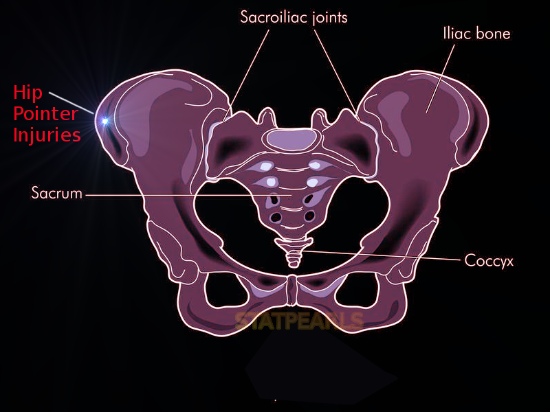
Hip Pointer Injuries.
Contributed by S Bhimji, MD
References
Hall M, Anderson J. Hip pointers. Clinics in sports medicine. 2013 Apr:32(2):325-30. doi: 10.1016/j.csm.2012.12.010. Epub 2013 Feb 8 [PubMed PMID: 23522513]
Frank RM, Slabaugh MA, Grumet RC, Bush-Joseph CA, Virkus WW, Nho SJ. Hip pain in active patients: what you may be missing. The Journal of family practice. 2012 Dec:61(12):736-44 [PubMed PMID: 23313991]
Level 3 (low-level) evidenceKuhn AW, Noonan BC, Kelly BT, Larson CM, Bedi A. The Hip in Ice Hockey: A Current Concepts Review. Arthroscopy : the journal of arthroscopic & related surgery : official publication of the Arthroscopy Association of North America and the International Arthroscopy Association. 2016 Sep:32(9):1928-38. doi: 10.1016/j.arthro.2016.04.029. Epub 2016 Jun 16 [PubMed PMID: 27318779]
Toney-Butler TJ, Varacallo MA. Motor Vehicle Collisions. StatPearls. 2026 Jan:(): [PubMed PMID: 28722984]
Bordoni B, Varacallo MA. Anatomy, Bony Pelvis and Lower Limb: Thigh Quadriceps Muscle. StatPearls. 2026 Jan:(): [PubMed PMID: 30020706]
Smith JM, Varacallo MA. Osgood-Schlatter Disease. StatPearls. 2026 Jan:(): [PubMed PMID: 28723024]
Raj MA, Mabrouk A, Varacallo MA. Posterior Cruciate Ligament Knee Injuries. StatPearls. 2026 Jan:(): [PubMed PMID: 28613477]
Feeley BT, Powell JW, Muller MS, Barnes RP, Warren RF, Kelly BT. Hip injuries and labral tears in the national football league. The American journal of sports medicine. 2008 Nov:36(11):2187-95. doi: 10.1177/0363546508319898. Epub 2008 Jul 18 [PubMed PMID: 18641370]
Level 2 (mid-level) evidenceWalters BB, Varacallo MA. Anatomy, Bony Pelvis and Lower Limb: Thigh Sartorius Muscle. StatPearls. 2026 Jan:(): [PubMed PMID: 30422484]
Attum B, Varacallo MA. Anatomy, Bony Pelvis and Lower Limb: Thigh Muscles. StatPearls. 2026 Jan:(): [PubMed PMID: 29494052]
Raj MA, Ampat G, Varacallo MA. Sacroiliac Joint Pain. StatPearls. 2026 Jan:(): [PubMed PMID: 29261980]
Buchanan BK, Varacallo MA. Sacroiliitis. StatPearls. 2026 Jan:(): [PubMed PMID: 28846269]
Musick SR, Varacallo MA. Snapping Hip Syndrome. StatPearls. 2026 Jan:(): [PubMed PMID: 28846235]