 Tools
Tools
 Herpangina
Introduction
Herpangina is an acute viral infection that primarily affects infants and young children and is most commonly caused by coxsackievirus A and other enteroviruses. Herpangina is characterized by the sudden onset of fever, sore throat, and small vesicular or ulcerative lesions in the posterior oropharynx, particularly the soft palate and tonsillar pillars. The disease occurs most frequently during the summer and early fall and spreads through fecal-oral and respiratory routes, especially in childcare and school settings. Although herpangina is usually self-limited, early recognition is important to ensure appropriate supportive treatment and to prevent unnecessary antibiotic use. Although herpangina is primarily a pediatric disease, rare cases have also been reported in neonates, adolescents, and adults.[1][2][3]
Etiology
Register For Free And Read The Full Article
 Search engine and full access to all medical articles
- 10 free questions in your specialty
- Free CME/CE Activities
- Free daily question in your email
- Save favorite articles to your dashboard
- Emails offering discounts
Learn more about a Subscription to StatPearls Point-of-Care
Etiology
Herpangina is primarily caused by viruses belonging to the genus Enterovirus within the family Picornaviridae. The most common causative agents are coxsackievirus A, particularly serotypes A1 to A10 and A16, although other enteroviruses such as coxsackievirus B and echovirus have also been implicated. An enterovirus strain, EV-A71, has been associated with neurologic complications.
These viruses are nonenveloped, single-stranded RNA viruses that replicate in the gastrointestinal tract and are transmitted primarily through fecal–oral and respiratory routes. Following entry into the host, viral replication occurs in the lymphoid tissues of the oropharynx and intestinal mucosa, leading to viremia and the development of the characteristic vesicular lesions in the posterior oropharynx.[4] Enterovirus B group serotypes, including B3, have also been implicated in epidemic clusters, demonstrating that multiple enterovirus lineages can produce the characteristic vesicular pharyngitis seen in herpangina.[5]
Epidemiology
Herpangina occurs worldwide and is most commonly reported among infants and young children, particularly children younger than 10. In the United States, enterovirus infections generally follow a seasonal pattern, with cases peaking during the summer and early fall. Transmission occurs through the fecal-oral route, respiratory droplets, and direct contact, facilitating outbreaks in childcare centers, schools, and other community settings. Results from surveillance studies conducted in the US and other countries identified several circulating serotypes, including coxsackievirus A serotypes 2, 4, 6, and 10, coxsackievirus B serotypes 2 and 4, and other nonpolio enteroviruses, with patterns varying by year and geographic region.[6] Recent surveillance data suggest that patterns of enterovirus circulation fluctuated after changes in public health measures during the COVID-19 pandemic.[7][8][9]
Pathophysiology
Herpangina is caused by nonpolio enteroviruses, primarily coxsackievirus A serotypes, although other enteroviruses, echoviruses, and parechoviruses may cause similar clinical findings. The viruses enter the host via the oral or respiratory route. Viral replication then occurs in the lymphoid tissue of the oropharynx and intestinal mucosa, leading to viremia and the subsequent formation of characteristic vesicular or ulcerative lesions in the posterior oropharynx. Infection also triggers a host immune response, resulting in systemic symptoms such as fever, malaise, and sore throat.[7][8][9] Because viremia contributes to the pathophysiology, more severe disease may occur, particularly in individuals with immunocompromised conditions.
The viruses causing herpangina likely attach to heparan sulfate glycosaminoglycans on host cell surfaces.[10] After attachment, the virus binds to functional cellular receptors, including the scavenger receptor B2 (SCARB2) or the coxsackie-adenovirus receptor (CAR), triggering viral membrane penetration and subsequent uncoating within the cell. These steps facilitate viral entry and intracellular replication. Herpangina has an incubation period of 4 to 14 days. After inoculation and local viral replication, viremia occurs, leading to involvement of secondary sites and the development of typical clinical features, including bilateral cervical lymphadenopathy and characteristic posterior pharyngeal lesions. The resulting clinical presentation reflects both local mucosal infection and systemic viral spread.
History and Physical
History
Children with herpangina usually present with a sudden onset of fever, sore throat, irritability, and decreased appetite. Preverbal children may push away a bottle containing an acidic liquid, such as orange juice, or refuse to drink altogether because of oral ulcerations. Verbal children may also report headache, malaise, or mild abdominal discomfort. The illness often develops rapidly over 1 to 2 days, and outbreaks commonly occur during the summer and early fall.
A recent history of exposure to other children with similar symptoms in childcare or school settings is common. Older children may report back pain and headache, and some patients may develop anorexia, dehydration, or abdominal pain. Depending on disease severity and organ system involvement, patients may also report headache, neck stiffness, confusion, seizures, muscle weakness, or difficulty breathing. Severe manifestations are rare. Adults generally have immunity from past infections but may occasionally present with more severe disease because of immunocompromised status or genetic susceptibility.[11][12]
Physical Examination
On physical examination, herpangina is generally straightforward to diagnose. The examiner will find small vesicles or shallow ulcers in the posterior oropharynx, including the soft palate, tonsillar pillars, and uvula, while the anterior oral cavity is usually spared. Cervical lymphadenopathy may be mild, and patients may appear irritable or lethargic because of fever. Other findings are generally minimal, and systemic involvement is rare, making the diagnosis largely clinical and based on the characteristic lesions and seasonal presentation.[11]
Even with the classic features of herpangina, the clinician should be aware that severe disease can occur in individuals without immunocompromised status. A persistently high fever accompanied by circulatory or neurologic abnormalities and elevated white blood cell counts, blood glucose levels, and lactate levels may signal the onset of more severe disease that can lead to pulmonary edema and death within 1 to 5 days. Severe disease is most often associated with enterovirus A71.[12]
The occurrence and characteristics of the rash may vary by viral subtype. Neurologic findings, such as neck stiffness or paralysis, may be present in patients with complications such as meningitis, acute flaccid paralysis, encephalitis, or encephalomyelitis. Dehydration is a common complication, with signs such as dry mouth and decreased skin turgor. The history and physical examination should also focus on excluding other serious febrile exanthems, including Kawasaki disease, Rocky Mountain spotted fever, eczema herpeticum, and toxic shock syndrome, which can present similarly but may evolve over time.
Evaluation
The diagnosis of herpangina is primarily clinical, based on the medical history and characteristic posterior oropharyngeal vesicular or ulcerative lesions; routine laboratory tests are often unnecessary in uncomplicated cases. Laboratory evaluation may include a complete blood count and basic metabolic panel to assess for dehydration or alternative diagnoses, but test results are usually normal. Confirmatory testing is reserved for complicated cases, outbreaks, or epidemiologic purposes; reverse transcription polymerase chain reaction testing for nonpolio enteroviruses using throat swabs, stool, cerebrospinal fluid, or vesicular fluid is the preferred method because of its high sensitivity and rapid turnaround time. Viral culture and paired serology, which demonstrate a 4-fold rise in antibody titers, are used less commonly. Imaging and additional studies (eg, lumbar puncture or central nervous system imaging) may be indicated if clinicians are concerned about complications such as aseptic meningitis, encephalitis, or acute flaccid paralysis, although these complications are rare.
Treatment / Management
Herpangina is typically a self-limited viral illness, and treatment is primarily supportive, focusing on symptom relief, hydration, and prevention of complications. Treatment can be broadly categorized into general care, symptomatic treatment, and antiviral therapy considerations.
General
Patients should have access to a well-ventilated, clean environment to reduce the risk of transmission. General supportive care includes maintaining adequate hydration and proper nutrition. Children should be encouraged to consume light, soft, or semiliquid foods to ensure adequate caloric intake, while avoiding hot, spicy, or irritating foods that may aggravate oral lesions.
Maintaining good oral hygiene is also important. Patients may rinse their mouths with normal saline after meals, while younger children who are unable to rinse may have their oral cavity gently wiped with saline-soaked gauze. If normal saline is unavailable, mildly salted water can be used as an alternative. Adequate oral fluid intake should be emphasized, particularly in children with fever or reduced oral intake. For those with significant feeding difficulties or at risk of dehydration, electrolyte-containing oral rehydration solutions are recommended. Patients should be monitored for signs of dehydration or worsening symptoms during the illness.[13](B3)
Symptomatic
Fever is one of the most common symptoms associated with herpangina. For children with temperatures of 38.5 °C (101.3 °F) or higher, antipyretic medications such as acetaminophen or ibuprofen may be administered according to age-appropriate dosing recommendations. Adequate hydration should be maintained while using these medications. Patients should avoid acidic or spicy foods and beverages until the lesions heal.
Nonpharmacologic measures such as cool compresses or tepid sponging may also provide additional comfort. Topical anesthetic preparations containing lidocaine or diphenhydramine are generally not recommended for the treatment of oral lesions in herpangina because of limited evidence of benefit and the potential risk of systemic toxicity in young children. In rare cases, younger children with high fever may develop febrile seizures, which require prompt treatment according to standard pediatric seizure protocols. Benzodiazepines such as intravenous midazolam may be used when clinically indicated.[13](B3)
Antiviral
Currently, no specific antiviral therapy is recommended for the routine treatment of herpangina. Supportive care remains the primary treatment approach. Results from some studies suggested that interferon alfa spray may provide benefit through local immunomodulatory and antiviral effects on mucosal immunity. However, its routine use remains limited, and the United States Food and Drug Administration has not approved this treatment for herpangina.[7][14](A1)
Differential Diagnosis
Herpangina may present with symptoms that overlap with those of several infectious and inflammatory pediatric conditions. Careful clinical evaluation, including the distribution of oral lesions, associated systemic findings, and rash characteristics, is important for distinguishing herpangina from other illnesses (see Image. Differential Diagnoses for Pediatric Oral Lesions). Diseases and conditions that should be considered in the differential diagnosis include:
- Eczema herpeticum
- Toxic shock syndrome
- Measles
- Varicella
- Kawasaki disease
- Insect bites or hypersensitivity reactions
- Rocky Mountain spotted fever
- Drug eruption or medication-related hypersensitivity reactions
- Erythema multiforme major
These conditions may present with fever, mucocutaneous lesions, or a rash resembling herpangina. However, distinguishing features, such as lesion distribution, systemic involvement, epidemiologic exposure, and laboratory findings, help establish the correct diagnosis. A large review of oral ulcerations lists more than 48 entities, including herpangina, and may serve as a useful diagnostic reference. In addition to infectious causes, noninfectious etiologies include rheumatologic, gastrointestinal, and autoimmune disorders, as well as medication and toxin ingestion.[15]
Prognosis
Herpangina is usually a mild illness that resolves spontaneously. With proper care, appropriate infection-control measures, good nutrition, and adequate hydration, most children recover within about 10 days. Treatment mainly focuses on relieving fever and oral pain, and young children with high fever should be monitored closely for febrile seizures. In rare cases, serious complications, such as acute flaccid paralysis, meningitis, encephalitis, or myocarditis, can occur, so patients should be monitored carefully for warning signs of severe disease.
Complications
Cases of herpangina generally resolve spontaneously without complications. However, clinicians should recognize the potential clinical overlap with other enterovirus-associated illnesses, including coxsackieviruses. Herpangina is usually mild, but some causative viruses, particularly enterovirus A71, can lead to serious complications. Complications may include brainstem encephalitis, acute flaccid paralysis, aseptic meningitis, and myocarditis. Patients who develop these complications can become critically ill and may require hospitalization, including admission to an intensive care unit.
Deterrence and Patient Education
Families should be informed that herpangina is usually a mild illness that resolves spontaneously. Because herpangina commonly affects children and may cause concern among parents, reassurance and clear guidance are important. Caregivers should be advised to keep the child at home, separated from others when possible, until symptoms improve, and to ensure adequate hydration and proper nutrition.
Good hygiene is essential to prevent the spread of infection. Frequent handwashing should be encouraged, especially after changing diapers, feeding the child, or handling items the child has used. Cleaning and disinfecting surfaces and objects the child frequently touches are also recommended. Although most children can be treated at home, caregivers should maintain communication with the clinician and monitor for worsening symptoms. Follow-up visits are usually unnecessary because the illness typically resolves quickly.
Enhancing Healthcare Team Outcomes
Herpangina is usually a mild viral infection, but rare cases may cause serious complications. Diagnosis is primarily clinical, with additional testing reserved for patients with atypical presentations, severe disease, or suspected complications. Effective treatment of herpangina relies on strong teamwork among healthcare professionals.
Clinicians, nurses, and pharmacists must communicate clearly about the patient’s condition, treatment plan, and warning signs of possible complications. Coordinating care ensures that families receive consistent guidance on symptom relief, hydration, nutrition, and infection prevention. Ethical practice, attention to patient safety, and shared responsibility can improve outcomes and reduce stress for patients and families. Regular updates and collaboration among the interprofessional team support coordinated, patient-centered care and improve team performance.
Media
(Click Image to Enlarge)
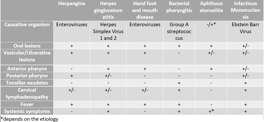
Differential Diagnoses for Pediatric Oral Lesions. This table outlines the causative agents and characteristic clinical features used to differentiate between common pediatric conditions presenting with oral lesions.
Contributed by R Ali, MD
References
Esposito S, Principi N. Hand, foot and mouth disease: current knowledge on clinical manifestations, epidemiology, aetiology and prevention. European journal of clinical microbiology & infectious diseases : official publication of the European Society of Clinical Microbiology. 2018 Mar:37(3):391-398. doi: 10.1007/s10096-018-3206-x. Epub 2018 Feb 6 [PubMed PMID: 29411190]
Freeman MC, Messacar K. Enterovirus and Parechovirus Neurologic Infections in Children: Clinical Presentations and Neuropathogenesis. Journal of the Pediatric Infectious Diseases Society. 2025 Jan 20:14(1):. pii: piae069. doi: 10.1093/jpids/piae069. Epub [PubMed PMID: 39776161]
Solomon T, Lewthwaite P, Perera D, Cardosa MJ, McMinn P, Ooi MH. Virology, epidemiology, pathogenesis, and control of enterovirus 71. The Lancet. Infectious diseases. 2010 Nov:10(11):778-90. doi: 10.1016/S1473-3099(10)70194-8. Epub 2010 Oct 18 [PubMed PMID: 20961813]
Xing B, Hu L, Hu X, Sun Y, Liang Z, Fu J, Liang Y, Chen Y, Li Y, Chen L, Wang L, Wang W, Lu Y, Wang S. Epidemiological, clinical, and molecular characteristics of coxsackievirus A2 infections during a herpangina outbreak in children on an Island in Eastern China in 2024: a surveillance-based study. Gut pathogens. 2025 Nov 4:17(1):85. doi: 10.1186/s13099-025-00762-9. Epub 2025 Nov 4 [PubMed PMID: 41189016]
Level 2 (mid-level) evidenceNakayama T, Urano T, Osano M, Hayashi Y, Sekine S, Ando T, Makinom S. Outbreak of herpangina associated with Coxsackievirus B3 infection. The Pediatric infectious disease journal. 1989 Aug:8(8):495-8 [PubMed PMID: 2549494]
Li W, Gao HH, Zhang Q, Liu YJ, Tao R, Cheng YP, Shu Q, Shang SQ. Large outbreak of herpangina in children caused by enterovirus in summer of 2015 in Hangzhou, China. Scientific reports. 2016 Oct 18:6():35388. doi: 10.1038/srep35388. Epub 2016 Oct 18 [PubMed PMID: 27752104]
Abzug MJ. The enteroviruses: problems in need of treatments. The Journal of infection. 2014 Jan:68 Suppl 1():S108-14. doi: 10.1016/j.jinf.2013.09.020. Epub 2013 Oct 8 [PubMed PMID: 24119825]
Pons-Salort M, Grassly NC. Serotype-specific immunity explains the incidence of diseases caused by human enteroviruses. Science (New York, N.Y.). 2018 Aug 24:361(6404):800-803. doi: 10.1126/science.aat6777. Epub [PubMed PMID: 30139872]
Chen J, You P, Li Y, Chen X, Zhang N, Ma C, Zhang Y, Lv T. Molecular epidemiology and genetic characteristics of enterovirus A identified in pediatric patients with hand, foot, and mouth disease and herpangina in Linyi, China, 2023-2024, following the relaxation of COVID-19 policies. BMC infectious diseases. 2026 Jan 29:26(1):. doi: 10.1186/s12879-026-12710-2. Epub 2026 Jan 29 [PubMed PMID: 41612251]
Zhang X, Shi J, Ye X, Ku Z, Zhang C, Liu Q, Huang Z. Coxsackievirus A16 utilizes cell surface heparan sulfate glycosaminoglycans as its attachment receptor. Emerging microbes & infections. 2017 Jul 26:6(7):e65. doi: 10.1038/emi.2017.55. Epub 2017 Jul 26 [PubMed PMID: 28745308]
Huang CY, Su SB, Chen KT. A review of enterovirus-associated hand-foot and mouth disease: preventive strategies and the need for a global enterovirus surveillance network. Pathogens and global health. 2024 Oct-Dec:118(7-8):538-548. doi: 10.1080/20477724.2024.2400424. Epub 2024 Sep 4 [PubMed PMID: 39229797]
Wang X, Liu H, Li Y, Su R, Liu Y, Qiao K. Relationship between polymorphism of receptor SCARB2 gene and clinical severity of enterovirus-71 associated hand-foot-mouth disease. Virology journal. 2021 Jun 30:18(1):132. doi: 10.1186/s12985-021-01605-0. Epub 2021 Jun 30 [PubMed PMID: 34193186]
Level 2 (mid-level) evidenceSawyer MH. Enterovirus infections: diagnosis and treatment. Current opinion in pediatrics. 2001 Feb:13(1):65-9 [PubMed PMID: 11176247]
Level 3 (low-level) evidenceYe YZ, Dou YL, Hao JH, Zhou L, Lin AW, Wang SN, Deng JK, Lei M, Luo RP, Liao YN, Chen Y, Long YY, Chen BQ, Yang Z, Gan L, Nong GM, Yan WL, Yu H. Efficacy and safety of interferon α-2b spray for herpangina in children: A randomized, controlled trial. International journal of infectious diseases : IJID : official publication of the International Society for Infectious Diseases. 2021 Jun:107():62-68. doi: 10.1016/j.ijid.2021.04.049. Epub 2021 Apr 17 [PubMed PMID: 33878461]
Level 1 (high-level) evidenceLégeret C, Furlano R. Oral ulcers in children- a clinical narrative overview. Italian journal of pediatrics. 2021 Jun 30:47(1):144. doi: 10.1186/s13052-021-01097-2. Epub 2021 Jun 30 [PubMed PMID: 34193212]
Level 3 (low-level) evidence