 Tools
Tools
 Hemarthrosis
Introduction
Hemarthrosis is defined as bleeding into the joint cavity, a condition that can occur following trauma, invasive procedures, or as a complication of bleeding disorders such as hemophilia. The knee is the most commonly affected joint, though hemarthrosis may involve the ankle, elbow, hip, or shoulder, depending on underlying pathology. Patients typically present with acute pain, swelling, warmth, and a restricted range of motion, reflecting the inflammatory response to intra-articular bleeding. If unrecognized or untreated, recurrent bleeding episodes can initiate a destructive cycle of synovial inflammation, fibrosis, and cartilage degradation, ultimately predisposing the joint to chronic arthropathy and early osteoarthritis.
Diagnosis begins with a thorough patient history and physical examination, supported by imaging modalities such as ultrasound, computed tomography, or magnetic resonance imaging to identify fluid collections and associated structural injury. However, arthrocentesis with synovial fluid analysis remains the gold standard for confirming hemarthrosis. Special attention should be given to identifying lipohemarthrosis, which indicates the presence of fat and blood within the joint cavity and is most often associated with intra-articular fracture, particularly of the knee. Understanding the natural history and progression of hemarthrosis, as well as recognizing characteristic patterns of presentation and spread, is essential for timely intervention and prevention of long-term disability.
Etiology
Register For Free And Read The Full Article
 Search engine and full access to all medical articles
- 10 free questions in your specialty
- Free CME/CE Activities
- Free daily question in your email
- Save favorite articles to your dashboard
- Emails offering discounts
Learn more about a Subscription to StatPearls Point-of-Care
Etiology
Hemarthrosis can occur in a variety of diseases and disorders, but it is most commonly classified into 3 categories: traumatic, nontraumatic, and postoperative. Traumatic injury is the most common cause of hemarthrosis. Nontraumatic hemarthrosis can be caused by a variety of bleeding disorders that are either hereditary or acquired. Hereditary bleeding disorders include hemophilia and other inherited coagulation factor deficiencies, such as factor XI deficiency.[1][2] Examples of acquired bleeding disorders include advanced liver or renal disease, vitamin K deficiency, disseminated intravascular coagulation, or anticoagulation medication use. Other, rarer causes of nontraumatic hemarthrosis include:
- Neurologic: Reflex sympathetic dystrophy, diabetic neuropathic arthropathy
- Infectious: Septic bacterial arthritis
- Vascular: Vitamin C deficiency, ruptured peripheral artery aneurysms, osteoarthritis (degenerative tears of peripheral arteries associated with a posterior horn of the lateral meniscus)
- Neoplasms: Benign synovial hemangiomas, pigmented villonodular synovitis, or any malignant tumor arising near a joint cavity or metastatic [3][4]
Postoperative recurrent hemarthrosis is frequently associated with total knee arthroplasty and has also been described as an uncommon complication following arthroscopy.[5]
Epidemiology
The exact prevalence of hemarthrosis among individuals with hemophilia is unknown; however, reports suggest that up to 50% experience hemarthrosis at some point in their lifetime. Joint trauma increases the chance of developing hemarthrosis in this population.[6] Based on a cohort study of 1145 consecutive patients who developed a traumatic hemarthrosis of the knee, approximately half had an anterior cruciate ligament (ACL) injury, and the annual incidence of ACL injury was 77 per 100,000 inhabitants, with a significant difference between men (91 per 100,000) and women (63 per 100,000). In those 16 years of age or younger, lateral patellar dislocation was the most frequent structural injury associated with traumatic hemarthrosis of the knee both in boys (39%) and girls (43%); in this age group, the annual incidence of lateral patellar dislocation was 88 per 100,000 and higher in boys (113 per 100,000) than girls (62 per 100,000).[7]
Pathophysiology
The cascade of inflammatory events that contribute to chronic osteoarthritic injury is complex.[8][9][10] An impaction or traumatic event can trigger a cyclical inflammatory response progressing to arthrosis.[8][9] This process involves damage to the extracellular matrix of the cartilage and irreversible chondrocyte necrosis. Matrix disruption leads to joint derangement, including collagen fibrillation, release of glycosaminoglycans (GAGs; sugar chains in connective tissue), and other pro-degenerative substances. Altogether, this cartilaginous detritus, this debris, interacts with chondrocytes, osteoblasts, macrophages, fibroblasts, and immune cells (eg, primarily T-cells).
Subsequently, there is a release of an inflammatory cytokine flurry of interleukin (IL)1, IL4, IL6, IL8, IL17, tumor necrosis factor (TNF)-α, cathepsins (involved with bone resorption), and complement factors. Also released are proangiogenic factors (eg, vascular endothelial growth factor) by CD69+ cells (macrophages/monocytes), thereby promoting synovial vascularization.[11] These compounds further assault the synovial cells and chondrocytes in the area, leading to further cartilage degradation. Synovitis is characterized by hyperplasia, vascularization, villi formation, and fibrin deposition, which, along with the presence of macrophages, releases transforming growth factor-β and other proteins, thereby setting the area up for rebleeding. The result is the formation of osteophytes.
Additionally, erythrocyte degradation from hemarthrosis contributes to joint pathology. With the dissolution of the joint blood, heme is released via macrophages and monocytes. The heme permeates the synovium, overwhelming its capacity to remove it and thus leaving the synovium laden with hemosiderin. This predisposes the cartilage to breakdown and causes synovial hyperplasia with neoangiogenic vessels that are frail and predisposed to bleeding at the site. There is increased joint effusion with capsular distension, thus, pain. Concurrent with the red cell/heme damage, monocytes and macrophages also create toxic matrix metalloproteases and hydroxy radicals. These substances are caustic to chondrocytes, causing their apoptosis. There is also a significant cytokine release. There is a cumulative and irreversible cartilaginous and articular damage due, in part, to a progressive and often irreversible impairment of proteoglycan synthesis. Contractures may occur, and arthrofibrosis is likely to occur.
Traumatic hemarthrosis typically occurs in the setting of intra-articular injury with ligamentous, osseous, and/or cartilage damage leading to a bloody synovial fluid collection.[12] Postoperative hemarthrosis from total knee arthroplasty has been attributed to the development of intra-capsular vascular tissue, which subsequently causes bleeding into the joint after joint replacement surgery. Lipohemarthrosis develops from marrow fat leakage into the synovial fluid, usually resulting from intracapsular fractures or extensive intra-articular soft tissue injury (see Images). Lateral Knee Showing Lipohemarthrosis, Radiograph; Lateral Knee Fracture With Lipohemarthrosis, Radiograph; and Knee Showing Nondisplaced Patellar Fracture With Lipohemarthrosis, Computed Tomography).[13]
Histopathology
The histopathology of hemophilic arthropathy can be divided into early and late disease characteristics. In early disease, there is synovial hypertrophy and fibrosis, which displays changes similar to rheumatoid arthritis. Progression of the disease shows disruption of cartilage and subchondral cyst formation from interosseous bleeding. The intra-articular bleeding has direct toxic effects, leading to the destruction of cartilage and bone. Iron in the form of ferric citrate stimulates human fibroblast cells to proliferate. Studies involving mice imply hemarthrosis leads to blood vessel hyperplasia, which may explain the predisposition for hemophiliacs to experience recurrent intra-articular bleeding.[14][15]
Toxicokinetics
Rodenticides are superwarfarins that, upon ingestion, can cause spontaneous bleeding in multiple sites, including acute hemarthrosis. Patients can be asymptomatic up to 72 hours after ingestion. Gastrointestinal symptoms can be present early in the course of poisoning, and spontaneous bleeding can present as early as 8 hours after exposure. Treatment involves the intensive administration of Vitamin K1.[16]
History and Physical
In most patients, particularly those without underlying neurologic sensory deficits or bleeding disorders, the cause of hemarthrosis is usually apparent from the history, as it often follows a clearly recognized traumatic event. Following an injury with intra-articular damage, the swelling from bloody synovial fluid accumulation is typically rapid within a few hours. Pain is a significant component; however, it may take time to develop, or may be minimal or absent in patients with preexisting impaired sensation. The range of motion of the affected joint is typically significantly reduced.
Certain locations and types of injury following a traumatic incident are more likely to develop a hemarthrosis. Intra-articular elbow fractures, including those of the radial head, are consistently associated with hemarthrosis. The knee is a frequent site of hemarthrosis, with the most common mechanism being forced twisting of the loaded joint. The majority of these posttraumatic hemarthroses are from ligamentous and meniscal damage. Anterior cruciate ligament (ACL) tears are the most common, representing 70% of posttraumatic hemarthrosis of the knee, followed by 10% to 15% patellar subluxation/dislocation injuries, 10% from meniscal tears, 2% to 5% from osteochondral fracture fragments, and 5% represented by other injuries, including posterior cruciate ligament or capsule tears. Depending on the site and kind of injury, tenderness to palpation of the joint lines or patella may be present, and clinical tests for joint laxity may be positive.[17]
Lipohemarthrosis indicates a fracture with intra-articular extension or significant intra-articular soft tissue injury, such as ligament or meniscus. If a lipohemarthrosis of the knee is identified, suspect in order of likelihood:
- Tibial plateau fracture: Lateral plateau fractures are often associated with ACL and the medial collateral ligament injuries.
- Tibial spine avulsion: The anterior attachment point of ACL, which is more common in young patients who have tougher ligaments than bones.
- Femoral condyle fracture: This is associated with popliteal artery and fibular nerve injury, although uncommon.
If hemarthrosis occurs spontaneously or after minimal trauma, bleeding disorders such as hemophilia should be suspected. Hemarthrosis is the most common musculoskeletal manifestation of hemophilia. Severe hemophilia (less than 1% of normal factor activity) causes hemarthrosis in 75% to 90% of patients, with the first attack typically occurring between the ages of 2 and 3. Any joint can be affected; however, the knee is the most common site. Patient history regarding family history, extensive or prolonged bleeding during or after surgery, use of anticoagulant medications, and diet habits should be elicited to help determine whether the presence of a bleeding diathesis exists.[18]
Clinical presentation of hemophilia with hemarthrosis can vary by age. In adults and older children, a prodromal stiffness or tingling typically precedes pain and swelling. In infants, signs of hemarthrosis may include irritability and decreased use of the affected limb. Bleeding into the hip joint is concerning due to the greater risk of increased intra-articular pressure and osteonecrosis of the femoral head. In hemophilic hemarthrosis, one joint is typically affected at a time; however, bilateral joint involvement is possible.[18][19]
Evaluation
A suspected hemarthrosis can be evaluated by plain radiographs, computed tomography scan, or MRI. The preferred initial imaging modality is film radiology of the involved joint and surrounding bones. Further evaluation with a computed tomography scan is warranted if a fracture is not seen but strongly suspected. MRI may be able to diagnose ligamentous or other soft tissue injuries as the cause of hemarthrosis. Ultrasound in the right clinical setting can help identify and characterize intra-articular fluid collections.[20]
Arthrocentesis and joint aspirate fluid analysis may be required for a definitive diagnosis. The synovial fluid from a hemarthrosis may appear red, pink, or brown, depending on the cause of bleeding. Joint aspiration and subsequent analysis can differentiate simple effusion, hemarthrosis, lipohemarthrosis, and septic arthritis. Arthrocentesis can also significantly relieve pain. The presence of lipid globules in the aspirate represents a lipohemarthrosis.
If the initial synovial fluid aspirate is nonbloody but bright red blood soon appears after withdrawing fluid, this indicates a traumatic hemarthrosis. A true hemarthrosis typically does not clot due to fibrinolysis, compared to a bloody aspirate from traumatic aspiration that does coagulate. Centrifugation of the synovial fluid aspirate and examination of the supernatant may also help discern the difference between true hemarthrosis and traumatic aspiration. A true hemarthrosis will display a serous appearance of the synovial fluid supernatant with xanthochromia.[21]
Lipohemarthrosis can be detected using arthrocentesis, plain radiographs, computed tomography scans, MRI, and, in a minority of patients, ultrasound. A linear interface between 2 different densities with an effusion suggests lipohemarthrosis. This feature results from the entry of marrow fat into the joint cavity and is highly suggestive of an intra-articular fracture. One series demonstrated that 65% of patients with intra-articular fractures did not have fat-fluid levels on plain radiographs; however, all patients with lipohemarthrosis visible on plain radiographs were found to have intra-articular fractures. On plain radiographs, a single fluid-fluid level, representing layering of blood and fat, can suggest lipohemarthrosis, although it may occur even without visible fat globules. A double fluid-fluid level, showing layering of fat, blood, and serum, is specific for lipohemarthrosis and can be observed on ultrasound, computed tomography, or MRI.
The sonographic finding of lipohemarthrosis may be used as a sensitive surrogate marker for intra-articular fracture. When used alone for the evaluation of acute knee trauma, plain radiographs can miss up to 21% of fractures. Suppose a lipohemarthrosis is identified on either plain film or ultrasound. In that case, there should be a high suspicion for an occult fracture, which should prompt further evaluation with a computed tomography scan.[22][23]
Treatment / Management
Treatment of hemarthrosis involves general measures applicable to any acute episode and interventions targeting the underlying cause. Initial management of acute hemarthrosis includes immobilization, ice, and compression. Analgesics may be used for pain control, particularly to facilitate diagnostic imaging. Arthrocentesis serves a dual role, providing diagnostic information and therapeutic relief by reducing joint pressure from the effusion.[24]
Clinically significant hemarthrosis is rare in patients receiving therapeutic anticoagulation, and these patients typically tolerate arthrocentesis. In the uncommon cases of persistent or recurrent hemarthrosis, treatment options may include clotting factor repletion with fresh frozen plasma or reversal of anticoagulation using the appropriate pharmacologic agent. For recurrent postoperative hemarthrosis, revision angioplasty may be necessary, while ruptured aneurysms may require vascular surgical intervention or arterial embolization. Recurrent hemarthrosis often originates from periarticular hypertrophy of a vascular synovium.[25][26][27] Angioplasty with embolization is considered when conservative measures—joint aspiration, immobilization, and cessation of anticoagulants—fail. Synovectomy may also be an option.(B2)
Recurrence after embolization can exceed 25%, often due to vessel collateralization or recanalization; however, even if embolization is unsuccessful, it can help map or define the surgical field for subsequent intervention.[28] Compared with surgery, angioplasty with embolization is generally less invasive. For benign tumors, arthroscopic or open synovectomy remains the treatment of choice to prevent recurrent hemarthrosis.(B3)
Special treatment considerations are required for hemophiliacs presenting with acute hemarthrosis. Coagulation factor replacement should be administered promptly at the first sign of joint bleeding—including the prodromal phase of stiffness or tingling before pain and swelling—ideally within 2 hours of bleed identification. For bleeding into the hip, target joints, or trauma-associated hemarthrosis, higher factor activity levels of 80% to 100% are recommended. The recommended dose is 50 units/kg of factor VIII for hemophilia A and 100 to 120 units/kg of factor IX for hemophilia B. For hemarthrosis in peripheral joints, such as knees, elbows, or ankles, factor activity should be raised to at least 40% to 50% (25 units/kg of factor VIII for hemophilia A and 50–60 units/kg of factor IX for hemophilia B).
Emicizumab, a monoclonal antibody, is a newer therapeutic alternative for hemophilia A.[29][30][31][32] This bridges factor IXa and factor X, mimicking factor VIII activity, and is effective as prophylaxis and in patients with factor VIII inhibitors. Medications with antiplatelet activity, such as nonsteroidal anti-inflammatory drugs, should be avoided. Arthrocentesis is generally unnecessary when diagnosing joint bleeding in patients with known hemophilia. If required to rule out septic arthritis or relieve pressure from accumulated blood, it should only be performed after factor replacement has raised specific factor levels.[33] In hemophiliacs with target joints experiencing repeated hemorrhage, a short course of glucocorticoids may help reduce pain and swelling from synovial inflammation, provided there are no signs of infection. Surgical intervention may be indicated for chronic synovitis or arthropathy, including synovectomy for recurrent bleeding or joint replacement for severe joint damage.[34][35](A1)
Differential Diagnosis
The differential diagnosis of an acute joint effusion includes other notable disease processes. Conditions which can be mistaken for an acute hemarthrosis include:
- Septic arthritis
- Lyme disease
- Tuberculosis
- Gout (monosodium urate crystals)
- Pseudogout (calcium pyrophosphate crystals)
- Rheumatoid arthritis
- Systemic lupus erythematosus
- Leukemia
- Seronegative spondyloarthropathies (psoriatic arthritis, reactive arthritis, inflammatory bowel disease arthritis)
- Osteoarthritis
This may be especially true if the association with trauma is remote, unclear, or unknown. Arthrocentesis with synovial fluid analysis, diagnostic imaging, and further laboratory workup specific to these other disease processes may be required to differentiate between the causes of an acute joint effusion.
Prognosis
With appropriate treatment, including arthrocentesis when indicated, the acute swelling and pain of hemarthrosis can improve substantially within days, although complete resolution may take several weeks. In cases caused by intra-articular fractures or significant soft tissue injury, orthopedic surgery may be necessary. After the hemarthrosis resolves, a structured rehabilitation program aimed at restoring range of motion, weight-bearing capacity, and joint strength is recommended. For patients with hemophilia, a physical therapist experienced in hemophilia management is ideal.[19]
Complications
Severe or recurrent hemarthrosis can destroy intra-articular cartilage and lead to degenerative arthritis. The toxic effects of blood within the joint lead to synovial hypertrophy and fibrosis. In hemophiliacs, joints that undergo chronic inflammatory changes from repeated hemarthrosis are referred to as ‘target joints.’ Recurrent or prolonged bleeding can cause chronic, disabling arthropathy due to internal joint derangement and impaired movement. Chronic arthropathy occurs in approximately 20% of hemophiliacs, but recurrent hemarthrosis can be prevented with prophylactic administration of coagulation factors.[13][19]
Consultations
In the setting of an acute hemarthrosis, if there is evidence or high suspicion of an intra-articular fracture or soft tissue injury (ligamentous, capsular, or meniscal tear), orthopedic consultation should be considered. Although not all intra-articular fractures or soft tissue injuries require operative intervention, the patient's case, including radiographic findings, should be discussed with an orthopedic surgeon.
Enhancing Healthcare Team Outcomes
The lifelong management of hemophilia, including treating and preventing hemarthrosis and disabling arthropathy, takes an interprofessional approach. The World Federation of Hemophilia has published guidelines for managing hemophilia based on an extensive literature review. The interprofessional approach encourages treatment contributions from all areas of medicine, including physical therapists, hematologists, physiologists, and orthopedic surgeons. Physical therapy promotes physical fitness and normal neuromuscular development with specific attention to muscle strengthening, coordination, physical functioning, healthy body weight, and self-esteem.[36] For patients with significant musculoskeletal dysfunction, weight-bearing activities promote the development and maintenance of bone density.[37][38]
The primary aim is to prevent and treat bleeding with the deficient clotting factor. The specific factor deficiency should be treated with a specific factor concentrate. Acute bleeds should be treated immediately, ideally within 2 hours. Arthrocentesis should be performed when the hemarthrosis is tense, painful, and shows no improvement after 24 hours of conservative treatment or when there is a neurovascular compromise of the limb from hemarthrosis or evidence of infection.[39][40][41]
Synovectomy should be performed if chronic synovitis persists along with frequent recurrent bleeding that is not controlled by other measures. Options for synovectomy include chemical, radioisotopic, arthroscopic, or open surgical synovectomy.[42] Nonsurgical synovectomy is the preferred approach. Radioisotopic synovectomy using a pure beta emitter such as phosphorus-32 or yttrium-90 is highly effective, has minimal side effects, and can be performed outpatient.[43][44] Pain should be controlled with appropriate analgesics such as COX-2 inhibitors.[45][46] Surgical intervention may be considered if conservative measures fail to relieve pain and improve joint function. A patient with hemophilia who requires surgery should be managed at a comprehensive hemophilia treatment center, including an anesthesiologist who has experience treating patients with bleeding disorders.[47][48]
Media
(Click Image to Enlarge)
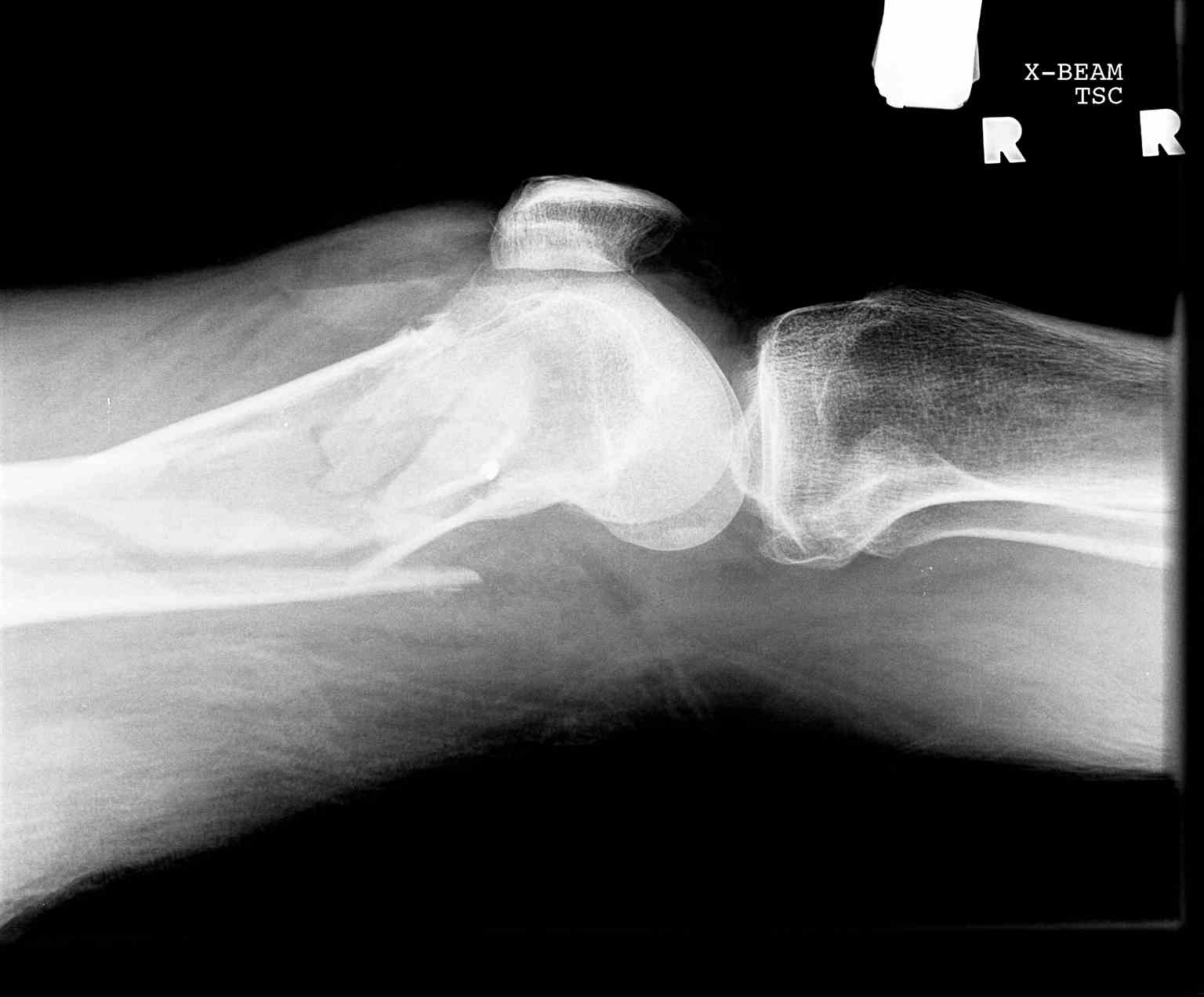
Lateral Knee Fracture With Lipohemarthrosis, Radiograph. A lateral knee fracture with lipohemarthrosis indicates the presence of an intraarticular fracture, where fat and blood from bone marrow enter the joint space, producing a characteristic fat–fluid level on imaging that confirms communication between the fracture and the joint capsule.
Contributed by S Dulebohn, MD
(Click Image to Enlarge)
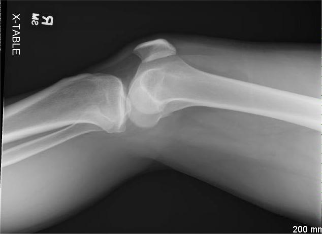
Lateral Knee Showing Lipohemarthrosis, Radiograph. This image is a horizontal cross-table lateral view plain radiograph of the knee, showing a fat-fluid level in the suprapatellar recess, compatible with lipohemarthrosis.
Contributed by A Cardenas, MD
(Click Image to Enlarge)
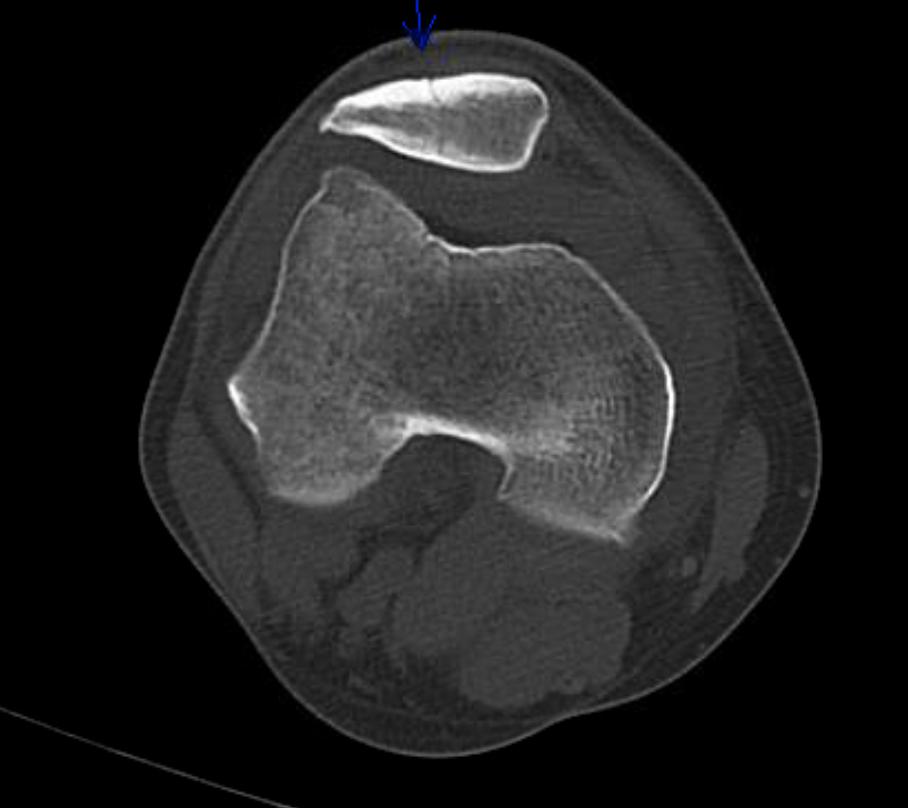
Knee Showing Nondisplaced Patellar Fracture With Lipohemarthrosis, Computed Tomography. This image is a computed tomography scan revealing an incomplete, nondisplaced fracture in the anterior cortex of the patella, along with lipohemarthrosis.
Contributed by Al Cardenas, MD
References
Souabni L, Meddeb N, Ajlani H, Ben Romdhane N, Sellami S. Hemarthrosis revealing congenital factor XI deficiency. Joint bone spine. 2008 May:75(3):348-9. doi: 10.1016/j.jbspin.2007.06.013. Epub 2008 Feb 22 [PubMed PMID: 18378181]
Levy BJ, Sperling K, Davila J, Levy IM. A Novel Case of Recurrent Hemarthrosis Following Knee Arthroscopy in a Patient with Undiagnosed Hemophilia. Arthroscopy, sports medicine, and rehabilitation. 2020 Jun:2(3):e289-e294. doi: 10.1016/j.asmr.2020.03.001. Epub 2020 May 14 [PubMed PMID: 32548594]
Level 3 (low-level) evidenceBaker CL. Acute hemarthrosis of the knee. Journal of the Medical Association of Georgia. 1992 Jun:81(6):301-5 [PubMed PMID: 1607844]
Kawamura H, Ogata K, Miura H, Arizono T, Sugioka Y. Spontaneous hemarthrosis of the knee in the elderly: etiology and treatment. Arthroscopy : the journal of arthroscopic & related surgery : official publication of the Arthroscopy Association of North America and the International Arthroscopy Association. 1994 Apr:10(2):171-5 [PubMed PMID: 8003144]
Level 3 (low-level) evidenceWorland RL, Jessup DE. Recurrent hemarthrosis after total knee arthroplasty. The Journal of arthroplasty. 1996 Dec:11(8):977-8 [PubMed PMID: 8986579]
Manners PJ, Price P, Buurman D, Lewin B, Smith B, Cole CH. Joint Aspiration for Acute Hemarthrosis in Children Receiving Factor VIII Prophylaxis for Severe Hemophilia: 11-year Safety Data. The Journal of rheumatology. 2015 May:42(5):885-90. doi: 10.3899/jrheum.141236. Epub 2015 Mar 1 [PubMed PMID: 25729030]
Olsson O, Isacsson A, Englund M, Frobell RB. Epidemiology of intra- and peri-articular structural injuries in traumatic knee joint hemarthrosis - data from 1145 consecutive knees with subacute MRI. Osteoarthritis and cartilage. 2016 Nov:24(11):1890-1897. doi: 10.1016/j.joca.2016.06.006. Epub 2016 Jun 29 [PubMed PMID: 27374877]
Potpally N, Rodeo S, So P, Mautner K, Baria M, Malanga GA. A Review of Current Management of Knee Hemarthrosis in the Non-Hemophilic Population. Cartilage. 2021 Dec:13(1_suppl):116S-121S. doi: 10.1177/1947603520942937. Epub 2020 Jul 22 [PubMed PMID: 32698601]
Evers BJ, Van Den Bosch MHJ, Blom AB, van der Kraan PM, Koëter S, Thurlings RM. Post-traumatic knee osteoarthritis; the role of inflammation and hemarthrosis on disease progression. Frontiers in medicine. 2022:9():973870. doi: 10.3389/fmed.2022.973870. Epub 2022 Aug 22 [PubMed PMID: 36072956]
Melchiorre D, Manetti M, Matucci-Cerinic M. Pathophysiology of Hemophilic Arthropathy. Journal of clinical medicine. 2017 Jun 25:6(7):. doi: 10.3390/jcm6070063. Epub 2017 Jun 25 [PubMed PMID: 28672826]
Lyons LP, Weinberg JB, Wittstein JR, McNulty AL. Blood in the joint: effects of hemarthrosis on meniscus health and repair techniques. Osteoarthritis and cartilage. 2021 Apr:29(4):471-479. doi: 10.1016/j.joca.2020.11.008. Epub 2020 Dec 8 [PubMed PMID: 33307179]
Knobe K, Berntorp E. Haemophilia and joint disease: pathophysiology, evaluation, and management. Journal of comorbidity. 2011:1():51-59 [PubMed PMID: 29090136]
Roosendaal G, Vianen ME, Marx JJ, van den Berg HM, Lafeber FP, Bijlsma JW. Blood-induced joint damage: a human in vitro study. Arthritis and rheumatism. 1999 May:42(5):1025-32 [PubMed PMID: 10323460]
Wen FQ, Jabbar AA, Chen YX, Kazarian T, Patel DA, Valentino LA. c-myc proto-oncogene expression in hemophilic synovitis: in vitro studies of the effects of iron and ceramide. Blood. 2002 Aug 1:100(3):912-6 [PubMed PMID: 12130502]
Rodriguez-Merchan EC. The destructive capabilities of the synovium in the haemophilic joint. Haemophilia : the official journal of the World Federation of Hemophilia. 1998 Jul:4(4):506-10 [PubMed PMID: 9873783]
Feinstein DL, Akpa BS, Ayee MA, Boullerne AI, Braun D, Brodsky SV, Gidalevitz D, Hauck Z, Kalinin S, Kowal K, Kuzmenko I, Lis K, Marangoni N, Martynowycz MW, Rubinstein I, van Breemen R, Ware K, Weinberg G. The emerging threat of superwarfarins: history, detection, mechanisms, and countermeasures. Annals of the New York Academy of Sciences. 2016 Jun:1374(1):111-22. doi: 10.1111/nyas.13085. Epub 2016 May 31 [PubMed PMID: 27244102]
Maffulli N, Binfield PM, King JB, Good CJ. Acute haemarthrosis of the knee in athletes. A prospective study of 106 cases. The Journal of bone and joint surgery. British volume. 1993 Nov:75(6):945-9 [PubMed PMID: 8245089]
Level 3 (low-level) evidenceLan HH, Eustace SJ, Dorfman D. Hemophilic arthropathy. Radiologic clinics of North America. 1996 Mar:34(2):446-50 [PubMed PMID: 8633126]
Level 3 (low-level) evidenceSoucie JM, Cianfrini C, Janco RL, Kulkarni R, Hambleton J, Evatt B, Forsyth A, Geraghty S, Hoots K, Abshire T, Curtis R, Forsberg A, Huszti H, Wagner M, White GC 2nd. Joint range-of-motion limitations among young males with hemophilia: prevalence and risk factors. Blood. 2004 Apr 1:103(7):2467-73 [PubMed PMID: 14615381]
Level 2 (mid-level) evidenceAponte EM, Novik JI. Identification of lipohemarthrosis with point-of-care emergency ultrasonography: case report and brief literature review. The Journal of emergency medicine. 2013 Feb:44(2):453-6. doi: 10.1016/j.jemermed.2012.07.062. Epub 2012 Sep 13 [PubMed PMID: 22981316]
Level 3 (low-level) evidenceDougados M. Synovial fluid cell analysis. Bailliere's clinical rheumatology. 1996 Aug:10(3):519-34 [PubMed PMID: 8876957]
Ryu KN, Jaovisidha S, De Maeseneer M, Jacobson J, Sartoris DJ, Resnick D. Evolving stages of lipohemarthrosis of the knee. Sequential magnetic resonance imaging findings in cadavers with clinical correlation. Investigative radiology. 1997 Jan:32(1):7-11 [PubMed PMID: 9007642]
Lugo-Olivieri CH, Scott WW Jr, Zerhouni EA. Fluid-fluid levels in injured knees: do they always represent lipohemarthrosis? Radiology. 1996 Feb:198(2):499-502 [PubMed PMID: 8596856]
Level 2 (mid-level) evidenceAllum R. The management of acute traumatic haemarthrosis of the knee. British journal of hospital medicine. 1997 Aug 20-Sep 2:58(4):138-41 [PubMed PMID: 9373401]
Weidner ZD, Hamilton WG, Smirniotopoulos J, Bagla S. Recurrent Hemarthrosis Following Knee Arthroplasty Treated with Arterial Embolization. The Journal of arthroplasty. 2015 Nov:30(11):2004-7. doi: 10.1016/j.arth.2015.05.028. Epub 2015 May 23 [PubMed PMID: 26092252]
Pow R, Fritsch B, Waugh R, Rogan C. Endovascular management of recurrent hemarthrosis of the knee: a case series. CVIR endovascular. 2020 Aug 30:3(1):43. doi: 10.1186/s42155-020-00135-0. Epub 2020 Aug 30 [PubMed PMID: 32864716]
Level 2 (mid-level) evidenceKolber MK, Shukla PA, Kumar A, Zybulewski A, Markowitz T, Silberzweig JE. Endovascular Management of Recurrent Spontaneous Hemarthrosis After Arthroplasty. Cardiovascular and interventional radiology. 2017 Feb:40(2):216-222. doi: 10.1007/s00270-016-1511-2. Epub 2016 Nov 23 [PubMed PMID: 27882429]
Geng X, Li Y, He X, Tian H. Traditional investigation and management for recurrent hemarthrosis after total knee arthroplasty: A case report. World journal of clinical cases. 2020 May 26:8(10):1966-1972. doi: 10.12998/wjcc.v8.i10.1966. Epub [PubMed PMID: 32518788]
Level 3 (low-level) evidenceBlair HA. Emicizumab: A Review in Haemophilia A. Drugs. 2019 Oct:79(15):1697-1707. doi: 10.1007/s40265-019-01200-2. Epub [PubMed PMID: 31542880]
Shima M, Suzuki N, Nishikii H, Amano K, Ogawa Y, Kobayashi R, Ozaki R, Yoneyama K, Mizuno N, Sakaida E, Saito M, Okamura T, Ito T, Hattori N, Higasa S, Seki Y, Nogami K. Final Analysis Results from the AGEHA Study: Emicizumab Prophylaxis for Acquired Hemophilia A with or without Immunosuppressive Therapy. Thrombosis and haemostasis. 2025 May:125(5):449-459. doi: 10.1055/a-2384-3585. Epub 2024 Aug 12 [PubMed PMID: 39134043]
Alcedo Andrade PE, Mannucci PM, Kessler CM. Emicizumab: the hemophilia A game-changer. Haematologica. 2024 May 1:109(5):1334-1347. doi: 10.3324/haematol.2022.282099. Epub 2024 May 1 [PubMed PMID: 37916312]
Gualtierotti R, Solimeno LP, Peyvandi F. Hemophilic arthropathy: Current knowledge and future perspectives. Journal of thrombosis and haemostasis : JTH. 2021 Sep:19(9):2112-2121. doi: 10.1111/jth.15444. Epub 2021 Jul 27 [PubMed PMID: 34197690]
Level 3 (low-level) evidenceDe la Corte-Rodriguez H, Rodriguez-Merchan EC, Alvarez-Roman MT, Martin-Salces M, Romero-Garrido JA, Jimenez-Yuste V. Accelerating recovery from acute hemarthrosis in patients with hemophilia: the role of joint aspiration. Blood coagulation & fibrinolysis : an international journal in haemostasis and thrombosis. 2019 Apr:30(3):111-119. doi: 10.1097/MBC.0000000000000803. Epub [PubMed PMID: 30958454]
Simpson ML, Valentino LA. Management of joint bleeding in hemophilia. Expert review of hematology. 2012 Aug:5(4):459-68 [PubMed PMID: 22992238]
Kisker CT, Burke C. Double-blind studies on the use of steroids in the treatment of acute hemarthrosis in patients with hemophilia. The New England journal of medicine. 1970 Mar 19:282(12):639-42 [PubMed PMID: 4907066]
Level 1 (high-level) evidenceGomis M, Querol F, Gallach JE, González LM, Aznar JA. Exercise and sport in the treatment of haemophilic patients: a systematic review. Haemophilia : the official journal of the World Federation of Hemophilia. 2009 Jan:15(1):43-54. doi: 10.1111/j.1365-2516.2008.01867.x. Epub 2008 Aug 21 [PubMed PMID: 18721151]
Level 1 (high-level) evidenceIorio A, Fabbriciani G, Marcucci M, Brozzetti M, Filipponi P. Bone mineral density in haemophilia patients. A meta-analysis. Thrombosis and haemostasis. 2010 Mar:103(3):596-603. doi: 10.1160/TH09-09-0629. Epub 2010 Jan 13 [PubMed PMID: 20076854]
Level 2 (mid-level) evidenceBlamey G, Forsyth A, Zourikian N, Short L, Jankovic N, De Kleijn P, Flannery T. Comprehensive elements of a physiotherapy exercise programme in haemophilia--a global perspective. Haemophilia : the official journal of the World Federation of Hemophilia. 2010 Jul:16 Suppl 5():136-45. doi: 10.1111/j.1365-2516.2010.02312.x. Epub [PubMed PMID: 20590873]
Level 3 (low-level) evidencevan Velzen AS, Eckhardt CL, Streefkerk N, Peters M, Hart DP, Hamulyak K, Klamroth R, Meijer K, Nijziel M, Schinco P, Yee TT, van der Bom JG, Fijnvandraat K, INSIGHT study group. The incidence and treatment of bleeding episodes in non-severe haemophilia A patients with inhibitors. Thrombosis and haemostasis. 2016 Mar:115(3):543-50. doi: 10.1160/TH15-03-0212. Epub 2015 Nov 19 [PubMed PMID: 26582077]
Rodriguez-Merchan EC. Peripheral nerve injuries in haemophilia. Blood transfusion = Trasfusione del sangue. 2014 Jan:12 Suppl 1(Suppl 1):s313-8. doi: 10.2450/2012.0111-12. Epub 2012 Dec 5 [PubMed PMID: 23245720]
Ingram GI, Mathews JA, Bennett AE. Controlled trial of joint aspiration in acute haemophilic haemarthrosis. Annals of the rheumatic diseases. 1972 Sep:31(5):423 [PubMed PMID: 5071639]
Bernal-Lagunas R, Aguilera-Soriano JL, Berges-Garcia A, Luna-Pizarro D, Perez-Hernandez E. Haemophilic arthropathy: the usefulness of intra-articular oxytetracycline (synoviorthesis) in the treatment of chronic synovitis in children. Haemophilia : the official journal of the World Federation of Hemophilia. 2011 Mar:17(2):296-9. doi: 10.1111/j.1365-2516.2010.02402.x. Epub 2010 Nov 11 [PubMed PMID: 21070486]
Thomas S, Gabriel MB, Assi PE, Barboza M, Perri ML, Land MG, Da Costa ES, Brazilian Hemophilia Centers. Radioactive synovectomy with Yttrium⁹⁰ citrate in haemophilic synovitis: Brazilian experience. Haemophilia : the official journal of the World Federation of Hemophilia. 2011 Jan:17(1):e211-6. doi: 10.1111/j.1365-2516.2010.02379.x. Epub 2010 Aug 22 [PubMed PMID: 20731723]
van Kasteren ME, Nováková IR, Boerbooms AM, Lemmens JA. Long term follow up of radiosynovectomy with yttrium-90 silicate in haemophilic haemarthrosis. Annals of the rheumatic diseases. 1993 Jul:52(7):548-50 [PubMed PMID: 8346985]
Rattray B, Nugent DJ, Young G. Celecoxib in the treatment of haemophilic synovitis, target joints, and pain in adults and children with haemophilia. Haemophilia : the official journal of the World Federation of Hemophilia. 2006 Sep:12(5):514-7 [PubMed PMID: 16919082]
Level 2 (mid-level) evidenceTsoukas C, Eyster ME, Shingo S, Mukhopadhyay S, Giallella KM, Curtis SP, Reicin AS, Melian A. Evaluation of the efficacy and safety of etoricoxib in the treatment of hemophilic arthropathy. Blood. 2006 Mar 1:107(5):1785-90 [PubMed PMID: 16291600]
Level 1 (high-level) evidenceSchild FJ, Mauser-Bunschoten EP, Verbout AJ, Van Rinsum AC, Roosendaal G. Total knee arthroplasty in hemophilic arthropathy: efficiency of clotting factor usage in multijoint procedures. Journal of thrombosis and haemostasis : JTH. 2009 Oct:7(10):1741-3. doi: 10.1111/j.1538-7836.2009.03569.x. Epub 2009 Aug 11 [PubMed PMID: 19682237]
Level 3 (low-level) evidenceStoffman J, Andersson NG, Branchford B, Batt K, D'Oiron R, Escuriola Ettingshausen C, Hart DP, Jiménez Yuste V, Kavakli K, Mancuso ME, Nogami K, Ramírez C, Wu R. Common themes and challenges in hemophilia care: a multinational perspective. Hematology (Amsterdam, Netherlands). 2019 Dec:24(1):39-48. doi: 10.1080/10245332.2018.1505225. Epub 2018 Aug 3 [PubMed PMID: 30073913]
Level 3 (low-level) evidence