 Tools
Tools
 Hairy Leukoplakia
Introduction
Hairy leukoplakia was first described in 1984 and is a mucosal disease.[1] It is associated with Epstein-Barr virus (EBV), also known as human herpesvirus 4. It occurs most commonly in people infected with HIV, although it can also be seen in people who do not have HIV. Such patients usually have had an organ or bone marrow transplant or some immunocompromised disease or hematological malignancy.[2] However, it has also been reported in individuals who are not immunocompromised.[3] Such immunocompetent patients have been shown to be on inhaled, topical, or systemic corticosteroids for the long term, which occurs as a risk factor for developing the condition.[4] The Centre for Disease Control and Prevention has classified this condition as a Category-B clinical marker of HIV disease since it has a clear prognostic value in the subsequent development of AIDS.[5]
Etiology
Register For Free And Read The Full Article
 Search engine and full access to all medical articles
- 10 free questions in your specialty
- Free CME/CE Activities
- Free daily question in your email
- Save favorite articles to your dashboard
- Emails offering discounts
Learn more about a Subscription to StatPearls Point-of-Care
Etiology
Hairy leukoplakia is commonly found in association with HIV infection and/or immunosuppression. It has been observed that the risk of developing hairy leukoplakia increases by almost 2-fold for every 300-unit decrease in CD4 count. It has also been seen in patients with other forms of severe immunodeficiencies, such as those on chemotherapy or who have had an organ transplant or leukemia. It has rarely been seen in immunocompetent patients. Moreover, hairy leukoplakia has also been seen associated with Behcet syndrome and ulcerative colitis. In HIV-positive men, smoking more than a pack of cigarettes/day has been correlated positively with the development of hairy leukoplakia.
Epidemiology
Hairy leukoplakia is commonly found in HIV-positive homosexual men, particularly those who smoke. Studies have shown that hairy leukoplakia can occur in up to 50% of patients who are not under any treatment for HIV, especially in those patients in whom the CD4 count has reached less than 0.3 × 10/L. No racial or age predilection has been observed for hairy leukoplakia.
Pathophysiology
The pathophysiology of this condition is complex, involving an interplay of factors such as EBV coinfection, productive replication, virulence, and genetic evolution, as well as the expression of specific EBV genes in a latent state. All these factors, enhanced by local and systemic deficiencies in the host immune system, lead to hairy leucoplakia.[6] The virus initially infects the basal cells of the pharyngeal epithelium. Here, it enters a replicative state, releasing the infectious virus into the saliva. This process continues throughout the infected person's lifespan. In the pharynx, the virus may also enter B cells, where it remains indefinitely in a latent state. Although eliminating EBV from the body is not possible with cytotoxic T lymphocytes, they are still required to maintain the infection in its latent state. The latent virus in B lymphocytes, upon reactivation, produces virions. The release of these virions infects circulating monocytes, which then migrate to the lamina propria of the oral mucosa and subsequently differentiate into macrophages and Langerhans cell precursors. These then migrate into the oral epithelium, where they lie mainly in the basal and /or the parabasal layer. Upon reactivation of the virus, it migrates to the keratinocytes in the spinosum and granulosum layer via the dendritic processes of the Langerhans cells. Upon reaching these layers, the virus starts replicating.
Moreover, a marked reduction or absence of Langerhans cells (antigen-presenting immune cells necessary for the immune system's response to viral infection) has also been found in hairy leukoplakia biopsy specimens.[7][8] Their deficiency may allow EBV to replicate persistently and escape immune recognition. Since the lateral border of the tongue is more susceptible to mechanical trauma, access to EBV to the prickle cell receptors is easy. Impaired immune status of the host, along with a marked reduction of Langerhans cells. then contribute to the successful establishment of EBV in the oral epithelium.[7]
Histopathology
The histopathological picture of the condition shows 5 major features:
- Hyperkeratosis of the superficial layer of the epithelium: This is due to an alteration in the pattern of expression of keratin in the squamous cells of the epithelium. This hyperkeratotic, thick layer may separate from the underlying cells, resulting in projections that produce the lesion's typical "hairy" appearance. This hyper-keratinized epithelium may get superficially infected with bacteria and/or Candida.
- Hyperparakeratosis of the superficial layer of the epithelium: The abnormal persistence of nuclei of the cell in this layer of the epithelium represents incomplete squamous differentiation. This layer may show numerous Candida hyphae.
- Acanthosis of the upper stratum spinosum/prickle cell layer: This abnormal expansion of cells occurs with layers of koilocyte-like cells or ballooned cells. The nuclei may show a ground-glass appearance throughout and may contain eosinophilic intranuclear inclusions with a halo (Cowdry type A).
- Absence of inflammation in the epithelium and minimal to zero inflammation in the lamina propria, and even the absence of inflammatory mononuclear cells infiltrate.
- Normal basal cells of the epithelium histologically: The gene BZLF1 is required to bring about the intracellular transition from the latent state to the productive infectious state. In hairy leukoplakia, the BZLF1 gene is restricted to the cells of the stratum spinosum and stratum granulosum. The cells of these layers express the gene Blimp1, which acts as a transcription factor during the terminal differentiation of keratinocytes. Neither Blimp1 nor BZLF1 can be detected in the basal cell layer of the oral epithelium, due to which it is assumed that the basal cell layer is not involved in hairy leukoplakia.[9][10]
Although the above features indicate hairy leukoplakia, none of the above histologic features is typical and found only in this lesion. Therefore, the definitive diagnosis of hairy leukoplakia requires histologic evidence, along with the demonstration of Epstein-Barr virus DNA, RNA, or protein within epithelial cells.
History and Physical
History
Hairy leukoplakia is often asymptomatic, so many patients are unaware of its presence. They may report a non-tender whitish plaque along the lateral border of the tongue. The lesion may appear and disappear spontaneously. Some patients may experience mild pain, dysesthesia, altered sensitivity to food temperature, altered taste sensation due to changes in taste buds, and the psychological impact of its unsightly cosmetic appearance.
Physical Examination
Intraorally, unilateral or bilateral non-painful white lesions are seen mostly on the lateral margin of the tongue.[11] The lesion may vary in appearance from a smooth, flat, and small lesion to an irregular "hairy" or "feathery" lesion with prominent folds or projections. It may occur as either a continuous or discontinuous lesion along the lateral border of the tongue and is often not symmetrical bilaterally. The lesions may vary in size, severity, and surface characteristics.[10] The lesion is adherent to the surface and cannot be removed by scraping. The surrounding tissue shows no signs of erythema or edema. Hairy leukoplakia may also involve other surfaces of the tongue, the buccal mucosa, and/or the gingiva. Here, it may appear flat and smooth, lacking the lesion's typical "hairy" appearance.
Evaluation
Generally, the diagnosis of hairy leukoplakia is established on a clinical basis. Still, the definitive diagnosis requires histological examination and demonstration of Epstein-Barr virus DNA, RNA, or protein within epithelial cells. For this purpose, several kits are commercially available.[12] Various techniques can be employed to detect EBV, such as polymerase chain reaction, electron microscopy, immunohistochemistry, and in situ hybridization. Among these techniques, in situ hybridization is considered the gold standard. A tissue biopsy can be done, but it is indicated only if the lesion shows an unusual appearance or is ulcerated, suggestive of cancer.[13]
Treatment / Management
As hairy leukoplakia is a benign condition with a low morbidity rate and a tendency to resolve spontaneously, every case does not need to be specifically treated. Treatment is provided to relieve symptoms caused by the condition or to treat the condition for aesthetic reasons. Treatment options include anti-retroviral drugs. Highly active antiretroviral therapy drugs usually reduce hairy leukoplakia, but the condition may recur when the drug dosage is reduced.[14]
Systemic antiviral therapy usually resolves the condition within 7 to 14 days of treatment. Oral antiviral medication, such as acyclovir, should be administered at a high dose of about 4000 mg per day, divided, for at least 7 days to achieve the required therapeutic levels.[15] Other drugs, such as valacyclovir and famciclovir, can also be used. These are new antiviral drugs with relatively higher oral bioavailability than acyclovir and require a relatively lower dosage. They are used at 3000 mg and 1500 mg per day, divided. The mechanism of action of these drugs is to inhibit the replication of productive EBV, but they do not eradicate the latent infection. But the disadvantage is that the condition often recurs a few weeks after the cessation of these drugs.
Topical treatment can be done using podophyllin resin. This resin solution is used at a concentration of 25%, which usually resolves the condition after a few applications.[16] Podophyllin has cytotoxic effects on cells, but its exact mechanism of action in resolving the condition is not yet known. But, as with other drugs, this treatment also has chances of recurrence after cessation. Also, the treatment may temporarily cause pain and discomfort at the application sites and dysgeusia.[17] Retinoic acid can also be used for topical treatment. Tretinoin (eg, 0.1% vitamin A) is generally applied 2 or 3 times daily until the patches have disappeared. Retinoic acids inhibit viral replication. However, the condition recurs several weeks after successful treatment with retinoic acid. Cryotherapy (liquid nitrogen) has also been reported to resolve hairy leukoplakia, but it is not a widely used treatment modality.[18](A1)
Differential Diagnosis
The differential diagnoses of hairy leukoplakia include:
- Candidiasis
- Oral leukoplakia
- White sponge nevus
- Oral frictional keratosis
- Smoker keratosis
- Proliferative verrucous leukoplakia
- Lichen planus
- Lichenoid reactions
- Condyloma acuminatum [19]
Prognosis
Most patients presenting with hairy leukoplakia tend to have significant immune suppression. The condition occurs relatively soon after seroconversion of HIV, ie, before AIDS. The CD4 count, on average, at the time of first detection may range from 235 to 468/microL. Studies have found that patients with AIDS in association with hairy leukoplakia have a shorter lifespan than those who do not show signs of this lesion. Moreover, if these patients get co-infected with the hepatitis B virus, their condition may worsen, resulting in early progression to AIDS.
Complications
Hairy leukoplakia may be complicated by an occasional candidal superinfection, resulting in glossopyrosis (burning tongue). The altered taste sensation is another, but rare complication. Topical retinoids, when used for prolonged use, may cause a burning sensation.
Deterrence and Patient Education
The presence of oral lesions significantly impacts the patient's health-related quality of life due to altered speech, difficulty with mastication and deglutition, and/or pain. This might exacerbate nutrition-related problems that further compromise the patient's health. Thus, patients should be provided with the proper knowledge of HIV-associated oral lesions and oral health. Prevention of hairy leukoplakia starts with having a healthy immune system. Patients should be educated to follow the prescribed HIV treatment plan and dental hygiene routine. Also, steps should be taken to maintain a healthy lifestyle. As people with HIV who have the habit of smoking are also at a greater risk of getting it in comparison to those who do not smoke, the patient should be educated in this regard.
Enhancing Healthcare Team Outcomes
The treatment outcome, in general, can be enhanced if the dentists have thorough knowledge about the treatment of oral conditions related to HIV infection, such as hairy leukoplakia. Based on the patient’s history, physical, and laboratory findings, the dentist and his interprofessional health care team should formulate a plan of adequate follow-up care. The role of health care professionals goes beyond basic education and training. It extends to providing patients with appropriate knowledge and information about oral lesions, along with developing reinforcing educational programs, as early diagnosis and management of these lesions play a vital role in reducing the impact of the disease on patients. Specialty-trained nurses in AIDS and otolaryngology educate patients, arrange for follow-up, and facilitate communication among the team. Pharmacists consult with clinicians to determine the appropriate agent(s) and dosing, review prescriptions, check for interactions, and inform patients. These interprofessional interventions improve patient outcomes.
Media
(Click Image to Enlarge)
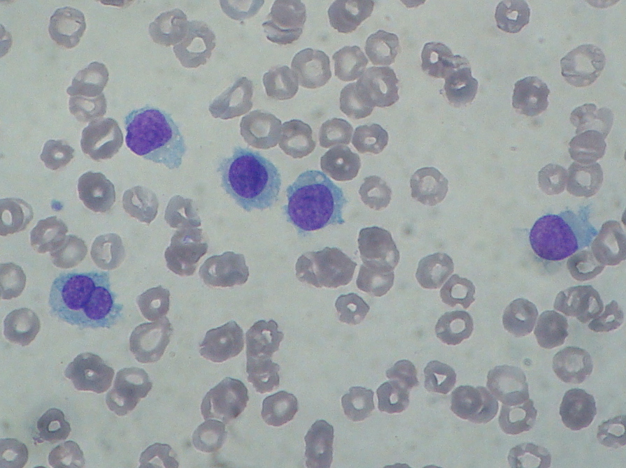
Hairy cell leukemia: abnormal B cells look "hairy" under a microscope because of radial projections from their surface. This picture is a contribution of Paulo Henrique Orlandi Mourao and is licensed to be shared under the Creative Commons Attribution-Share Alike 3.0 Unported license (https://creativecommons.org/licenses/by-sa/3.0/deed.en). The picture is obtained from the following link (https://commons.wikimedia.org/wiki/File:Hairy_cell_leukemia_smear_2009-08-20.JPG)
(Click Image to Enlarge)
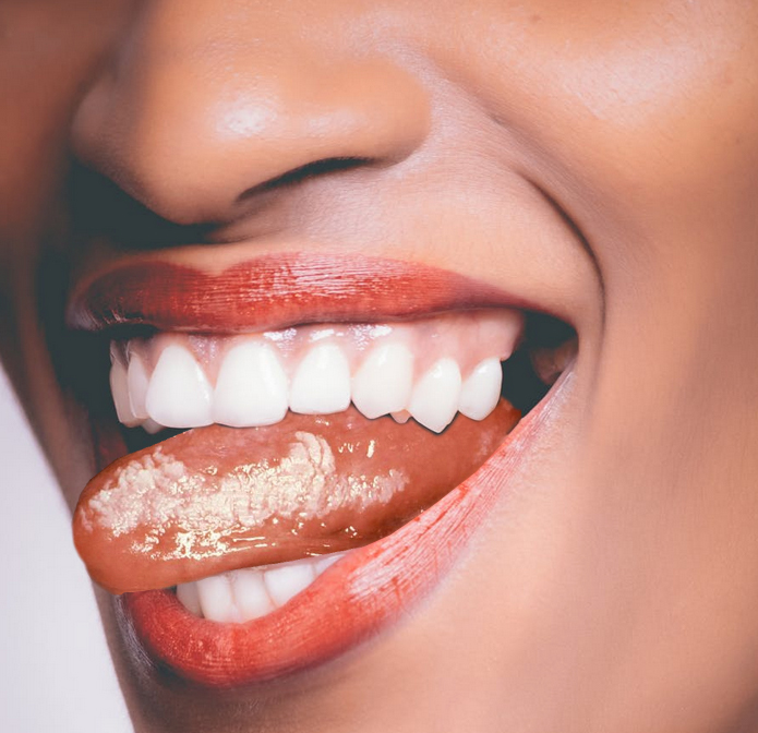
Hairy Cell Leukoplakia.
Contributed by K Humphreys
References
Kreuter A, Wieland U. Oral hairy leukoplakia: a clinical indicator of immunosuppression. CMAJ : Canadian Medical Association journal = journal de l'Association medicale canadienne. 2011 May 17:183(8):932. doi: 10.1503/cmaj.100841. Epub 2011 Mar 7 [PubMed PMID: 21398239]
Level 3 (low-level) evidenceKhammissa RA, Fourie J, Chandran R, Lemmer J, Feller L. Epstein-Barr Virus and Its Association with Oral Hairy Leukoplakia: A Short Review. International journal of dentistry. 2016:2016():4941783. doi: 10.1155/2016/4941783. Epub 2016 Mar 7 [PubMed PMID: 27047546]
Darling MR, Alkhasawneh M, Mascarenhas W, Chirila A, Copete M. Oral Hairy Leukoplakia in Patients With No Evidence of Immunosuppression: A Case Series and Review of the Literature. Journal (Canadian Dental Association). 2018 May:84():i4 [PubMed PMID: 31199724]
Level 2 (mid-level) evidencePiperi E, Omlie J, Koutlas IG, Pambuccian S. Oral hairy leukoplakia in HIV-negative patients: report of 10 cases. International journal of surgical pathology. 2010 Jun:18(3):177-83. doi: 10.1177/1066896908327865. Epub 2008 Nov 25 [PubMed PMID: 19033322]
Level 3 (low-level) evidence. 1993 revised classification system for HIV infection and expanded surveillance case definition for AIDS among adolescents and adults. MMWR. Recommendations and reports : Morbidity and mortality weekly report. Recommendations and reports. 1992 Dec 18:41(RR-17):1-19 [PubMed PMID: 1361652]
Level 3 (low-level) evidenceSachithanandham J, Kannangai R, Pulimood SA, Desai A, Abraham AM, Abraham OC, Ravi V, Samuel P, Sridharan G. Significance of Epstein-Barr virus (HHV-4) and CMV (HHV-5) infection among subtype-C human immunodeficiency virus-infected individuals. Indian journal of medical microbiology. 2014 Jul-Sep:32(3):261-9. doi: 10.4103/0255-0857.136558. Epub [PubMed PMID: 25008818]
Daniels TE, Greenspan D, Greenspan JS, Lennette E, Schiødt M, Petersen V, de Souza Y. Absence of Langerhans cells in oral hairy leukoplakia, an AIDS-associated lesion. The Journal of investigative dermatology. 1987 Aug:89(2):178-82 [PubMed PMID: 3110300]
Gondak RO, Alves DB, Silva LF, Mauad T, Vargas PA. Depletion of Langerhans cells in the tongue from patients with advanced-stage acquired immune deficiency syndrome: relation to opportunistic infections. Histopathology. 2012 Feb:60(3):497-503. doi: 10.1111/j.1365-2559.2011.04068.x. Epub 2011 Dec 14 [PubMed PMID: 22168427]
Dias EP, Spyrides KS, Silva Júnior AS, Rocha ML, da Fonseca EC. [Oral hairy leukoplakia: histopathologic features of subclinical stage]. Pesquisa odontologica brasileira = Brazilian oral research. 2001 Apr-Jun:15(2):104-11 [PubMed PMID: 11705191]
Level 2 (mid-level) evidenceSoutham JC, Felix DH, Wray D, Cubie HA. Hairy leukoplakia--a histological study. Histopathology. 1991 Jul:19(1):63-7 [PubMed PMID: 1655610]
Jones KB, Jordan R. White lesions in the oral cavity: clinical presentation, diagnosis, and treatment. Seminars in cutaneous medicine and surgery. 2015 Dec:34(4):161-70. doi: 10.12788/j.sder.2015.0180. Epub [PubMed PMID: 26650693]
De Souza YG, Freese UK, Greenspan D, Greenspan JS. Diagnosis of Epstein-Barr virus infection in hairy leukoplakia by using nucleic acid hybridization and noninvasive techniques. Journal of clinical microbiology. 1990 Dec:28(12):2775-8 [PubMed PMID: 2177752]
Martins LL, Rosseto JHF, Andrade NS, Franco JB, Braz-Silva PH, Ortega KL. Diagnosis of Oral Hairy Leukoplakia: The Importance of EBV In Situ Hybridization. International journal of dentistry. 2017:2017():3457479. doi: 10.1155/2017/3457479. Epub 2017 Jul 17 [PubMed PMID: 28798771]
Greenspan JS, Greenspan D, Webster-Cyriaque J. Hairy leukoplakia; lessons learned: 30-plus years. Oral diseases. 2016 Apr:22 Suppl 1():120-7. doi: 10.1111/odi.12393. Epub [PubMed PMID: 27109280]
Resnick L, Herbst JS, Ablashi DV, Atherton S, Frank B, Rosen L, Horwitz SN. Regression of oral hairy leukoplakia after orally administered acyclovir therapy. JAMA. 1988 Jan 15:259(3):384-8 [PubMed PMID: 2826830]
Gowdey G, Lee RK, Carpenter WM. Treatment of HIV-related hairy leukoplakia with podophyllum resin 25% solution. Oral surgery, oral medicine, oral pathology, oral radiology, and endodontics. 1995 Jan:79(1):64-7 [PubMed PMID: 7614164]
Level 1 (high-level) evidenceBrasileiro CB, Abreu MH, Mesquita RA. Critical review of topical management of oral hairy leukoplakia. World journal of clinical cases. 2014 Jul 16:2(7):253-6. doi: 10.12998/wjcc.v2.i7.253. Epub [PubMed PMID: 25032199]
Level 3 (low-level) evidenceGoh BT, Lau RK. Treatment of AIDS-associated oral hairy leukoplakia with cryotherapy. International journal of STD & AIDS. 1994 Jan-Feb:5(1):60-2 [PubMed PMID: 8142532]
Level 3 (low-level) evidenceTriantos D, Porter SR, Scully C, Teo CG. Oral hairy leukoplakia: clinicopathologic features, pathogenesis, diagnosis, and clinical significance. Clinical infectious diseases : an official publication of the Infectious Diseases Society of America. 1997 Dec:25(6):1392-6 [PubMed PMID: 9431384]